18 Myelodysplastic syndromes
MDSs are heterogeneous and have a multitude of expressions; however, two morphologic findings are common to all types of MDS: the presence of progressive cytopenias in spite of a cellular bone marrow and dyspoiesis in one or more cell lines. Subtypes of the 2008 World Health Organization classification of MDSs are listed in Box 18-1.
BOX 18-1 World Health Organization Classification of Myelodysplastic Syndromes (2008)
Refractory cytopenia with unilineage dysplasia
Refractory anemia with ringed sideroblasts
Refractory cytopenia with multilineage dysplasia
Refractory anemia with excess blasts
Myelodysplastic syndrome with isolated del(5q)
From Swerdlow SH, Campo E, Harris NL, et al, editors: WHO classification of tumours of haematopoietic and lymphoid tissues, ed 4, Lyon, France, 2008, IARC Press.
Dyserythropoiesis
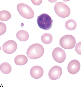
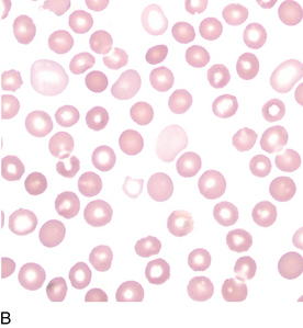
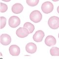
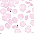
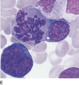
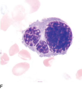
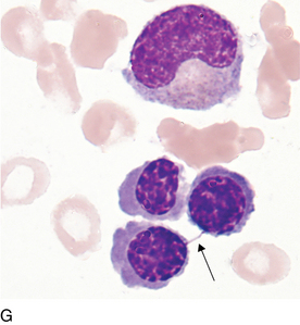
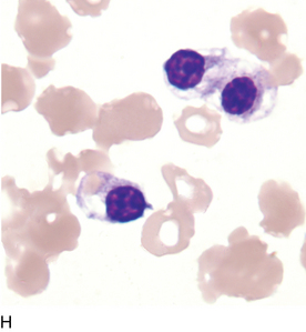
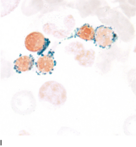
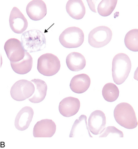
Evidence of dyserythropoiesis (Figure 18-1, A-I) may include any or all of the following: oval macrocytes, hypochromic microcytes, dimorphic erythrocyte population, erythrocyte precursors with more than one nucleus, abnormal nuclear shapes, nuclear bridging, uneven cytoplasmic staining, and/or ringed sideroblasts.
Dysmyelopoiesis

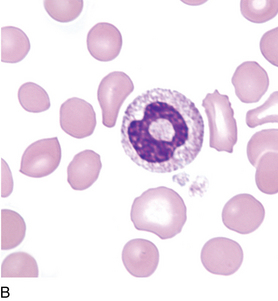
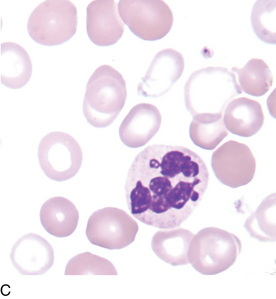
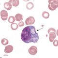
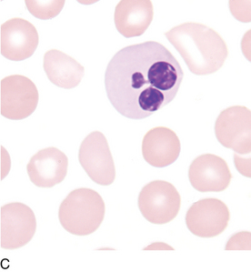
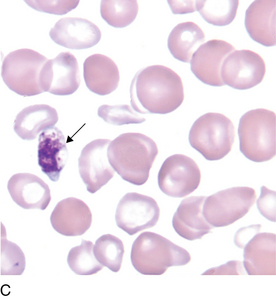
FIGURE 18–2C Abnormal nuclear shapes, neutrophil with hypersegmented nucleus; also exhibits hypogranulation.
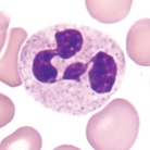
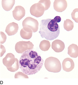
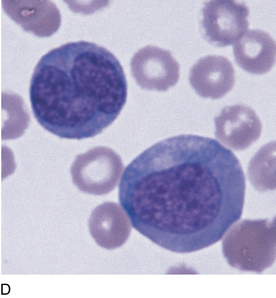
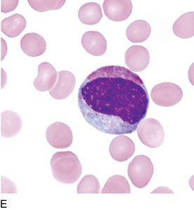
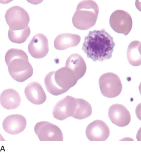
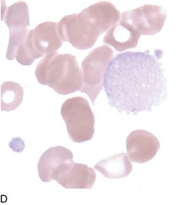
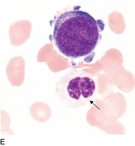
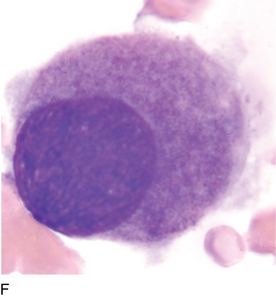
Evidence of dysmyelopoiesis (Figure 18-2, A-E) may include any or all of the following: abnormal granulation, abnormal nuclear shapes, persistent basophilic cytoplasm, and/or uneven cytoplasmic staining and pseudo-Pelger-Hüet cells (see Figure 14-1).
Dysmegakaryopoiesis
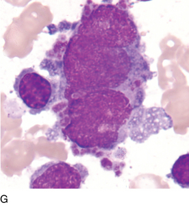
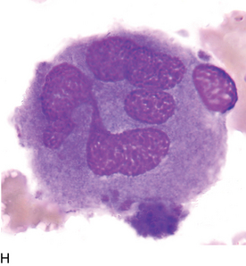
Evidence of dysmegakaryopoiesis (Figure 18-3, A-H) may include any or all of the following: giant platelets, platelets with abnormal granulation, circulating micromegakaryocytes, large mononuclear megakaryocytes, and abnormal nuclear shapes.
All photomicrographs are ×1000 original magnification with Wright-Giemsa staining unless stated otherwise.