Mycobacteria

Published on 08/02/2015 by admin

Filed under Basic Science

Last modified 22/04/2025

Print this page

rate 1 star rate 2 star rate 3 star rate 4 star rate 5 star
Your rating: none, Average: 2 (1 votes)

This article have been viewed 3352 times

Mycobacteria

Objectives

1. Describe the general characteristics of the Mycobacterium spp., including oxygen requirements, staining patterns and cell morphology, artificial media required for cultivation and growth, and pigmentation.

2. Explain the chemical composition of the bacterial cell wall.

3. Explain the microscopic staining characteristics of Mycobacterium spp. using the Gram stain and acid-fast staining methods.

4. List the most common pathogenic species in the Mycobacterium genus and state the natural habitat, mode of transmission, and reservoir for each.

5. Differentiate M. tuberculosis clinical infections based on the signs and symptoms of the following: primary infection, latent infection, disseminated infection, and reactivation.

6. Compare the current safety and containment methods recommended for handling mycobacterial infectious materials and routine bacteriology in a diagnostic laboratory.

7. Describe the purified protein derivative (PPD; also referred to as the tuberculin skin test). What is the significance of a positive result?

8. List the clinical specimens acceptable for recovery of mycobacteria and describe the limitations of recovery from each type of specimen.

9. Justify the use of DNA probes and molecular sequencing or amplification methods to identify Mycobacterium spp.

10. Evaluate the effectiveness of the staining procedures—Kinyoun, Ziehl-Neelsen, and fluorescent staining (auramine-rhodamine or acridine orange)—for identifying mycobacteria.

11. Describe the requirements for using digestion and decontamination procedures to improve the recovery of Mycobacterium spp.

12. Explain the limitations of digestion and decontamination procedures.

13. Explain the methods commonly used for biochemical identification of Mycobacterium spp. (i.e., niacin, nitrate, urease, modified catalase, Tween 80, tellurite, arylsulfatase, thiophene-2-carboxylic acid hydrazide [TCH], and 5% NaCl tests), including the purpose, principle, and control organisms used for each.

14. Describe the role of the human immunodeficiency virus (HIV) and acquired immunodeficiency syndrome (AIDS) in the dissemination and/or pathogenesis of infections with Mycobacterium spp.

15. Explain the recommended susceptibility testing methods and state when susceptibility testing is required or recommended for Mycobacterium spp.

Traditionally, Mycobacterium spp. have been classified according to phenotypic characteristics. However, since the late 1980s, molecular diagnostics have been used to shift the characterization of these organisms to genotypic studies. This chapter discusses both the phenotypic characterization and the new taxonomy based on molecular genetic data.

The organisms that belong to the genus Mycobacterium are aerobic (although some may grow in reduced oxygen concentrations), non–spore forming (except for M. marinum), nonmotile, very thin, slightly curved or straight rods (0.2 to 0.6 × 1 to 10 µm). Some species may display a branching morphology. Mycobacterium is the only genus in the Mycobacteriaceae family (Actinomycetales order, Actinomycetes class). Genera that are closely related to Mycobacterium include Nocardia, Rhodococcus, Tsukamurella and Gordonia.

Mycobacterium spp. have an unusual cell wall structure. The cell wall contains N-glycolylmuramic acid instead of N-acetylmuramic acid, and it has a very high lipid content, which creates a hydrophobic permeability barrier. Because of this cell wall structure, mycobacteria are difficult to stain with commonly used basic aniline dyes, such as those used in Gram staining. Although these organisms cannot be readily Gram stained, they generally are considered gram positive. However, they resist decolorization with acidified alcohol (3% hydrochloric acid) after prolonged application of a basic fuchsin dye or with heating of this dye after its application. This important property of mycobacteria, which derives from their cell wall structure, is referred to as acid fastness; this characteristic distinguishes mycobacteria from other genera. Rapid-growing mycobacteria (RGMs) may partially or completely lose this characteristic as a result of their growth characteristics.

Another important feature of many species is that they grow more slowly than most other human pathogenic bacteria because of their hydrophobic cell surface. Because of this hydrophobicity, organisms tend to clump, so that nutrients are not easily allowed into the cell. A single cell’s generation time (the time required for a cell to divide into two independent cells) may range from approximately 20 hours to 36 hours for Mycobacterium ulcerans. This slow growth results in the formation of visible colonies in 2 to 60 days at optimum temperature.

Currently, the genus Mycobacterium includes more than 100 recognized or proposed species. These organisms produce a spectrum of infections in humans and animals ranging from localized lesions to disseminated disease. Some species cause only human infections, and others have been isolated from a wide variety of animals. Many species are also found in water and soil.

For the most part, mycobacteria can be divided into two major groups, based on fundamental differences in epidemiology and association with disease: those belonging to the Mycobacterium tuberculosis complex and those referred to as nontuberculous mycobacteria (NTM) (Box 43-1).

Box 43-1   Major Groupings of Organisms Belonging to the Genus Mycobacterium*

Nontuberculous Mycobacteria


*This box is not inclusive; rather, it lists only the prominent mycobacteria isolated from humans.

Mycobacterium Tuberculosis Complex

Tuberculosis was endemic in animals in the Paleolithic period, long before it ever affected humans. This disease (also called consumption) has been known in all ages and climates. For example, tuberculosis was the subject of a hymn in a sacred text from India dating from 2500 BC, and DNA unique to Mycobacterium tuberculosis was identified in lesions from the lung in 1000-year-old human remains found in Peru.

Epidemiology and Pathogenesis

Epidemiology

M. tuberculosis is the cause of most cases of human tuberculosis, particularly in developed countries. An estimated 1.7 billion people, or one third of the world’s population, are infected with M. tuberculosis. This reservoir of infected individuals results in 8 million new cases of tuberculosis and 2.9 million deaths annually. Tuberculosis continues to be a public health problem in the United States. An additional complicating factor in the management of tuberculosis is the increasing incidence of co-infection with the human immunodeficiency virus (HIV). HIV-associated tuberculosis remains a significant challenge to world health, with an estimated 1.1 million individuals living with HIV-associated tuberculosis. In the United States, tuberculosis typically is found among the poor, homeless, intravenous (IV) drug users, alcoholics, the elderly, or medically underserved populations. Although the organisms belonging to the M. tuberculosis complex have numerous characteristics in common, including extreme genetic homogeneity, they differ in certain epidemiologic aspects (Table 43-1).

TABLE 43-1

Epidemiology of Organisms Belonging to M. tuberculosis Complex That Cause Human Infections

Organism Habitat Primary Route of Transmission Distribution
M. tuberculosis Patients with cavitary disease are primary reservoir Person to person by inhalation of droplet nuclei: droplet nuclei containing the organism (infectious aerosols, 1 to 5 µm) are produced when people with pulmonary tuberculosis cough, sneeze, speak, or sing; infectious aerosols may also be produced by manipulation of lesions or processing of clinical specimens in the laboratory. Droplets are so small that air currents keep them airborne for long periods; once inhaled, they are small enough to reach the lungs’ alveoli* Worldwide
M. bovis Humans and a wide range of host animals, such as cattle, nonhuman primates, goats, cats, buffalo, badgers, possums, dogs, pigs, and deer Ingestion of contaminated milk from infected cows; airborne transmission Worldwide
M. africanum Humans§ Inhalation of droplet nuclei East and West tropical Africa; some cases have been identified in the United States
M. caprae Humans rarely; predominately infects a wide range of animals Inhalation of droplet nuclei Europe
M. microti Humans rarely; small animals (e.g., voles and other wild rodents) Inhalation of droplet nuclei Europe; Great Britain, Netherlands
M. canettii Natural reservoir has not been clearly defined. Rarely infects humans. Unclear Africa
M. pinnipedii Humans rarely; predominantly infects a wide range of animals Unclear Europe

image

*Infection occasionally can occur through the gastrointestinal tract or skin.

The incidence has decreased significantly in developed countries since the introduction of universal pasteurization of milk and milk products and the institution of effective control programs for cattle.

Can be transmitted human to human, animal to human, and human to animal.

§Infections in animals have not been totally excluded.

Pathogenesis

The pathogenesis of tuberculosis caused by organisms of the M. tuberculosis complex is discussed in Chapter 69. Inhalation of a single viable organism has been shown to lead to infection, although close contact is usually necessary. Of those who become infected with M. tuberculosis, 15% to 20% develop disease. The disease usually occurs some years after the initial infection, when the patient’s immune system breaks down for some reason other than the presence of tuberculosis bacilli in the lung. In a small percentage of infected hosts, the disease becomes systemic, affecting a variety of organs.

After ingestion of milk from infected cows, Mycobacterium bovis may penetrate the gastrointestinal mucosa or invade the lymphatic tissue of the oropharynx. An attenuated strain of M. bovis, bacillus Calmette-Guérin (BCG), has been used extensively in many parts of the world to immunize susceptible individuals against tuberculosis. Because mycobacteria are the classic examples of intracellular pathogens and the body’s response to BCG hinges on cell-mediated immunoreactivity, immunized individuals are expected to react more aggressively against all antigens that elicit cell-mediated immunity. In rare cases, an unfortunate individual’s immune system is so compromised that it cannot handle the BCG, and systemic BCG infection may develop.

Spectrum of Disease

Tuberculosis may mimic other diseases, such as pneumonia, neoplasm, or fungal infections. In addition, clinical manifestations in patients infected with M. tuberculosis complex may range from asymptomatic to acutely symptomatic. Patients who are symptomatic can have systemic symptoms, pulmonary signs and symptoms, signs and symptoms related to other organ involvement (e.g., the kidneys), or a combination of these features. Cases of pulmonary disease caused by M. tuberculosis complex organisms are clinically, radiologically, and pathologically indistinguishable.

Primary tuberculosis typically is considered a disease of the respiratory tract. Common presenting symptoms include low-grade fever, night sweats, fatigue, anorexia (loss of appetite), and weight loss. A patient who presents with pulmonary tuberculosis usually has a productive cough, along with low-grade fever, chills, myalgias (aches), and sweating; however, these signs and symptoms are similar for influenza, acute bronchitis, and pneumonia.

Upon respiratory infection with M. tuberculosis complex organisms, the cellular immune system T cells and macrophages migrate to the lungs, and the organisms are phagocytized by the macrophages. However, these organisms are capable of intracellular multiplication in the macrophages. Often the host is unable to eliminate the organisms, and the result is a systemic hypersensitivity to Mycobacterium antigens. Granulomas or a hard tubercle forms in the lung from the lymphocytes, macrophages, and cellular pathology, including giant cell formation (cellular fusion displaying multiple nuclei). If the Mycobacterium antigen concentration is high, the hypersensitivity reaction may result in tissue necrosis, caused by enzymes released from the macrophages. In this case no granuloma forms, and a solid or semisolid, caseous material is left at the primary lesion site.

In some patients infected with primary active tuberculosis, the disease may spread via the lymph system or hematogenously, leading to meningeal or miliary (disseminated) tuberculosis. This most often occurs in patients with depressed or ineffective cellular immunity.

As previously mentioned, in a small percentage of patients, organs besides the lungs can become involved after infection with M. tuberculosis complex organisms. These organs include the following:

Disseminated tuberculosis may be diagnosed by a positive tuberculin skin test (described later in the chapter).

Patients also may have latent disease (i.e., they have no apparent signs, symptoms, or pathologic condition). A patient with latent tuberculosis is not infectious and does not have active disease, although the organism is present in granulomas. Patients with latent tuberculosis may progress to active disease (also referred to as reactivation of tuberculosis) at any time. Reactivation tuberculosis typically occurs after an incident in which cellular immunity is suppressed or damaged as a result of a change in life style or other health condition.

Individuals infected with HIV are particularly susceptible to developing active tuberculosis. These patients are likely to have rapidly progressive primary disease instead of a subclinical infection.

Diagnosing tuberculosis is more difficult in people infected with HIV, because chest radiographs of the pulmonary disease often lack specificity, and patients frequently are anergic (lack a biologic response) to tuberculin skin testing, a primary means of identifying individuals infected with M. tuberculosis. The tuberculin skin test, or purified protein derivative (PPD) test, is based on the premise that after infection with M. tuberculosis, an individual develops a delayed hypersensitivity cell-mediated immunity to certain antigenic components of the organism. To determine whether a person has been infected with M. tuberculosis, a culture extract of M. tuberculosis (i.e., PPD of tuberculin) is injected intracutaneously. After 48 to 72 hours, an infected individual shows a delayed hypersensitivity reaction to the PPD, characterized by erythema (redness) and, most important, induration (firmness as a result of influx of immune cells). The diameter of induration is measured and then interpreted as to whether the patient has been infected with M. tuberculosis; different interpretative criteria are used for different patient populations (e.g., immunosuppressed individuals, such as those infected with HIV). More recently, the T-Spot TB test (Oxford, Immunotec, United Kingdom) offers next-day results and does not require a follow-up visit with a physician. The assay measures T cells that have been activated by Mycobacterium tuberculosis antigens. Peripheral blood mononuclear cells are incubated with M. tuberculosis-specific antigens stimulating any sensitized T cells in the patient sample. T cell cytokines released in the sample are measured using antibody to capture them and then detected with a secondary antibody conjugated to alkaline phosphatase. This assay should be interpreted in correlation with the patient’s signs and symptoms.

The PPD test is not 100% sensitive or specific, and a positive reaction to the skin test does not necessarily signify the presence of disease. Because of these issues, a new test approved by the U.S. Food and Drug Administration (FDA) has become available. It is an enzyme-linked immunosorbent assay (ELISA) called QuantiFERON-TB Gold (Cellestis Limited, Carnegie, Victoria, Australia). The assay measures a component of the cell-mediated immune response to M. tuberculosis to diagnose latent tuberculosis infection and tuberculosis disease. It is based on the quantification of interferon-gamma released from sensitized lymphocytes in heparinized whole blood that has been incubated overnight with a mixture of synthetic peptides simulating two proteins in M. tuberculosis. The test assesses responses to multiple antigens; it can be performed in a single patient visit; and it is less subject to reader bias and error. An important feature is that the results of the assay are unaffected by previous BCG vaccination. Guidelines published by the Centers for Disease Control and Prevention (CDC) recommend the use of this assay in all circumstances in which the tuberculin skin test currently is used (e.g., contact investigations and evaluation of recent immigrants). The guidelines also provide specific cautions for interpreting negative results in individuals from selected populations.

Nontuberculous Mycobacteria

The NTM include all mycobacterial species that do not belong to M. tuberculosis complex. Currently, approximately 130 species of nontuberculous mycobacteria have been recognized. The members of this large group of mycobacteria have been known by several names (Box 43-2). Significant geographic variability is seen both in the prevalence of and the species responsible for NTM disease. As previously mentioned, NTM are present everywhere in the environment and sometimes colonize the skin and respiratory and gastrointestinal tracts of healthy individuals. Little is known about how infection is acquired, but some mechanisms appear to be trauma, inhalation of infectious aerosols, and ingestion; a few diseases are nosocomial or are acquired as an iatrogenic infection. In contrast to M. tuberculosis complex, NTM are not usually transmitted from person to person, nor does isolation of these organisms necessarily mean they are associated with a disease process. Interpretation of a positive NTM culture is complicated, because these organisms are widely distributed in nature, their pathogenic potential varies greatly from one species to another, and humans can be colonized by these mycobacteria without necessarily developing infection or disease. With few exceptions, little is known about the pathogenesis of infections caused by these bacterial agents.

Box 43-2

Other Names That Have Been Used to Designate the Nontuberculous Mycobacteria

From Debrunner M et al: Epidemiology and clinical significance of nontuberculous mycobacteria in patients negative for human immunodeficiency virus in Switzerland, Clin Infect Dis 15:330, 1992.

In 1959 Runyon1 classified NTM into four groups (Runyon groups I to IV) based on the phenotypic characteristics of the various species, most notably the growth rate and colonial pigmentation (Table 43-2). Runyon’s system first categorizes the slow-growing NTM (Runyon groups I to III) and then the rapid-growers (Runyon group IV). One other NTM, M. leprae, which cannot be cultivated on artificial media, is also reviewed. (As with many classification schemes, the Runyon classification does not always hold true. For example, some NTM can be either a photochromogen or a nonphotochromogen.)

TABLE 43-2

Runyon Classification of Nontuberculous Mycobacteria (NTM)

Runyon Group Number Group Name Description
I Photochromogens NTM colonies that develop pigment on exposure to light after being grown in the dark and take longer than 7 days to appear on solid media
II Scotochromogens NTM colonies that develop pigment in the dark or light and take longer than 7 days to appear on solid media
III Nonphotochromogens NTM colonies that are nonpigmented regardless of whether they are grown in the dark or light and take longer than 7 days to appear on solid media
IV Rapid growers NTM colonies that grow on solid media and take fewer than 7 days to appear

Because determining the clinical significance of isolating NTM from a clinical sample is difficult, several clinical classification schemes also have been proposed. One such scheme classifies NTM recovered from humans into four major groups (pulmonary, lymphadenitis, cutaneous, or disseminated) based on the clinical disease they cause. Other NTM classifications are based on the pathogenic potential of a species.

Slow-Growing Nontuberculous Mycobacteria

The slow-growing NTM can be subdivided into three groups based on the phenotypic characteristics of the species. Mycobacterium spp. synthesize carotenoids (a group of yellow to red pigments) in varying amounts and thus can be categorized into three groups based on the production of these pigments: photochromogens, scotochromogens, and nonphotochromogens. Some of these NTM are considered potentially pathogenic for humans, whereas others are rarely associated with disease.

Photochromogens

The photochromogens (Table 43-3) are slow-growing NTM that produce colonies that require light to form pigment.

TABLE 43-3

Characteristics of Nontuberculous Mycobacteria—Photochromogens

Organism Epidemiology Pathogenicity Type of Infection
M. kansasii Infection more common in white males; natural reservoir is tap water; aerosols are involved in transmission Potentially pathogenic Chronic pulmonary disease; extrapulmonary diseases, such as cervical lymphadenitis and cutaneous disease
M. asiaticum Not commonly encountered (primarily seen in Australia) Potentially pathogenic Pulmonary disease
M. marinum Natural reservoirs are freshwater and saltwater as a result of contamination from infected fish and other marine life. Transmission is by contact with contaminated water and organism entry by means of trauma or small breaks in the skin; associated with aquatic activity usually involving fish Potentially pathogenic Cutaneous disease; bacteremia
M. intermedium Unknown Potentially pathogenic Pulmonary disease
M. novocastrense Unknown Potentially pathogenic Cutaneous disease

image

Scotochromogens

The scotochromogens (Table 43-4) are slow-growing NTM that produce pigmented colonies whether grown in the dark or the light. The epidemiology of the potentially pathogenic scotochromogens has not been definitively described. In contrast to potentially pathogenic nonphotochromogens, these agents are rarely recovered in the clinical laboratory.

TABLE 43-4

Characteristics of Nontuberculous Mycobacteria—Scotochromogens

Organism Epidemiology/Habitat Pathogenicity Type of Infection
M. szulgai Water and soil Potentially pathogenic Pulmonary disease, predominantly in middle-aged men; cervical adenitis; bursitis
M. scrofulaceum Raw milk, soil, water, dairy products Potentially pathogenic Cervical adenitis in children, bacteremia, pulmonary disease, skin infections
M. interjectum Unknown Potentially pathogenic Chronic lymphadenitis, pulmonary disease
M. heckeshornense Unknown Potentially pathogenic Pulmonary disease (rare)
M. tusciae Unknown—isolated from tap water Potentially pathogenic Cervical lymphadenitis (rare)
M. kubicae Unknown Potentially pathogenic Pulmonary disease
M. gordonae Tap water, water, soil Nonpathogenic* NA
M. cookie Sphagnum moss, surface waters in New Zealand Nonpathogenic* NA
M. hiberniae Sphagnum moss, soil in Ireland Nonpathogenic* NA

image

NA, Not applicable.

*Rarely, if ever, causes disease.

Nonphotochromogens

The nonphotochromogens (Table 43-5) are slow-growing NTM that produce unpigmented colonies whether grown in the dark or the light. Of the organisms in this group, M. terrae complex (M. terrae, M. triviale, and M. nonchromogenicum) and M. gastri are considered nonpathogenic for humans. The other nonphotochromogens are considered potentially pathogenic, and many are frequently recovered in the clinical laboratory. The nonphotochromogens belonging to Mycobacterium avium complex are frequently isolated in the clinical laboratory and are able to cause infection in the human host.

TABLE 43-5

Characteristics of the Nontuberculous Mycobacteria—Nonphotochromogens and Species Considered Potential Pathogens

Organism Epidemiology Type of Infection
M. avium complex Environmental sources, including natural waters, and soil Patients without AIDS: Pulmonary infections in patients with preexisting pulmonary disease; cervical lymphadenitis; and disseminated disease* in immunocompromised patients who are HIV negative
Patients with AIDS: Disseminated disease
M. xenopi Water, especially hot water taps in hospitals; believed to be transmitted in aerosols Primarily pulmonary infections in adults; less common, extrapulmonary infections (bone, lymph nodes, sinus tract) and disseminated disease
M. ulcerans Stagnant tropical waters; also harbored in an aquatic insect’s salivary glands; infections occur in tropical or temperate climates Indolent cutaneous and subcutaneous infections (African Buruli ulcer or Australian Bairnsdale ulcer)
M. malmoense Most cases from England, Wales, and Sweden. Rarely isolated from patients infected with HIV. Little is known about epidemiology; to date, isolated only from humans and captured armadillos Chronic pulmonary infections, primarily in patients with preexisting disease; cervical lymphadenitis in children; less common, infections of the skin or bursae
M. genovense Isolated from pet birds and dogs. Mode of acquisition unknown Disseminated disease in patients with AIDS (wasting disease characterized by fever, weight loss, hepatosplenomegaly, anemia)
M. haemophilum Unknown Disseminated disease; cutaneous infections in immunosuppressed adults; mild and limited skin infections in preadolescence or early adolescence; cervical lymphadenitis in children
M. heidelbergense Unknown Lymphadenitis in children; also isolated from sputum, urine, and gastric aspirate
M. shimoidei To date has not been isolated from environmental sources; few case reports, but widespread geographically Tuberculosis-like pulmonary infection; disseminated disease
M. simiae Tap water and hospital water tanks; rarely isolated Tuberculosis-like pulmonary infection

AIDS, Acquired immunodeficiency syndrome; HIV, human immunodeficiency virus.

*Disseminated disease can involve multiple sites, such as bone marrow, lungs, liver, lymph nodes.

Can be either nonphotochromogenic or scotochromogenic.

Mycobacterium avium Complex (MAC).

Largely because of the increasing populations of immunosuppressed patients, the incidence of infection caused by M. avium complex spp., as well as these organisms’ clinical significance, has changed significantly since they were first recognized as human pathogens in the 1950s. The introduction of highly active antiretroviral therapy (HAART) has dramatically reduced the infections caused by these organisms in patients with acquired immunodeficiency syndrome (AIDS).

General Characteristics.

Taxonomically, M. avium complex comprises M. avium, M. intracellulare, M. avium subsp. avium, M. avium subsp. paratuberculosis, M. avium subsp. silvaticum (wood pigeon bacillus), M. vulneris, M. marseillense, M. bouchedurhonense, and M. timonense. The name M. avium subsp. hominissuis has been proposed for another subspecies capable of infecting humans. Unfortunately, the nomenclature is somewhat confusing. Although M. avium and M. intracellulare are clearly different organisms, they so closely resemble each other that the distinction cannot be made by routine laboratory determinations or on clinical grounds. As a result, these organisms sometimes are referred to as M. avium-intracellulare. Furthermore, because isolation of M. avium subsp. paratuberculosis in a routine laboratory setting is exceedingly rare, the term M. avium complex is most commonly used to report the isolation of M. avium-intracellulare.

Epidemiology and Pathogenesis.

MAC is an important pathogen in both immunocompromised and immunocompetent populations. These are among the most commonly isolated NTM species in the United States. MAC is particularly noteworthy for its potentially pathogenic role in pulmonary infections in patients with AIDS and also in patients who are not infected with HIV. The organisms are ubiquitous in the environment and have been isolated from natural water, soil, dairy products, pigs, chickens, cats, and dogs. As a result of extensive studies, it is generally accepted that natural waters serve as the major reservoir for most human infections.

Infections caused by MAC are acquired by inhalation or ingestion. The pathogenesis of MAC infections is not clearly understood. The organisms are commonly associated with respiratory disease clinically similar to tuberculosis in adults, lymphadenitis in children, and disseminated infection in patients with HIV. However, these organisms and other environmental NTM have extraordinary starvation survival. They can persist well over a year in tap water, and MAC tolerates temperature extremes. In addition, similar to legionellae, M. avium can infect and replicate in protozoa. Amoebae-grown M. avium is more invasive toward human epithelial and macrophage cells.

MAC cultures can have an opaque, a translucent, or a transparent colony morphology. Studies suggest that transparent colonies are more virulent because they are more drug resistant, are isolated more frequently from the blood of patients with AIDS, and appear more virulent in macrophage and animal models.

M. avium subsp. paratuberculosis is known to cause an inflammatory bowel disease (known as Johne’s disease) in cattle, sheep, and goats. It also has been isolated from the bowel mucosa of patients with Crohn’s disease, a chronic inflammatory bowel disease of humans. The organism is extremely fastidious, seems to require a growth factor (mycobactin, produced by other species of mycobacteria, such as M. phlei, a saprophytic strain) and may take as long as 6 to 18 months for primary isolation. Whether these and other mycobacteria actually contribute to development of Crohn’s disease or are simply colonizing an environmental niche in the bowel of these patients remains to be elucidated.

Other Nonphotochromogens.

Several other mycobacterial species that are considered nonphotochromogens are potentially pathogenic in humans. The epidemiology and spectrum of disease for these organisms are summarized in Table 43-5. In addition to the species in this table, other, newer species of mycobacteria that are nonphotochromogens have been described, such as M. celatum and M. conspicuum. These newer agents appear to be potentially pathogenic in humans.

Rapidly Growing Nontuberculous Mycobacteria (RGM)

Mycobacteria that produce colonies on solid media in 7 days or earlier constitute the second major group of NTM. Currently, approximately 70 species have been classified into this group.

General Characteristics

The large group of organisms that constitute the RGM is divided into six major groups of potentially pathogenic species, based on pigmentation and molecular studies (see Box 43-1). Unlike the majority of other mycobacteria, most rapid-growers can grow on routine bacteriologic media and on media specific for cultivation of mycobacteria. On Gram staining, these organisms appear as weakly gram-positive rods resembling diphtheroids.

Epidemiology and Pathogenesis

The rapidly growing mycobacteria considered potentially pathogenic can cause disease in either healthy or immunocompromised patients. Like many other NTM, these organisms are ubiquitous in the environment and are present worldwide. They have been found in soil, marshes, rivers, and municipal water supplies (tap water) and in marine and terrestrial life forms. Infections caused by rapidly growing mycobacteria can be acquired in the community from environmental sources. They also can be nosocomial infections, resulting from medical interventions (including bone marrow transplantation), wound infections, and catheter sepsis. These organisms may be commensals on the skin. They gain entry into the host by inoculation into the skin and subcutaneous tissues as a result of trauma, injections, or surgery, or through animal contact.

The RGM also can cause disseminated cutaneous infections. The description of chronic pulmonary infections caused by rapidly growing mycobacteria suggests a possible respiratory route for acquisition of organisms present in the environment. Of the potentially pathogenic, rapidly growing NTM, M. fortuitum, M. chelonae, and M. abscessus are commonly encountered; these three species account for approximately 90% of clinical disease. Little is known about the pathogenesis of these organisms.

Spectrum of Disease

The spectrum of disease caused by the most commonly encountered rapid-growers is summarized in Table 43-6. The most common infection associated with RGM is posttraumatic wound infection. An increase in wound infections has been associated with planktonic M. abscessus, which can be identified as a rough colonial phenotype on artificial media; these organisms are capable of infecting macrophages. The smooth colonial phenotype typically is identified in biofilms and lacks infectivity.

TABLE 43-6

Common Types of Infections Caused by Rapidly Growing Mycobacteria

Organism Common Types of Infection
M. abscessus subsp. abscessus Disseminated disease, primarily in immunocompromised individuals; skin and soft tissue infections; pulmonary infections; postoperative infections
M. fortuitum Postoperative infections in breast augmentation and median sternotomy; skin and soft tissue infections; pulmonary infections, usually single. localized lesions.
Central nervous system (CNS) disease is rare but has high morbidity and mortality
M. chelonae Skin and soft tissue infections, postoperative wound infections, keratitis
Less Common Types of Infection (More Than 10 Cases)  
M. peregrinum Skin and soft tissue infections; bacteremia
M. mucogenicum Posttraumatic wound infections, catheter-related sepsis, health care associated
M. smegmatis Skin or soft tissue infections; less frequently, pulmonary infections
M. abscessus subsp. bolletii Health care–associated infections, skin and soft tissue infections, pulmonary infections
M. boenickei Bone and joint infections
M. canariasense Bacteremia
M. cosmeticum Pulmonary and urosepsis
M. goodii Bone and joint infections, osteomyelitis
M. houstonense Bone and joint infections
M. immunogenum Hypersensitivity pneumonitis
M. neoaurum (closely related to M. lacticola) Catheter-related sepsis
M. porcinum Surgical site infection
M. senegalense Catheter-related sepsis
Rare Infections (Fewer Than 10 Cases)  
M. aubagnense Various opportunistic health care–associated infections
M. brisbanense Various opportunistic health care–associated infections
M. brumae Various opportunistic health care–associated infections
M. elephantis Various opportunistic health care–associated infections
M. mageritense Skin and soft tissue infections
M. monacense Various opportunistic health care–associated infections
M. moriokaense Various opportunistic health care–associated infections
M. neworleansense Various opportunistic health care–associated infections
M. novocastrense Various types of opportunistic health care–associated infections
M. phocaicum Catheter-related sepsis
M. septicum Various opportunistic health care–associated infections
M. setense Bone and joint infections
M. wolinskyi Skin and soft tissue infections, bone infection, osteomyelitis

Noncultivatable Nontuberculous Mycobacteria—mycobacterium Leprae

The nontuberculous mycobacterium M. leprae is a close relative of M. tuberculosis. This organism causes leprosy (also called Hansen’s disease). Leprosy is a chronic disease of the skin, mucous membranes, and nerve tissue. Leprosy remains a worldwide public health concern as a result of the development of drug-resistant isolates.

Epidemiology and Pathogenesis

Understanding of the epidemiology and pathogenesis of leprosy is hampered by the inability to grow the organism in culture. In tropical countries, where the disease is most prevalent, it may be acquired from infected humans; however, infectivity is very low. Prolonged close contact and the host’s immunologic status play roles in infectivity.

Spectrum of Disease

Based on the host’s response, the spectrum of disease caused by M. leprae ranges from subclinical infection to intermediate stages of disease to full-blown and serious clinical manifestations involving the skin, upper respiratory system, testes, and peripheral nerves. The two major forms of the disease are a localized form, called tuberculoid leprosy, and a more disseminated form, called lepromatous leprosy. Patients with lepromatous leprosy are anergic to M. leprae because of a defect in their cell-mediated immunity. Because the organisms’ growth is unimpeded, these individuals develop extensive skin lesions containing numerous acid-fast bacilli; the organisms can spill over into the blood and disseminate. In contrast, individuals with tuberculoid leprosy do not have an immune defect, so the disease is localized to the skin and nerves; few organisms are observed in skin lesions. Most of the serious sequelae associated with leprosy are the result of this organism’s tropism for peripheral nerves.

Laboratory Diagnosis of Mycobacterial Infections

Specimens received by the laboratory for mycobacterial smear and culture must be handled in a safe manner. Tuberculosis ranks high among laboratory-acquired infections; therefore, laboratory and hospital administrators must provide laboratory personnel with facilities, equipment, and supplies that reduce this risk to a minimum. M. tuberculosis has a very low infective dose for humans (i.e., an infection rate of approximately 50% with exposure to fewer than 10 acid-fast bacilli). All tuberculin-negative personnel should have a skin test at least annually. The CDC recommends Biosafety Level 2 practices, containment equipment, and facilities for preparing acid-fast smears and culture for nonaerosolizing manipulations. If M. tuberculosis is grown and then propagated and manipulated, biologic safety cabinet (BSC) class II safety precautions are required; however, Biosafety Level 3 practices are recommended. BSC Level 3 practices are recommended for opening centrifuge vials, adding reagents to biochemical testing medias, and sonication; these practices include restricted laboratory access, negative pressure airflow, and special personal protective equipment (e.g., certified respirators). Respiratory devices should be certified through the National Institute for Occupational Safety and Health (NIOSH).

Specimen Collection and Transport

Acid-fast bacilli can infect almost any tissue or organ of the body. Successful isolation of these organisms depends on the quality of the specimen obtained and the use of appropriate processing and culture techniques by the mycobacteriology laboratory. In suspected mycobacterial disease, as in all other infectious diseases, the diagnostic procedure begins at the patient’s bedside. Collection of proper clinical specimens requires careful attention to detail by health care professionals. Most specimens are respiratory samples, such as sputum, tracheal or bronchial aspirates, and specimens obtained by bronchial alveolar lavage. Other samples may include urine, gastric aspirates, tissue (biopsy) specimens, cerebrospinal fluid (CSF), and pleural and pericardial fluid. Blood or fecal specimens may be collected from immunocompromised patients. Specimens should be collected in sterile, leak-proof, disposable, and appropriately labeled containers without fixatives and placed in bags to contain leakage. If transport and processing will be delayed longer than 1 hour, all specimens except blood should be refrigerated at 4° C until processed.

Pulmonary Specimens

Pulmonary secretions may be obtained by any of the following methods: spontaneously produced or induced sputum, gastric lavage, transtracheal aspiration, bronchoscopy, and laryngeal swabbing. Most specimens submitted for examination are sputum, aerosol-induced sputum, bronchoscopic aspirations, or gastric lavage samples. Spontaneously produced sputum is the specimen of choice. To raise sputum, patients must be instructed to take a deep breath, hold it momentarily, and then cough deeply and vigorously. Patients must also be instructed to cover the mouth carefully while coughing and to discard tissues in an appropriate receptacle. Saliva and nasal secretions should not be collected, nor should the patient use oral antiseptics during the collection period. Sputum specimens must be free of food particles, residues, and other extraneous matter.

The aerosol (saline) induction procedure can best be done on ambulatory patients who are able to follow instructions. Aerosol-induced sputum specimens have been collected from children as young as 5 years of age. This procedure should be performed in an enclosed area with appropriate airflow. Operators should wear particulate respirators and take appropriate safety measures to prevent exposure. The patient is told that the procedure is being performed to induce coughing to raise sputum that the patient cannot raise spontaneously and that the salt solution is irritating. The patient is instructed to inhale slowly and deeply through the mouth and to cough at will, vigorously and deeply, coughing and expectorating into a collection tube. The procedure is discontinued if the patient fails to raise sputum after 10 minutes or feels any discomfort. Ten milliliters of sputum should be collected; if the patient continues to raise sputum, a second specimen should be collected and submitted. Specimens should be delivered promptly to the laboratory and refrigerated if processing is delayed.

Sputum collection guidelines recommend collection of an early morning specimen for 3 consecutive days. In many cases the third specimen demonstrates minimal recovery of organisms, and this collection may not be recommended in some laboratories. Pooled specimens are unacceptable because of an increased risk of contamination.

Gastric Lavage Specimens

Gastric lavage is used to collect sputum from patients who may have swallowed sputum during the night. The procedure is limited to senile, nonambulatory patients; children younger than 3 years of age (specimen of choice); and patients who fail to produce sputum by aerosol induction. The most desirable gastric lavage is collected at the patient’s bedside before the patient arises and before exertion empties the stomach. Gastric lavage cannot be performed as an office or clinic procedure.

The collector should wear a cap, gown, and particulate respirator mask and should stand beside (not in front of) the patient, who should sit up on the edge of the bed or in a chair, if possible. The Levine collection tube is inserted through a nostril, and the patient is instructed to swallow the tube. When the tube has been fully inserted, a syringe is attached to the end of the tube and filtered distilled water is injected into the tube. The syringe is then used to withdraw 5 to 10 mL of gastric secretions, which is expelled slowly down the sides of the 50-mL conical collecting tube. Samples should be adjusted to a neutral pH. The laboratory may choose to provide sterile receptacles containing 100 mg of sodium carbonate to reduce the acidity; this improves the recovery of organisms. The top of the collection tube is screwed on tightly, and the tube is held upright during prompt delivery to the laboratory. Three specimens should be collected over a period of consecutive days. Specimens should be processed within 4 hours.

Bronchial lavages, washings, and brushings are collected and submitted by medical personnel. These are the specimens of choice for detecting nontuberculous mycobacteria and other opportunistic pathogens in patients with immune dysfunction.

Urine Specimens

The incidence of urogenital infections shows little evidence of decreasing. About 2% to 3% of patients with pulmonary tuberculosis show urinary tract involvement, but 30% to 40% of patients with genitourinary disease have tuberculosis at some other site. The clinical manifestations of urinary tuberculosis, which are variable, include frequency of urination (most common), dysuria, hematuria, and flank pain. Definitive diagnosis requires recovery of acid-fast bacilli from the urine.

Early morning voided urine specimens (40 mL minimum) in sterile containers should be submitted daily for at least 3 days. The collection procedure is the same as for collecting a clean-catch midstream urine specimen (see Chapter 73). The 24-hour urine specimen is undesirable because of excessive dilution, higher contamination, and difficulty in concentrating. Catheterization should be used only if a midstream voided specimen cannot be collected.

Blood Specimens

Immunocompromised patients, particularly those infected with HIV, can have disseminated mycobacterial infection; most of these infections are caused by M. avium complex. A blood culture positive for MAC is always associated with clinical evidence of disease. Recovery of mycobacteria is improved with blood collection in either a broth or the Isolator lysis-centrifugation system (see Chapter 68). Some studies have indicated that the lysis-centrifugation system is advantageous, because quantitative data can be obtained with each blood culture; in patients with AIDS, quantitation of such organisms can be used to monitor therapy and determine the prognosis. However, the necessity of quantitative blood cultures remains unclear.

Blood for culture of mycobacteria should be collected as for routine blood cultures. Blood collected in regular phlebotomy procedures in anticoagulants such as sodium polyanethol sulfonate (SPS), heparin, and citrate may be used to inoculate cultures for the recovery of Mycobacterium species. Conventional blood culture collection systems are unacceptable for the isolation of Mycobacterium spp. However, specialized automated systems are available for growth of Mycobacterium spp., including the Bactec MGIT 960 system (Becton-Dickinson, Franklin Lakes, N.J.), and the BacT/ALERT 3D (Biomerieux, Durham, N.C.).

Specimen Processing

Processing to recover acid-fast bacilli from clinical specimens involves several complex steps, each of which must be carried out with precision. Specimens from sterile sites can be inoculated directly to media (small volume) or concentrated to reduce volume. Other specimens require decontamination and concentration. A processing scheme is shown in Figure 43-1, and the procedures are explored in detail in the following discussions.

Contaminated Specimens

Most specimens submitted for mycobacterial culture consist of organic debris, such as mucin, tissue, serum, and other proteinaceous material contaminated with organisms. A typical example of such a specimen is sputum. Laboratories must process these specimens to kill or reduce contaminating bacteria that can rapidly outgrow mycobacteria, and mycobacteria are released from mucin and/or cells. After decontamination, mycobacteria are concentrated, usually by centrifugation, to enhance their detection by acid-fast stain and culture. Unfortunately, there is no single ideal method for decontaminating and digesting clinical specimens. Although continuously faced with the inherent limitations of various methods, laboratories must strive to maximize the survival and detection of mycobacteria while maximizing the elimination of contaminating organisms. Rapidly growing mycobacteria are especially susceptible to high or prolonged exposure to greater than or equal to 2% sodium hydroxide (NaOH). Digestion-decontamination procedures should be as gentle as possible.

Inadequate Specimens and Rejection Criteria

Identification and detection of Mycobacterium spp. is costly and time consuming. It is essential that the laboratory have a detailed policy regarding the rejection of inadequate specimens for the identification of these organisms. Specimens should be rejected according to the following guidelines: (1) insufficient volume, (2) contamination with saliva, (3) dried swabs, (4) pooled sputum or urine, (5) container has been compromised, broken or leaking, and (6) length of time from collection to processing is too long.

Overview.

Commonly used digestion-decontamination methods are the NaOH method, the Zephiran-trisodium phosphate method, and the N-acetyl-L-cysteine (NALC)–2% NaOH method. The NALC-NaOH method is presented in detail in Procedure 43-1, which can be found on the Evolve site. Another decontaminating procedure that uses oxalic acid is very useful for treating specimens known to harbor gram-negative rods, particularly Pseudomonas and Proteus spp., which are extremely troublesome contaminants. It is important to note that oxalic acid, NaOH, and mild hydrogen chloride (HCl) may reduce the recovery of M. ulcerans.

NaOH, a commonly used decontaminant that is also mucolytic, should be used with caution. It not only reduces contamination, but also reduces recovery of Mycobacterium spp. as alkalinity increases, temperature rises, and exposure time increases. The sample should be homogenized by centrifugal swirling, minimizing physical agitation. The container then should be allowed to sit for 15 minutes so that aerosolized droplets can fall to the bottom, thus reducing the risk of infection for the laboratory professional.

Several agents can be used to liquefy a clinical specimen, including NALC, dithiothreitol (sputolysin), and enzymes. None of these agents are inhibitory to bacterial cells. In most procedures, liquefaction (release of the organisms from mucin or cells) is enhanced by vigorous mixing with a vortex-type mixer in a closed container. After mixing as previously described, the container should be allowed to stand for 15 minutes before opening, to prevent the dispersion of fine aerosols generated during mixing. Of utmost importance during processing is strict adherence to processing and laboratory safety protocols. All of these procedures should be carried out in a biologic safety cabinet (BSC).

After digestion and decontamination, specimens are concentrated by centrifugation at greater than or equal to 3000× g.

Procedure 43-1   N-Acetyl-L-Cysteine–Sodium Hydroxide Method for Liquefaction and Decontamination of Specimens

Method

1. Reagent preparation:

Use sterile distilled water to prepare solutions to minimize the chance of inadvertently adding acid-fast tap water contaminants to the specimens. Mix, sterilize, and store the NaOH and the citrate in sterile, screw-capped flasks for later use. This solution should be used within 24 hours after the NALC is added.

Add 50 mL of solution B to 50 mL of solution A and adjust the pH to 6.8.

2. Work in a biologic safety cabinet (BSC) and wear protective clothing, gloves, and a mask. Transfer a maximum of 10 mL of sputum, urine, or other fluid to be processed to a sterile, disposable, plastic, 50-mL conical centrifuge tube with a leak-proof and aerosol-free plastic screw cap. Tubes with easily visible volume indicator marks are best.

3. Add an equal volume of freshly prepared digestant to the tube; take great care when pouring the digestant not to touch the lip of the specimen container, because this might inadvertently transfer positive material to a negative specimen. Tighten the cap completely.

4. Vortex the specimen for approximately 15 seconds or for a maximum of 30 seconds; make sure to create a vortex in the liquid and not merely agitate the material. Check for homogeneity by inverting the tube. If clumps remain, vortex the specimen intermittently while the rest of the specimens are being digested. An extra pinch of NALC crystals may be necessary to liquefy mucoid sputa.

5. Start a 15-minute timer when the first specimen is finished being vortexed. Continue digesting the other specimens, noting the time required for the entire run. The digestant should remain on the specimens for a maximum exposure of 20 minutes.

6. After 15 minutes of digestion, add enough phosphate buffer to reach within 1 cm of the top, screw the cap tightly closed, and invert the tube to mix the solutions and stop the digestion process. Addition of this solution also reduces the specific gravity of the specimen, aiding sedimentation of the bacilli during centrifugation.

7. Centrifuge all tubes at 3600× g for 15 minutes, using aerosol-free, sealed centrifuge cups.

8. Carefully pour off the supernatant into a splash-proof container. To ensure that the specimen does not run down the outside of the tube after pouring, wipe the lip of the tube with Amphyl- or phenol-soaked gauze to absorb drips. Be careful not to touch the lip of any tube to another container. It is helpful to watch the sediment carefully as the supernatant is being decanted, because a very mucoid sediment may be loose and may pour out with the supernatant. If the sediment begins to slip, stop decanting and use a sterile capillary pipette to remove the supernatant without losing the sediment.

9. Suspend the sediment in 1 to 2 mL of phosphate buffer, pH 6.8 buffer (with bovine serum albumin [BSA]).

10. Inoculate the sediment to culture media and prepare slides.

Special Considerations.

Many specimen types besides respiratory samples contain normal flora and require decontamination and concentration.

Aerosol-induced sputum should be treated as sputum. Gastric lavages should be processed within 4 hours of collection or neutralized with 10% sodium carbonate (check with pH paper to make sure the specimen is at neutral pH) and refrigerated until processed as for sputum. If more than 10 mL of watery-appearing aspirate was obtained, the specimen can be centrifuged at 3600× g for 30 minutes, the supernatant decanted, and the sediment processed as for sputum.

Urine specimens should be divided into a maximum of four 50-mL centrifuge tubes and centrifuged at 3600× g for 30 minutes. The supernatant should be decanted, leaving approximately 2 mL of sediment in each tube. The tubes are vortexed to suspend the sediments, and sediments are combined. If necessary, distilled water can be added to a total volume of 10 mL. This urine concentrate is treated as for sputum or with the sputolysin–oxalic acid method.

For fecal specimens, approximately 0.2 g of stool (a portion about the size of a pea) is emulsified in 11 mL of sterile, filtered, distilled water. The suspension is vortexed thoroughly, and particulate matter is allowed to settle for 15 minutes. Ten milliliters of the supernatant is then transferred to a 50-mL conical centrifuge tube and decontaminated using the oxalic acid or NALC-NaOH method.

Swabs and wound aspirates should be transferred to a sterile, 50-mL conical centrifuge tube containing a liquid medium (Middlebrook 7H9, Dubos Tween albumin broth) at a ratio of 1 part specimen to 5 to 10 parts liquid medium. The specimen is vortexed vigorously and allowed to stand for 20 minutes. The swab is removed, and the resulting suspension is processed as for sputum.

Large pieces of tissue should be finely minced with a sterile scalpel and scissors. This material is homogenized in a sterile tissue grinder with a small amount of sterile saline (0.85%) or sterile 0.2% bovine albumin; the suspension then is processed as for sputum. If the tissue is not known to be sterile, it is homogenized, and half is directly inoculated to solid and liquid media. The other half is processed as for sputum. If the tissue is collected aseptically (i.e., it is sterile), it may be processed without being treated with NALC-NaOH.

Specimens Not Requiring Decontamination

Tissues or body fluids collected aseptically usually do not require the digestion and decontamination methods used with contaminated specimens. The processing of clinical specimens that do not routinely require decontamination for acid-fast culture is described here. If such a specimen appears contaminated because of color, cloudiness, or foul odor, Gram staining is performed to detect bacteria other than acid-fast bacilli. Specimens found to be contaminated should be processed as described in the preceding section.

CSF should be handled aseptically and centrifuged for 30 minutes at 3600× g to concentrate the bacteria. The supernatant is decanted, and the sediment is vortexed thoroughly before the smear is prepared and the media inoculated. If insufficient quantity of spinal fluid is received, the specimen should be used directly for smear and culture. Recovery of acid-fast bacilli from CSF is difficult, and additional solid or liquid media should be inoculated if material is available.

Pleural fluid should be collected in sterile anticoagulant (1 mg/mL ethylenediaminetetraacetic acid [EDTA] or 0.1 mg/mL heparin). If the fluid becomes clotted, it should be liquefied with an equal volume of sputolysin and vigorously mixed. To lower the specific gravity and density of pleural fluid, 20 mL is transferred to a sterile, 50-mL centrifuge tube, and the specimen is diluted by filling the tube with distilled water. The tube is inverted several times to mix the suspension and then centrifuged at 3600× g for 30 minutes. The supernatant should be removed, and the sediment should be suspended for smear and culture.

Joint fluid and other sterile exudates can be handled aseptically and inoculated directly to media. Bone marrow aspirates may be injected into Pediatric Isolator tubes (Alere, Waltham, MA), which help prevent clotting; the specimen can be removed with a needle and syringe for preparation of smears and cultures. As an alternative, these specimens are either inoculated directly to media or, if clotted, treated with sputolysin or glass beads and distilled water before concentration.

Direct Detection Methods

Microscopy

Microscopy is considered a reasonably sensitive and rapid procedure for the presumptive identification of Mycobacterium spp. in clinical specimens.

Acid-Fast Stains

The cell walls of mycobacteria contain long-chain, multiply cross-linked fatty acids, called mycolic acids. Mycolic acids probably complex basic dyes, contributing to the characteristic of acid-fastness that distinguishes mycobacteria from other bacteria. Mycobacteria are not the only group with this unique feature. Species of Nocardia and Rhodococcus are also partially acid-fast; Legionella micdadei, a causative agent in pneumonia, is partially acid-fast in tissue. Cysts of the genera Cryptosporidium and Isospora are distinctly acid-fast. The mycolic acids and lipids in the mycobacterial cell wall probably account for the unusual resistance of these organisms to the effects of drying and harsh decontaminating agents in addition to the property of acid-fastness.

When Gram stained, mycobacteria usually appear as slender, poorly stained, beaded, gram-positive bacilli (Figure 43-2); sometimes they appear as “gram neutral,” or “gram-ghosts,” by failing to take up either crystal violet or safranin. Acid-fastness is affected by the age of colonies, the medium on which growth occurs, and exposure to ultraviolet light. Rapidly growing species appear to be acid-fast variable.

Three types of staining procedures are used in the laboratory for rapid detection and confirmation of acid-fast bacilli: fluorochrome, Ziehl-Neelsen, and Kinyoun. Smears for all methods are prepared in the same way (see Procedure 43-2 on the Evolve site).

Visualization of acid-fast bacilli in sputum or other clinical material should be considered only presumptive evidence of tuberculosis, because staining does not specifically identify M. tuberculosis. The report form should indicate this. For example, M. gordonae, a nonpathogenic scotochromogen commonly found in tap water, has been a problem when tap water or deionized water has been used in the preparation of smears or even when patients have rinsed their mouths with tap water before using an aerosolized saline solution to induce sputum. However, the incidence of false-positive smears is very low when good quality control is maintained. Conversely, acid-fast–stained smears of clinical specimens require at least 104 acid-fast bacilli per milliliter for detection from concentrated specimens.

Methods

Fluorochrome Stain.

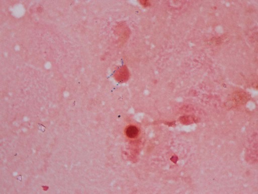
Fluorochrome staining is the screening procedure recommended for laboratories that have a fluorescent (ultraviolet) microscope (see Procedure 43-3 on the Evolve site). Fluorochrome stain is more sensitive than the conventional carbolfuchsin stains, because the fluorescent bacilli stand out brightly against the background (Figure 43-3). Because the smear can be examined initially at lower magnifications (×250 to ×400), more fields can be visualized in a short period. In addition, a positive fluorescent smear may be restained using the conventional Ziehl-Neelsen or Kinyoun procedure, thereby saving the time needed to make a fresh smear. Screening of specimens with rhodamine or rhodamine-auramine results in a higher yield of positive smears and substantially reduces the time needed to examine smears.

One drawback of the fluorochrome stains is that many rapid-growers may not appear fluorescent with these reagents. All positive fluorescent smears should be confirmed with a Ziehl-Neelsen stain or by examination by another technologist. It is important to wipe the immersion oil from the objective lens after examining a positive smear, because stained bacilli can float off the slide into the oil, possibly contributing to a false-positive reading for the next smear examined.

Fuchsin Acid-Fast Stains.

The classic carbolfuchsin stain (Ziehl-Neelsen) requires heating of the slide for better penetration of the stain into the mycobacterial cell wall; hence, it is also known as the hot stain procedure (see Procedure 6-3 on the Evolve site). With Ziehl-Neelsen staining, Mycobacterium spp. appear red or have a red-blue, beaded appearance, whereas nonmycobacteria appear blue.

Procedure 6-4, which can be found on the Evolve site, describes the Kinyoun acid-fast stain. The method is similar to Ziehl-Neelsen staining, but no heat is used (see Figure 43-3); this technique is known as the cold stain procedure. If present, typical acid-fast bacilli appear as purple to red, slightly curved, short or long rods (2 to 8 µm); they also may appear beaded or banded (M. kansasii). For some nontuberculous species, such as M. avium complex, they appear pleomorphic, usually coccoid.

Examination, Interpretation, and Reporting of Smears.

Before a smear is reported as negative, it should be examined carefully by scanning at least 300 oil immersion fields (magnification ×1000), equivalent to three full horizontal sweeps of a smear that is 2 cm long and 1 cm wide. Because the fluorescent stain can be examined using a lower magnification (×250 or ×450) than that required for a fuchsin-stained smear, the equivalent number of fields (30) can be examined in less time, which makes the fluorochrome stain the preferred method.

When acid-fast organisms are observed on a smear, the report should include information about the type of staining method used and the quantity of organisms. The recommended interpretations and ways to report smear results are shown in Table 43-7.

TABLE 43-7

Acid-Fast Smear Reporting

Number of AFB Seen Fuchsin Stain
(1000× Magnification)
Number of AFB Seen Fluorochrome Stain
(450× Magnification)
Number of AFB Seen Fluorochrome Stain
(250× Magnification)
Report
0 0 0 No AFB seen
1-2/300 fields 1-2/70 fields 1-2/30 fields Doubtful; request another specimen
1-9/100 fields 2-18/50 fields 1-9/10 fields 1+
1-9/10 fields 4-36/10 fields 1-9/field 2+
1-9/field 4-36/field 10-90/field 3+
>9/field >36/field >90/field 4+

image

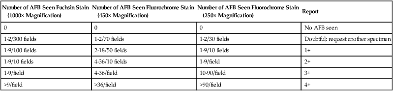
AFB, Acid-fast bacilli.

Modified from Kent PT, Kubica GP: Public health mycobacteriology: a guide for the level III laboratory, US Department of Health and Human Service, Public Health Service, Washington, DC, 1985, Centers for Disease Control and Prevention; and Versalovic J: Manual of clinical microbiology, ed 10, 2011, Washington, DC, ASM Press.

The overall sensitivity of an acid-fast smear ranges from 20% to 80%. Factors such as specimen type, staining method, and culture method can influence the acid-fast smear sensitivity. In general, specificity of acid-fast smear examination is very high. However, cross-contamination of slides during the staining process and use of water contaminated with saprophytic mycobacteria can lead to false-positive results. Staining receptacles should not be used; acid-fast bacilli can also be transferred from one slide to another in immersion oil. For these reasons, the best course is to confirm a positive result.

Although not without some limitations, because of its simplicity and speed, the stained smear is an important and useful test, particularly for detection of smear-positive patients (“infectious reservoirs”), who pose the greatest risk to others in their environment.

Antigen-Protein Detection

The detection of microbial products or components has been used in recent years to diagnose infections caused by M. tuberculosis. For example, tuberculostearic acid is a fatty acid that can be extracted from the cell wall of mycobacteria and detected by gas chromatography or mass spectrometry in clinical samples containing few mycobacteria. Because of the limited number of species that can cause meningitis and because M. tuberculosis appears to be the only one of these species that releases tuberculostearic acid into the surrounding environment, the presence of this substance in CSF is thought to be diagnostic of tuberculous meningitis. Performance of this assay is limited to a few laboratories. Various immunoassays for antigen detection directly in clinical specimens, including sputum and CSF, have been evaluated and show some promise.

Production of adenosine deaminase, a host enzyme, is increased in certain infections caused by M. tuberculosis. For example, elevated levels of this enzyme were found in most patients with tuberculous pleural effusions (98% sensitive); the test for the enzyme also was determined to be highly specific (96% specificity).

Immunodiagnostic Testing

As previously discussed, interferon-gamma release assays have become more widely used for the diagnosis of tuberculosis. The available test systems, T-SPOT-TB (Oxford Immunotec, Oxford, United Kingdom) and QuantiFERON Gold In-Tube (QFNG-IT; Cellestis, Chadstone, Victoria, Australia), do not typically cross react with nontuberculous mycobacterium, are not affected by the BCG vaccine, and are not as variable as the historical serologic tuberculin skin tests. The T-SPOT-TB assay is an enzyme-linked immunospot assay that requires isolation and incubation of peripheral blood mononuclear cells (PBMCs). It takes approximately 2 days and is technically complicated. The QFNG-IT assay measures the stimulation of T-cell interferon-gamma in whole blood in a tube precoated with M. tuberculosis antigens. It yields results in approximately 8 hours. Neither assay distinguishes between latent and active infections. In addition, specificity and sensitivity vary in the population tested, including immunocompromised patients and children. Variation is associated with the patient’s CD4 cell count; therefore, interpretations and results should be evaluated with caution.

Genetic Sequencing and Nucleic Acid Amplification

Subsequent to the introduction of commercially available hybridization assays, commercially available and in-house–developed nucleic acid amplification tests were used successfully for early identification of M. tuberculosis complex grown in liquid cultures. Currently, PCR-based sequencing for mycobacterial identification consists of PCR amplification of mycobacterial DNA with genus-specific primers and sequencing of the amplicons. The organism is identified by comparison of the nucleotide sequence with reference sequences. The most reliable sequence for identification of mycobacteria is the approximately 1500 bp 16S rRNA gene. However, only a 600 bp sequence at the 5′ end is required for identification. The sequence homogeneity in the M. tuberculosis complex prevents the use of this sequence to differentiate these species. This region contains both conserved and variable regions, which makes it an ideal target for identification purposes.

Despite the accuracy of PCR-based sequencing to identify mycobacteria, problems remain: the sequences in some databases are not accurate; no present consensus exists as to the quantitative definition of a genus or species based on 16S rRNA gene sequence data; and procedures are not standardized. In addition, the 16S rRNA 5′ region contains two hypervariable regions, A and B. The A region provides the signature sequences for species identification. However, M. chelonae and M. abscessus both require additional sequencing, because the A and B regions are identical and the 3′ end of the 16srRNA contains a 4-bp sequence difference.

Several other genes have also been used to identify mycobacterial species, including the 23S rRNA, ITS 1, hsp65, rpoB, and gyrB gene. The 23S rRNA sequence is 3100 bp in length, which limits accurate sequencing. ITS 1 is a spacer sequence located between the 16S and 23S rRNA genes. This sequence, which is only 200 to 330 bp, is more easily analyzed. The limitation of this sequence is that it is not a genus-specific sequence; therefore, results may be affected by contaminating bacteria. The 65 kDa heat shock protein, also referred to as the groEL2 gene, is a 440-bp fragment that can be amplified and analyzed with restriction digestion, followed by agarose electrophoresis. The hsp65 is highly conserved but contains a greater variation in polymorphisms than the 16S RNA, particularly in a 441-bp region referred to as the “Telanti fragment.” This allows for differentiation of Mycobacterium species based on the variation in restriction fragment length polymorphisms (RFLPs). Repetitive sequence–based PCR, Diversilab (Biomérieux, Durham, N.C.), demonstrates better species discrimination than RFLP. In addition, a commercially available system in which the 16S to 23S rRNA spacer region of mycobacterial species (INNO-LiPA Mycobacteria; Innogenetics, Ghent, Belgium) has been successfully used to directly detect and identify several of the most clinically relevant mycobacterial species in aliquots of positive liquid culture. However, caution should be used in interpretation of results, because some cross reactivity has occurred with closely related species.

Another commercial system, GenoType Mycobacterium (Hain Lifescience GmbH, Nehrin, Germany), which uses a similar format, has additional probes from M. celatum, M. malmoense, M. peregrinum, M. phlei, and two subgroups of M. fortuitum, in addition to a supplemental kit that allows for 16 additional mycobacterial species. Yet another commercial system, MicroSeq500 16S rRNA (Applied Biosystems, Foster City, California), sequences a 500-bp region and uses a comparative database for species identification.

The rpoB gene encodes the beta-subunit in the organism’s RNA polymerase. Mutations in this gene confer rifampin resistance to M. tuberculosis. Different regions in this gene have been used to identify rapid-growing isolates, but little data are available for the slow-growing species. Finally, the gyrB gene encodes the beta-subunit in the organism’s topoisomerase II. Several single nucleotide polymorphisms have been identified in this gene that are useful in distinguishing species in the M. tuberculosis complex. After amplification, identification and differentiation of species requires restriction analysis and gel electrophoresis.

Additional molecular techniques, such as conventional and real-time PCR, have been used to detect M. tuberculosis directly in clinical specimens. For example, the Amplicor Mycobacterium tuberculosis test (Roche Diagnostic Systems, Branchburg, New Jersey) uses PCR to detect M. tuberculosis directly in respiratory specimens. The Amplified Mycobacterium tuberculosis Direct Test (AMTD; Gen-Probe, San Diego, California) is based on ribosomal RNA amplification. The Roche assay currently is approved by the FDA for use only on acid-fast, smear-positive specimens, because numerous studies have demonstrated less than optimum sensitivity on smear-negative specimens. Because of subsequent kit modifications that improved sensitivity, Gen-Probe’s assay is approved on both smear-positive and smear-negative specimens. Ribosomal RNA is released from the mycobacteria by means of a lysing agent, sonication, and heat. The specific DNA probe is allowed to react with the extracted rRNA to form a stable DNA-RNA hybrid. Any nonhybridized DNA–acridinium ester probes are chemically degraded. When an alkaline hydrogen peroxide solution is added to elicit chemiluminescence, only the hybrid-bound acridinium ester is available to emit light; the amount of light emitted is directly related to the amount of hybridized probe. The light produced is measured on a chemiluminometer. Numerous laboratories have incorporated these tests into their routine procedures.

The Amplicor test (Roche Diagnostic Systems), which uses TaqMan technology and is a real-time PCR test, has received FDA approval for smear-positive respiratory specimens from patients suspected of having tuberculosis. These tests are limited in the number of species they are able to identify. Clinical laboratories have developed their own PCR assays to detect M. tuberculosis directly in clinical specimens.

Line probe assays (DNA strip assays) involve PCR amplification coupled with a reverse hybridization step. The target sequence is amplified using biotinylated primers. The amplicon is then hybridized to membrane-immobilized, sequence-specific probes for each species. The membrane is developed using an enzyme-mediated reaction and color indicator to analyze the banding pattern. Banding patterns are species specific based on the immobilized probe map on the membrane. A commercially available line probe assay (GenoType MTBC; Hain Lifescience, Nehren, Germany) enables the identification of M. tuberculosis complex organisms at the species level using the 23S rRNA.

In addition to assays developed in-house and the Genotype MTBC, five non-FDA-approved commercial amplification tests are widely used outside the United States. The Artus M. tuberculosis PCR kit (Qiagen GmbH, Hilden/Hamburg, Germany) assay uses real-time PCR for amplification of the 16S rRNA gene; the ProbeTec Direct TB energy transfer system (Becton Dickinson, Sparks, Maryland) uses strand displacement amplification technology; the RealArt M. tuberculosis TM PCR reagents (Abbott Laboratories, Abbott Park, Illinois) is a real-time PCR assay using the ABI Prism 7000 system; and the Loop-mediated isothermal amplification test (Eiken Chemical, Tokyo) uses isothermal amplification and UV light detection. These systems have sensitivities and specificities comparable to those of the FDA-approved amplification assays. The GeneXpert system (Cepheid, Sunnyvale, California), which is used for real-time PCR detection of M. tuberculosis complex and resistance to rifampin, uses amplification of the rpoB gene previously discussed in this section.

Currently no molecular assays are available for direct detection of nontuberculous mycobacteria. In 2004, the Centers for Disease Control established a national tuberculosis genotyping system. Details and updates are available at http://www.cdc.gov/tb/programs/default.htm.

Chromatographic Analysis

Analysis of mycobacterial lipids by chromatographic methods, including thin-layer chromatography, gas-liquid chromatography (GLC), capillary gas chromographic methods, and reverse-phase high-performance liquid chromatography (HPLC), has been used to identify mycobacteria. In HPLC, a liquid mobile phase is combined with various technical advances to separate large cellular metabolites and components. HPLC of extracted mycobacteria is a specific and rapid method for identifying species. Many state health departments and the CDC now use this method routinely. The long-chain mycolic acids are separated better by HPLC than by GLC, because they do not withstand the high temperatures needed for GLC. The patterns produced by different species are very easily reproducible, and a typical identification requires only a few hours.

Solid Media

Solid media, such as those listed in Box 43-3, are recommended because of the development of characteristic, reproducible colonial morphology, good growth from small inocula, and a low rate of contamination. Optimally, at least two solid media (a serum [albumin] agar base medium, [e.g., Middlebrook 7H10] and an egg-potato base medium [e.g., Löwenstein-Jensen, or L-J]) should be used for each specimen (these media are available from commercial sources). All specimens must be processed appropriately before inoculation. It is imperative to inoculate test organisms to commercially available products for quality control (see Procedure 43-4 on the Evolve site).

Box 43-3   Suggested Media for Cultivation of Mycobacteria from Clinical Specimens*

Media Comments
Solid
AGAR BASED—GROWTH WITHIN 10 TO 12 DAYS
Middlebrook Contains 2% glycerol, which enhances the growth of Mycobacterium avium complex (MAC).
Middlebrook 7H10
Middlebrook 7H10 selective
Supplemented with carbenicillin (for inhibition of pseudomonads), polymyxin B, trimethoprim lactate, and amphotericin B
Middlebrook 7H11
Middlebrook 7H11 selective
Contains 0.1% enzymatic hydrolysate of casein, which improves recovery of isoniazid-resistant M. tuberculosis)
Middlebrook 7H11 Supplemented with mycobactin J, which provides for growth of M. genovense
Middlebrook 7H11 thin pour plates, 10 × 90 mm (Remel, Lenexa, Kansas) Enhances visibility of colonies within 11 days
Middlebrook biplate (7H10/7H11S agar)  
EGG BASED—GROWTH WITHIN 18 TO 24 DAYS
Löwenstein-Jensen (L-J) Commonly used medium; good recovery of M. tuberculosis but poor recovery of many other species; M. genovense fails to grow
L-J Gruft Supplemented with penicillin and nalidixic acid
L-J Mycobactosel Supplemented with cycloheximide, lincomycin, and nalidixic acid
L-J with pyruvic acid Enhances recovery of M. bovis
L-J with glycerol Enhances recovery of M. ulcerans
Petragnani medium Contains twice the concentration of malachite than Lowenstein-Jensen green (an inhibitor of contaminating organisms); improves recovery from heavily contaminated specimens
Heme-supplemented media (egg or agar based) Supplemented with hemin, hemoglobin or ferric ammonium citrate increases recovery of M. haemophilum.
Liquid
BACTEC 12B medium Used in the MGIT960 system; PANTA is added before incubation; 14C-labeled palmitic acid is metabolized to produce 14CO2, which is detected by the instrument
Middlebrook 7H9 broth  
Dubos Tween albumin  
Septi-Chek AFB 20 mL of Middlebrook 7H9 broth is incubated in 20% CO2; solid phase contains three media: modified L-J, Middlebrook 7H11, and a chocolate agar slab
Media Used in Commercially Supplied Growth and Semiautomated or Fully Automated Systems
Mycobacteria Growth Indicator Tube [MGIT] (Becton Dickinson Microbiology Systems, Cockeysville, Md.) MGIT 460TB (semiautomated system) or MGIT 960 (fully automated system); MGIT is a modified Middlebrook 7H9 broth that incorporates a fluorescence-quenching–based oxygen sensor for detection
MB Redox (Heipha Diagnostica Biotest, Eppelheim, Germany) Nonradiometric medium; a modified Kirchner medium, enriched with additives and antibiotics, that uses tetrazolium salt as the redox indicator
ESP Culture System II and versa TREK Culture System II (Trek Diagnostic Systems, Cleveland, Ohio) Modified Middlebrook 7H9 broth
MB/BacT Alert 3D (bioMérieux, Durham, N.C.) Uses Middlebrook 7H9 broth
BACTEC 9000 MB (Becton Dickinson); recently discontinued by manufacturer Used Middlebrook 7H9 broth

image

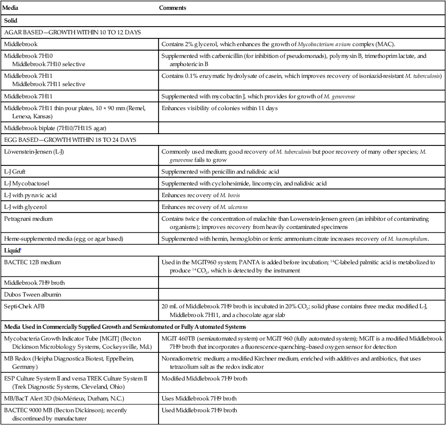
*For optimal recovery of mycobacteria, a minimum combination of liquid medium and solid media is recommended.

Tween 80 added to liquid media acts as a surfactant, breaking up clumps of organisms and increasing recovery rates.

Procedure 43-4   Quality Control for Mycobacteriology

Method

1. Suspend several colonies of H37Rv in a tube containing 3 mL of Middlebrook liquid medium and several plastic or glass beads. Mix vigorously on a test tube mixer, then allow large particles to settle for 15 minutes.

2. Prepare a dilution of approximately 106 organisms per milliliter (106 cells/mL) by adding the above cell suspension drop by drop to 1 mL of buffer until a barely turbid suspension occurs. Transfer 0.5 mL of the 106 cells/mL dilution to 4.5 mL of glycerol broth to give a suspension of 105 cells/mL. Repeat the procedure to make a 104 cells/mL suspension and a 103 cells/mL suspension.

3. Label fifteen 3-dram vials for each suspension (105, 104, and 103). Transfer 0.3 mL of the appropriate suspension to each vial. Store the vials at −70° C to use for future quality control testing.

4. Thaw one vial of each of the three dilutions each time the quality control procedure is performed.

5. Add 2.7 mL of autoclaved sputum to each cell suspension to effect a tenfold dilution, and inoculate three sets of the media used for primary isolation with each of the three dilutions of sputum. Inoculate 0.1 mL of sputum per bottle.

6. Decontaminate and concentrate the remainder as with sputum specimens. Reconstitute the sediments with sterile buffer to 2.6 mL, resuspend vigorously, and inoculate a second set of media with 0.1 mL of each of the concentrated and resuspended samples.

7. Incubate at 35° C in 5% to 10% carbon dioxide (CO2) for 21 days.

Interpreting and Recording Results

Egg media should have been inoculated with approximately 104, 103, and 102 organisms, respectively. The first dilution should produce semiconfluent growth, and the second and third dilutions should produce countable colonies in each bottle. Because of the retrospective nature of these determinations, close comparisons must be made between current and previous results to note trends or developing deficiencies. Failures may be the result of faulty media, lethal effects of decontamination and concentration procedures, improperly prepared reagents, or overexposure of specimens to these reagents. Should deficiencies become evident, techniques should be reviewed and attempts made to determine the source of the problem. New batches of media must be substituted for deficient media, and the latter rechecked to verify deficiencies. Personnel should be included in all discussions of problems and corrective measures. All deficiencies and corrective actions should be recorded in the appropriate section of the quality control records.

Example of Interpreting Quality Control Test Results of Decontamination and Concentration Procedure

  SPUTUM specimen  
Unprocessed-Quantification of Growth Processed Quantification of Growth
Sputum Sample 104 103 102 104 103 102 Interpretation
1 3+ 2+ 50-100 colonies 2+ 1+ or 2+ Approximately 10 colonies Media and decontamination procedures acceptable
2 3+ 2+ 50-100 colonies 1+ 0 0 Media acceptable; procedures too toxic
3 2+ or 1+ 2+ or 1+ 0 1+ or 0 1+ or 0 0 One or more of the media are not supporting growth of acid-fast bacilli (AFB) adequately

image

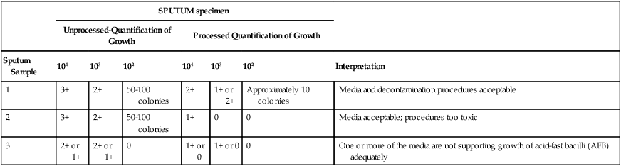
0, No growth; 1+, scanty, barely discernible countable colonies; 2+, dense, discrete growth, not countable; 3+, confluent, abundant growth.

Cultures are incubated at 35° C in the dark in an atmosphere of 5% to 10% carbon dioxide (CO2) and high humidity. Tube media are incubated in a slanted position with screw caps loose for at least 1 week to allow for evaporation of excess fluid and the entry of CO2; plated media are either placed in a CO2-permeable plastic bag or wrapped with CO2-permeable tape. If specimens obtained from the skin or superficial lesions are suspected to contain M. marinum or M. ulcerans, an additional set of solid media should be inoculated and incubated at 25° to 30° C. In addition, a chocolate agar plate (or placement of an X-factor [hemin] disk on conventional media) and incubation at 25° to 33° C is needed for recovery of M. haemophilum from these specimens. RGM optimally require incubation at 28° to 30° C.

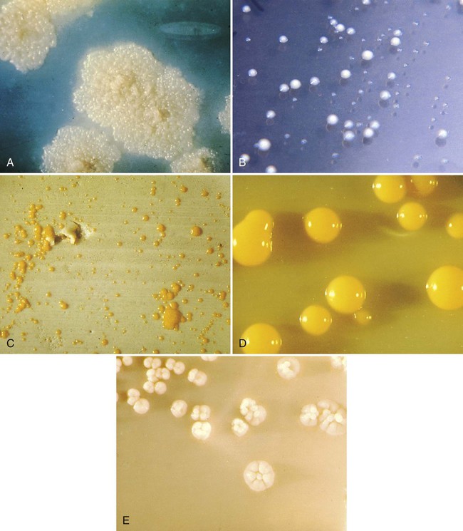
Cultures are examined weekly for growth. Contaminated cultures are discarded and reported as “contaminated, unable to detect presence of mycobacteria”; additional specimens are also requested. If available, sediment may be recultured after enhanced decontamination or by inoculating the sediment to a more selective medium. Most isolates appear between 3 and 6 weeks; a few isolates appear after 7 or 8 weeks of incubation. When growth appears, the rate of growth, pigmentation, and colonial morphology are recorded. The typical colonial appearance of M. tuberculosis and other mycobacteria is shown in Figure 43-4. After 8 weeks of incubation, negative cultures (those showing no growth) are reported, and the cultures are discarded.

Because of the resurgence of tuberculosis in the United States in the late 1980s and early 1990s, significant effort has been put into developing methods to provide more rapid diagnosis of tuberculosis. Welch et al.2 refined a method that reduced the time to detection of mycobacterial growth by half or more, compared with conventional culture methods, by using a thinly poured Middlebrook 7H11 plate. These plates are inoculated in a routine manner, sealed, incubated, and examined microscopically (×40) at regular intervals for the appearance of microcolonies. Presumptive identification of M. tuberculosis or M. avium complex could be made for about 83% of the isolates within 10 and 11 days after inoculation, respectively.

Liquid Media

In general, use of a liquid media system reduces the turnaround time for isolation of acid-fast bacilli to approximately 10 days, compared with 17 days or longer for conventional solid media. Several different systems are available for culturing and detecting the growth of mycobacteria in liquid media. The most commonly used systems are summarized in Table 43-8. Growth of mycobacteria in liquid media, regardless of the type, requires 5% to 10% CO2; CO2 is either already provided in the culture vials or is added according to the manufacturer’s instructions. When growth is detected in a liquid medium, acid-fast staining of a culture aliquot is performed to confirm the presence of acid-fast bacilli, and the material is subcultured to solid agar. Gram staining can also be performed if contamination is suspected.

TABLE 43-8

Liquid Media Systems Commonly Used to Culture and Detect the Growth of Mycobacteria

System Basic Principles of Detection
BACTEC 460 TB (Becton Dickinson Diagnostic Systems, Cockeysville, Md.) Culture medium contains 14C-labeled palmitic acid. If present in the broth, mycobacteria metabolize the 14C-labeled substrates and release radioactively labeled 14CO2 in the atmosphere, which collects above the broth in the bottle. The instrument withdraws this carbon dioxide (CO2)-containing atmosphere and measures the amount of radioactivity present. Bottles that yield a radioactive index, called a growth index, greater than or equal to 10 are considered positive.
Septi-Chek AFB System (Becton Dickinson) Biphasic culture system made up of a modified Middlebrook 7H9 broth with a three-sided paddle containing chocolate, egg-based, and modified 7H11 solid agars. After inoculation, a supplement is added to the liquid that includes glucose, glycerol, oleic acid, pyridoxal HCl, catalase, albumin, and antibiotics (PANTA). The bottle is inverted regularly to inoculate the solid media. Growth is detected by observing the three-sided paddle.
Mycobacteria Growth Indicator Tube (MGIT) (Becton Dickinson) A culture tube contains Middlebrook 7H9 broth and a fluorescent compound embedded in a silicone sensor. Growth is detected visually using an ultraviolet light. Oxygen (O2) diminishes the fluorescent output of the sensor; therefore, O2 consumption by organisms present in the medium is detected as an increase in fluorescence under ultraviolet (UV) light at 365 nm. The MGIT medium is supplemented with oleic acid-albumin-dextrose and PANTA before incubation.
Continuous Growth Monitoring Systems  
Versa TREK (TREK Diagnostic Systems, Cleveland, Ohio) Organisms are cultured in a modified Middlebrook 7H9 broth with enrichment and a cellulose sponge to increase the culture’s surface area. The instrument detects growth by monitoring pressure changes that occur as a result of O2 consumption or gas production by the organisms as they grow.
BacT/Alert System (bioMérieux, Durham, N.C.) Organisms are cultured in modified Middlebrook 7H9 broth. The instrument detects growth by monitoring CO2 production by means of a colorimetric CO2 sensor in each bottle.
BACTEC 9000 MB (Becton Dickinson- recently discontinued) Organisms are cultured in a modified Middlebrook 7H9 broth. The instrument detects growth by monitoring O2 consumption by means of a fluorescent sensor.
BACTEC MGIT 960 (Becton Dickinson) See above for basic principle (MGIT). The instrument detects growth by monitoring O2 consumption by means of a fluorescent sensor.
MB Redox (Heipha Diagnostica Biotest, Eppelheim, Germany) This is a nonradiometric medium. It is a modified Kirchner medium enriched with additives and antibiotics, and tetrazolium salt is the redox indicator. AFB are identified as pink to purple, pinhead-sized particles.

Approach to Identification

Regardless of the identification methods used, the first test always performed on organisms growing on solid or liquid mycobacterial media is acid-fast staining, to confirm that the organisms are indeed mycobacteria. Identification of species other than MAC and the more frequently isolated NTM (MAC, M. avium, M. intracellulare, M. gordonae, and M. kansasii) has become challenging for routine clinical microbiology laboratories, particularly in light of the ever-increasing number of new mycobacterial species. Traditional methods (i.e., phenotypic methods) for identifying mycobacteria, particularly the NTM, are based on growth parameters, biochemical characteristics, and analysis of cell wall lipids, all of which are slow, cumbersome, and often inconclusive procedures. Over the past decade, the rate of non-AIDS–associated infections has been increasing, and many of the newly identified NTM species have been associated with various diseases. As a result, identification of species is vital to selecting effective antimicrobial therapy and to deciding whether to perform susceptibility testing on accurately speciated NTM. Most of the newer species have been identified using nucleic acid sequencing with limited published phenotypic characteristics. Because of these issues and limitations with conventional phenotypic methods for identification, molecular and genetic investigations are becoming indispensable to identify the NTM accurately. Therefore, for timely and accurate identification of mycobacteria, molecular approaches in conjunction with some phenotypic characteristics should be used.

Regardless of whether molecular or phenotypic methods are used, when growth is detected, broth subcultures of colonies growing in liquid media or on solid media (several colonies inoculated to Middlebrook 7H9 broth [5 mL] and incubated at 35° C for 5 to 7 days with daily agitation to enhance growth) are then used to determine pigmentation and growth rate and to inoculate all test media for biochemical tests, if performed. Additional cultures may be inoculated and then incubated at different temperatures when more definitive identification is needed.

Conventional Phenotypic Tests

Growth Characteristics.

Preliminary identification of mycobacterial isolates depends on the organisms’ rate of growth, colonial morphology (see Figure 43-4), colonial texture, pigmentation and, in some instances, the permissive incubation temperatures of mycobacteria. Despite the limitations of phenotypic tests, the mycobacterial growth characteristics are helpful for determining a preliminary identification (e.g., an isolate appears as rapidly growing mycobacteria). To perform identification procedures, quality control organisms should be tested along with unknowns (Table 43-9). The commonly used quality control organisms can be maintained in broth at room temperature and transferred monthly. In this way they are always be available for inoculation to test media along with suspensions of the unknown mycobacteria being tested.

TABLE 43-9

Controls and Media Used for Biochemical Identification of Mycobacteria

CONTROL ORGANISMS RESULT
Biochemical Test Positive Negative Positive Negative Medium Used Duration Incubation Conditions
Niacin M. tuberculosis M. intracellulare Yellow No color change 0.5 mL DH2O 15-30 min Room temperature
Nitrate M. tuberculosis M. intracellulare Pink or red No color change 0.3 mL DH2O 2 hours 37°C bath
Urease M. fortuitum M. avium Pink or red No color change Urea broth for AFB 1, 3, and 5 days 37°C incubator (without CO2)
68°C Catalase M. fortuitum or M. gordonae M. tuberculosis Bubbles No bubbles 0.5 mL phosphate buffer (pH, 7.0) 20 min 68°C bath
SQ Catalase M. kansasii or M. gordonae M. avium >45 mm <45 mm Commercial medium 14 days 37°C incubator (with CO2)
Tween 80 M. kansasii M. intracellulare Pink or red No color change 1 mL DH2O 5 or 10 days 37°C incubator (in the dark, without CO2)
Tellurite M. avium M. tuberculosis Smooth, fine, black precipitate (smokelike action) Gray clumps (no smokelike action) Middlebrook 7H9 broth 7, then 3 additional days 37°C incubator (with CO2)
Arylsulfatase M. fortuitum M. intracellulare Pink or red No color change Wayne’s arylsulfatase medium 3 days 37°C incubator (without CO2)
5% NaCl M. fortuitum M. gordonae Substantial growth Little or no growth Commercial slant with and without 5% NaCl 28 days 37°C incubator (with CO2)
TCH M. bovis M. tuberculosis No growth (i.e., susceptible) Growth (i.e., resistant or ≥1% of colonies are resistant) TCH slant 3 weeks 37°C incubator (with CO2)

image

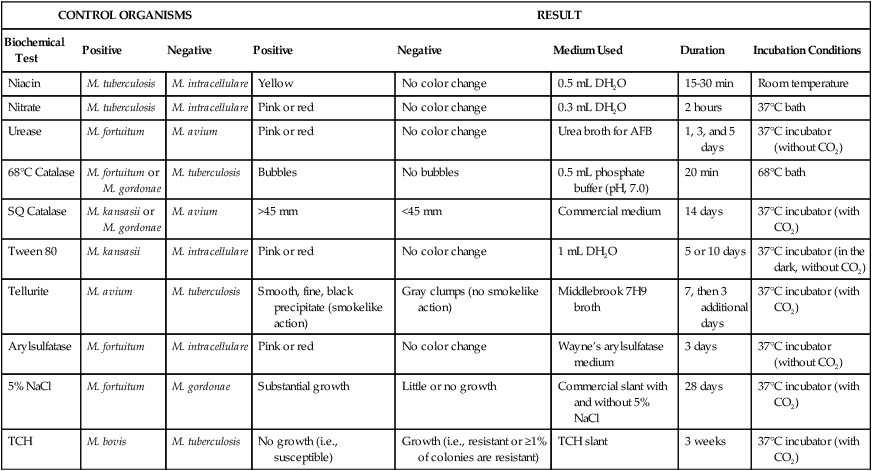
AFB, Acid-fast bacilli; CO2, carbon dioxide; DH2O, distilled water; NaCl, sodium chloride; SQ, semiquantitative; TCH, thiophene-2-carboxylic acid hydrazide.

Growth Rate.

The rate of growth is an important criterion for determining the initial category of an isolate. Rapid-growers usually produce colonies within 3 to 4 days after subculture. However, even a rapid-grower may take longer than 7 days to initially produce colonies because of inhibition by a harsh decontaminating procedure. Therefore, the growth rate (and pigment production) must be determined by subculture (see Procedure 43-5 on the Evolve site). The dilution of the organism used to assess the growth rate is critical. Even slow-growing mycobacteria appear to produce colonies in less than 7 days if the inoculum is too heavy. One organism particularly likely to exhibit false-positive rapid growth is M. flavescens. This species therefore serves as an excellent quality control organism for this procedure.

Procedure 43-5   Determination of Pigment Production and Growth Rate

Method

1. After the broth culture has incubated for 5 to 7 days, adjust the turbidity to that of a McFarland 0.5 standard. (104 concentration)

2. Inoculate 0.1 mL of the diluted broth to each of three tubes of Löwenstein-Jensen agar. Completely wrap two of the tubes in aluminum foil to block all light. If the isolate was obtained from a skin lesion or the initial colony was yellow pigmented (possible M. szulgai colony), inoculate six tubes. If six tubes are used, the second set of tubes, two of them also wrapped with aluminum foil, is incubated at 30° C or at room temperature if a 30° C incubator is not available.

3. Examine the cultures after 5 and 7 days for grossly visible colonies. Examine again at intervals of 3 days. Interpretation: Rapid-growers produce visible colonies in less than 7 days; slow-growers require more than 7 days.

4. When the colonies are mature, expose the growth from a foil-wrapped tube to a bright light, such as a desk lamp, for 2 hours. The cap must be loose during exposure, because pigment production is an oxygen-dependent reaction. Then rewrap the tube and return it to the incubator, leaving the cap loose.

5. Examine the three tubes 24 and 48 hours after light exposure. For tubes incubating at 30° C, pigment may require 72 hours for development.

Pigment Production.

As previously discussed, mycobacteria may be categorized into three groups based on pigment production. Procedure 43-5, which can be found on the Evolve site, describes how to determine pigment production. To achieve optimum photochromogenicity, colonies should be young, actively metabolizing, isolated, and well aerated. Although some species (e.g., M. kansasii) turn yellow after a few hours of light exposure, others (e.g., M. simiae) may take prolonged exposure to light. Scotochromogens produce pigmented colonies even in the absence of light, and colonies often become darker with prolonged exposure to light (Figure 43-5). One member of this group, M. szulgai, is peculiar in that it is a scotochromogen at 35° C and nonpigmented when grown at 25° to 30° C. For this reason, all pigmented colonies should be subcultured to test for photoactivated pigment at both 35° C and 25° to 30° C. Nonchromogens are not affected by light.

Biochemical Testing.

Once categorized into a preliminary subgroup based on its growth characteristics, an organism must be definitively identified to species or complex level. Although conventional biochemical tests can be used for this purpose, new methods (discussed later in this section) have replaced biochemical tests for identifying mycobacterial species because of the previously discussed limitations of phenotypic testing. Although key biochemical tests are still discussed in this edition, the reader must be aware that this approach to identification ultimately will be replaced by molecular methods. Table 43-10 summarizes distinctive properties of the more commonly cultivable mycobacteria isolated from clinical specimens; key biochemical tests for each of the major mycobacterial groupings, including M. tuberculosis complex, are listed in Table 43-11. The following sections address key biochemical tests.

TABLE 43-10

Distinctive Properties of Commonly Cultivable Mycobacteria Encountered in Clinical Specimens

Group/Complex Species Optimal Temp (°C) Usual Colonial Morphologya Niacin Growth on TCH (10 mg/mL)b Nitrate Reduction Semi quantitative Catalase (>45 mm) 68° C Catalase Tween Hydrolysis, 5 Days Tellurite Reduction Tolerance to 5% NaCl Arylsulfatase, 3 Days Iron Uptake Growth on MacConkey Agar Urease Pyrazinamidase, 4 Days
M. tuberculosis complex M. tuberculosis 37 R + + + c image ± +
M. bovis 37 Rt image ±
M. africanum 37 R V V V +
Photochromogens M. marinum 30 S/SR image + + image imaged −/+ +
M. kansasii 35 SR/S + + + + + image +
M. simiae 37 S ± + + + +   ± +
M. asiaticum 37 S + + + +  
Scotochromogens M. scrofulaceum 37 S + + + image V V ±
M. szulgai 37 S or R + + + + imagec ± V + +
M. gordonae 37 S + + + + V V ±
Nonphotochromogens M. avium complex 35-37 St/R + + ± + image +
M. genavensee 37 St + + +       + +
M. gastri 35 S/SR/R + + + image image
M. malmoense 30 S + ± + +   +
M. haemophilumf 30 R + +
M. shimoidei 37 R + +   +
M. ulcerans 30 R + +      
M. flavescensg 37 S + + + + + image + + +
M. xenopih 42 Sf + + image ± V
M. terrae complex
M. terrae
M. trivialei
M. nonchromogenicum
35 SR + + + + V V
Rapidly growing M. fortuitum group 28-30 Sf/Rf + + + + V + + + + + + +
M. chelonae 28-30 S/R −/+ + + V V + + + + +
M. abscessus 28-30 S/R   + V V + + + + +  
M. smegmatis 28-30 R/S + + + + + + + +    

image

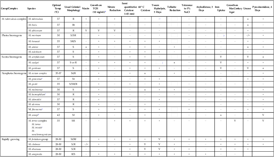
Plus and minus signs indicate the presence or absence, respectively, of the feature; blank spaces indicate either that the information is not currently available or that the property is unimportant.

V, Variable; ±, usually present; image, usually absent.

See Versalovic J: Manual of clinical microbiology, ed 10, Washington, DC, 2011, ASM Press, for biochemical reactions of other mycobacterial species and for additional biochemical reactions on the mycobacteria included in this table.

aR, Rough; S, smooth; SR, intermediate in roughness; t, thin or transparent; f, filamentous extensions.

bTCH, Thiophene-2-carboxylic acid hydrazide.

cTween hydrolysis may be positive at 10 days.

dArylsulfatase, 14 days, is positive.

eRequires mycobactin for growth on solid media.

fRequires hemin as a growth factor.

gYoung cultures may be nonchromogenic or have only pale pigment that may intensify with age.

hStrains of M. xenopi can be nonphotochromogenic or scotochromogenic.

iM. triviale is tolerant to 5% NaCl, and a rare isolate may grow on MacConkey agar.

TABLE 43-11

Key Biochemical Reactions to Help Differentiate Organisms Belonging to the Same Mycobacterial Group

Mycobacterial Group Key Biochemical Tests
M. tuberculosis complex Niacin, nitrate reduction; susceptibility to thiophene-2-carboxylic acid hydrazide (TCH) if M. bovis is suspected
Photochromogens Tween 80 hydrolysis, nitrate reduction, pyrazinamidase, 14-day arylsulfatase, urease, niacin
Scotochromogens Permissive growth temperature, Tween 80 hydrolysis, nitrate reduction, semiquantitative catalase, urease, 14-day arylsulfatase
Nonphotochromogens Heat-resistant and semiquantitative catalase activity, nitrate reduction, Tween 80 hydrolysis, urease, 14-day arylsulfatase, tellurite reduction, acid phosphatase activity
Rapidly growing Growth on MacConkey agar, nitrate reduction, Tween 80 hydrolysis, 3-day arylsulfatase, iron uptake

Niacin.

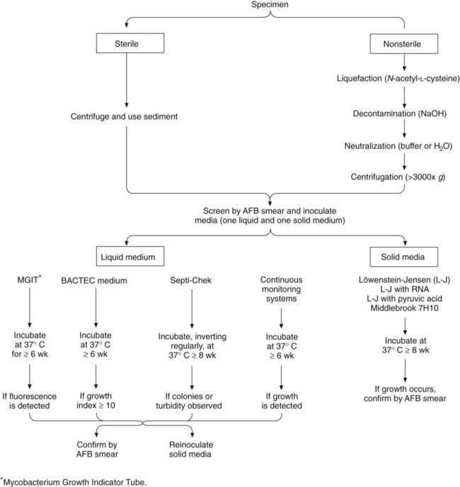
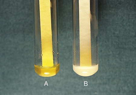
Niacin (nicotinic acid) plays an important role in the oxidation-reduction reactions that occur during mycobacterial metabolism. Although all species produce nicotinic acid, M. tuberculosis accumulates the largest amount. (M. simiae and some strains of M. chelonae also produce niacin.) Niacin therefore accumulates in the medium in which these organisms are growing. A positive niacin test (see Procedure 43-6 on the Evolve site) is preliminary evidence that an organism that exhibits a buff-colored, slow-growing, rough colony may be M. tuberculosis (Figure 43-6). However, this test is not sufficient to confirm identification. If sufficient growth is present on an initial L-J slant (the egg-base medium enhances accumulation of free niacin), a niacin test can be performed immediately. If growth on the initial culture is scant, the subculture used for growth rate determination can be used. If this culture yields only rare colonies, the colonies should be spread around with a sterile cotton swab (after the growth rate has been determined) to distribute the inoculum over the entire slant. The slant then is incubated until light growth over the surface of the medium is visible. For reliable results, the niacin test should be performed only from cultures on L-J medium that are at least 3 weeks old and show at least 50 colonies; otherwise, enough detectable niacin might not have been produced.

Procedure 43-6   Niacin Test Performed with Commercially Available Filter Paper Test Strips*

Method

1. Add 1 mL of sterile distilled water to the surface of the egg-based medium on which the colonies to be tested are growing.

2. Lay the tube horizontally so that the fluid is in contact with the entire surface. Using a pipette, scratch or lightly poke through the surface of the agar; this allows the niacin in the medium to dissolve in the water.

3. Allow the tube to sit for up to 30 minutes at room temperature. It can incubate longer to achieve a stronger reaction.

4. Remove 0.6 mL of the distilled water (which appears cloudy at this point) to a clean, 12 × 75 mm screw cap or snap top test tube. Insert a niacin test strip with the arrow down, following the manufacturer’s instructions.

5. Cap the tube tightly and incubate at room temperature, occasionally shaking the tube to mix the fluid with the reagent on the bottom of the strip.

6. After 20 minutes, observe the color of the liquid against a white background (see Figure 45-6).


*Manufactured by Remel, Lenexa, Kansas.

Nitrate Reduction.

This test is valuable for identifying M. tuberculosis, M. kansasii, M. szulgai, and M. fortuitum. The ability of acid-fast bacilli to reduce nitrate is influenced by the age of the colonies, temperature, pH, and enzyme inhibitors. Although rapid-growers can be tested within 2 weeks, slow-growers should be tested after 3 to 4 weeks of luxuriant growth. Commercially available nitrate strips yield acceptable results only with strongly nitrate-positive organisms, such as M. tuberculosis. This test may be tried first because of its ease of performance. The M. tuberculosis–positive control must be strongly positive in the strip test, or the test results are unreliable. If the paper strip test is negative or if the control test result is not strongly positive, the chemical procedure (see Procedure 43-7 on the Evolve site) must be carried out using strong and weakly positive controls.

Procedure 43-7   Nitrate Reduction Test Using Chemical Reagents

Method

Sulfanilic acid (Sigma Chemical Co., St Louis, Mo.) 1 part
N-(1-Naphthyl)ethylenediamine dihydrochloride (Eastman Chemical Co., Rochester, N.Y.) 1 part
L-tartaric acid (Sigma Chemical Co.) 10 parts

These crystals can be measured with any small scoop or tiny spoon, because the proportions are by volume, not weight. The mixture should be ground in a mortar and pestle to ensure adequate mixing, because the crystals have different textures. The reagent can be stored in a dark glass bottle at room temperature for at least 6 months.

2. Add 0.2 mL sterile distilled water to a 16 × 125 mm screw cap tube. Emulsify two very large clumps of growth from a 4-week culture on Löwenstein-Jensen agar in the water. The suspension should be milky.

3. Add 2 mL nitrate substrate broth (Becton-Dickinson, Franklin Lakes, N.J. or Remel, Lenexa, Kansas) to the suspension and cap tightly. Shake gently and incubate upright for 2 hours in a 35° C water bath.

4. Remove from water bath and add a small amount of the crystalline reagent. A wooden stick or a small spatula can be used to add crystals; the amount is not critical. Examine immediately.

Catalase.

Most species of mycobacteria, except for certain strains of M. tuberculosis complex (some isoniazid-resistant strains) and M. gastri, produce the intracellular enzyme catalase, which splits hydrogen peroxide into water and oxygen. Catalase can be assessed by using the semiquantitative catalase test or the heat-stable catalase test.

• The semiquantitative catalase test is based on the relative activity of the enzyme, as determined by the height of a column of bubbles of oxygen (Figure 43-7) formed by the action of untreated enzyme produced by the organism. Based on the semiquantitative catalase test, mycobacteria are divided into two groups: those that produce less than 45 mm of bubbles and those that produce more than 45 mm of bubbles.

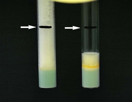
• The heat-stable catalase test is based on the ability of the catalase enzyme to remain active after heating (i.e., it is a measure of the enzyme’s heat stability). When heated to 68° C for 20 minutes, the catalase of M. tuberculosis, M. bovis, M. gastri, and M. haemophilum becomes inactivated.
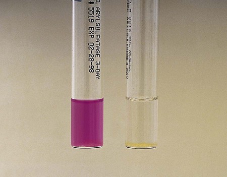
Other Tests.

Other tests are often performed to make more subtle distinctions between species (see Table 43-11). However, performing all the procedures necessary for definitive identification of mycobacteria is not cost-effective for routine clinical microbiology laboratories; therefore, specimens that require further testing can be forwarded to regional laboratories.

Antimicrobial Susceptibility Testing and Therapy

Drug-resistant tuberculosis is a major health threat; more than 500,000 cases of multidrug-resistant (MDR) tuberculosis occur each year. Multidrug-resistant tuberculosis is resistant to rifampin and isoniazid, the two drugs most often used as effective treatment against tuberculosis. In addition, strains of extensively drug-resistant tuberculosis (XDR TB) are emerging that are resistant not only to rifampin and isoniazid, but also to quinolones and other drugs, such as aminoglycosides and capreomycin.

Standardized methods for susceptibility testing, including direct and indirect testing and new molecular tools, currently are available for susceptibility testing.

M. Tuberculosis Complex

In vitro drug susceptibility testing should be performed on the first isolate of M. tuberculosis from all patients. Susceptibility testing of M. tuberculosis requires meticulous care in the preparation of the medium, selection of adequate samples of colonies, standardization of the inoculum, use of appropriate controls, and interpretation of results. Laboratories that see very few positive cultures should consider sending isolates to a reference laboratory for testing. Isolates must be saved in sterile 10% skim milk in distilled water at −70° C for possible future additional studies (e.g., susceptibilities if the patient does not respond well to treatment).

Conventional Methods

The development of primary drug resistance in tuberculosis represents an increase in the proportion of resistant organisms. This increase in resistant organisms results from a spontaneous mutation and subsequent selection to predominance of these drug-resistant mutants by the action of a single or ineffective drug therapy. A poor clinical outcome is predicted with an agent when more than 1% of bacilli in the test population are resistant. If an isolate is reported as resistant to a drug, treatment failure is likely if this drug is used for therapy.

Drug resistance is defined for M. tuberculosis complex in terms of the critical concentration of the drug. The critical concentration of a drug is the amount of drug required to prevent growth above the 1% threshold of the test population of tubercle bacilli.

Four general methods are used throughout the world to determine the susceptibility of M. tuberculosis isolates to various antituberculous agents (Table 43-12). Initial isolates of M. tuberculosis are tested against five antimicrobials, which are referred to as primary drugs (Box 43-4.) If resistance to any of the primary drugs is detected, a second battery of agents is tested (Box 43-4).

TABLE 43-12

Overview of Conventional Methods to Determine Susceptibility of M. tuberculosis Isolates to Antimycobacterial Agents

Method Principle
Absolute concentration For each drug tested, a standardized inoculum is inoculated to control (drug free) media and media containing several appropriately graded drug concentrations. Resistance is expressed as the lowest concentration of drug that inhibits all or almost all of the growth; that is, the minimum inhibitory concentration (MIC)
Resistance ratio The resistance of the test organism is compared with that of a standard laboratory strain. The two strains are tested in parallel by inoculating a standard inoculum to media containing twofold serial dilutions of the drug. Resistance is expressed as the ratio of the MIC of the test strain divided by the MIC for the standard strain for each drug
Proportion For each drug tested, several dilutions of standardized inoculum are inoculated onto control and drug-containing agar media. The extent of growth in the absence or presence of drug is compared and expressed as a percentage. If growth at the critical concentration of a drug is >1%, the isolate is considered clinically resistant. This is the standard method for all drugs except pyrazinamide.
Commercial systems approved for use by the FDA: BACTEC 460TB (Becton Dickinson, Sparks, Md.); BACTEC MGIT 960 (Becton Dickinson); VersaTREK (Trek Diagnostic Systems, Cleveland, Ohio); MB/BacT Alert 3D (bioMérieux, Durham, N.C.) Using the principles of the agar proportion method, these methods use liquid media. Growth is indicated by the amount of 14C-labeled carbon dioxide (CO2) released (as measured by the BACTEC 460 instrument) or the amount of fluorescence or gas produced (as measured by the MB/BacT Alert 3D and VersaTREK systems, respectively). For each drug tested, a standardized inoculum is inoculated into a drug-free and a drug-containing vial. The rate and amount of CO2 produced in the absence or (with the BACTEC MGIT 960) presence of drug are then compared. The BACTEC 460TB is being replaced in many laboratories by the MGIT 960 to avoid hazardous waste disposal of radioactive materials, in addition to the cross-contamination that may occur with the 460TB.
Alternate methods and molecular methods Several molecular methods have been developed that identify the mutations in the rifampin-resistant gene (rpoB). Greater than 96% of rifampin resistance correlates to mutations in an 81 bp segment.
Additional molecular methods have been developed for the identification of resistance to isoniazid, ethambutol, and pyrazinamide.
Molecular methods should be followed up with culture, especially for confirmation of second-line drug resistance and in XDR TB.

FDA, U.S. Food and Drug Administration. XDR TB, extensively drug-resistant tuberculosis.

New Approaches

Several technologies recently introduced show promise of being faster, more reliable, and/or easier to perform than most conventional methods of susceptibility testing. For example, mutations leading to rifampin resistance have been detected using molecular methods. One molecular method, the line probe assay (INNO-LiPA Rif TB; Innogenetics, Ghent, Belgium), is a commercially available, reverse hybridization–based probe assay for rapid detection of rifampin mutations leading to rifampin resistance in M. tuberculosis. Many different genotypic assays are currently available for drug susceptibility testing. Most are based on PCR amplification of a specific region of an M. tuberculosis gene, followed by analysis of the amplicon for specific mutations associated with resistance to a particular drug. The presence or absence of mutations can then be detected by several methods, such as automated sequencing.

As previously mentioned, high-density DNA probe assays (see Chapter 8) have been used to detect rifampin resistance and to identify mycobacterial species identification.

An innovative approach used by Jacobs et al.3 to perform susceptibility testing involved the use of a luciferase-reporter mycobacteriophage (bacterial viruses). The basis for this assay is simple: viable mycobacteria can become infected with and replicate the mycobacteriophage; dead tubercle bacilli cannot. The mycobacteriophage was constructed to have the firefly luciferase gene next to a mycobacterial promoter; therefore, the presence and growth of the mycobacteriophage is detected by chemiluminescence. In brief, the isolate of M. tuberculosis to be tested is grown in the presence and absence of drug, and the specially constructed mycobacteriophage is added. After infection, luciferin, a substrate of luciferase, is added. If organisms are viable (i.e., thereby allowing infection of the bacteriophage and subsequent transcription and translation of the luciferase gene), the luciferin is broken down and light is emitted that can be measured; the amount of light emitted is directly proportional to the number of viable M. tuberculosis organisms. Therefore, if an organism is resistant to the drug, light is emitted; organisms susceptible to the drug do not emit light. Another commercially available assay that uses mycobacteriophages is the FAST Plaque TB–RIF test (Bio Tec Laboratories, Ipswich, UK).

Susceptibility testing should be repeated if the patient remains culture positive after 3 months following appropriate therapy or fails to respond clinically to therapy.

Therapy

Therapy directed against M. tuberculosis depends on the susceptibility of the isolate to various antimicrobial agents. To prevent the selection of resistant mutants, treatment of tuberculosis requires four drugs: isoniazid, rifampin, ethambutol, and pyrazinamide. Initial therapy includes all four drugs for 8 weeks. However, if drug susceptibility is determined for isoniazid, rifampin, and pyrazinamide, ethambutol may be discontinued. This is the preferred therapy for initial treatment, followed by isoniazid and rifampin for an additional 18 weeks. The most common two-drug regimen is isoniazid (INH, also known as isonicotinylhydrazine) and rifampin. The combination is administered for 9 months in cases of uncomplicated tuberculosis; if pyrazinamide is added to this regimen during the first 2 months, the total duration of therapy can be shortened to 6 months. Ethambutol may also be added to the regimen. INH prophylaxis is recommended for individuals with a recent skin test conversion who are disease free.

Nontuberculous Mycobacteria

In general, the treatment of patients infected with NTM requires more individualization of therapy than does the treatment of patients with tuberculosis. This individualization is based on the species of mycobacteria recovered, the site and severity of infection, antimicrobial drug susceptibility results, concurrent diseases, and the patient’s general condition. Currently, sufficient data exist to allow general recommendations for susceptibility testing of MAC, M. kansasii, and M. marinum. Pulmonary infections with M. avium complex are often treated with clarithromycin, rifampin, and ethambutol (or streptomycin or amikacin for severe disease). If the infection is disseminated, clarithromycin, ethambutol, and rifabutin may be prescribed. Pulmonary infections with M. kansasii are treated with isoniazid, rifampin, and ethambutol. M. marinum skin and soft tissue infections may be treated with either clarithromycin and ethambutol, clarithromycin and rifampin, or rifampin and ethambutol.

Susceptibility testing should be performed on clinically significant, rapidly growing mycobacteria (Table 43-13). Skin and soft tissue infections, if susceptible, are treated with clarithromycin and at least one additional drug based on susceptibility testing. Pulmonary infections with M. abscessus should also be treated with a multidrug regimen that includes clarithromycin, if susceptible, and then additional drugs based on susceptibility testing.

Broth-based method;
microdilution or BACTEC Clarithromycin or azithromycin
Second line: Moxifloxacin or linezolid
Isolates typically are intrinsically resistant to isoniazid and pyrazinamide M. kansasii

Agar proportion
Broth based Rifampin
If rifampin resistant, test rifabutin, ethambutol, isoniazid, linezolid, moxifloxacin, streptomycin, clarithromycin, amikacin, ciprofloxacin, and trimethoprim-sulfamethoxazole M. marinum Susceptibility testing not recommended; should be done only if patient fails to respond clinically after several months of therapy and remains culture positive Agar proportion
Agar disk elution
Broth microdilution Rifampin, ethambutol, clarithromycin, doxycycline, minocycline or trimethoprim-sulfamethoxazole Rapidly growing mycobacteria Broth microdilution Amikacin, cefoxitin, ciprofloxacin, clarithromycin, doxycycline, imipenem, linezolid, trimethoprim- sulfamethoxazole, tobramycin, moxifloxacin

image

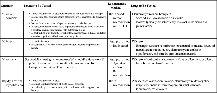
CLSI, Clinical and Laboratory Standards Institute.

Prevention

As previously mentioned, prophylactic chemotherapy with INH is used when known or suspected primary tuberculous infection poses a risk of clinical disease. At present, the BCG vaccine is the only vaccine available against tuberculosis. The effectiveness of this live vaccine is controversial, because studies have demonstrated ineffectiveness to 80% protection. The greatest potential value for this vaccine is in developing countries with high prevalence rates for tuberculosis. At this time, at least four types of antituberculosis vaccines are currently being evaluated in experimental studies in animals.

Case Study 43-1

A 40-year-old man who has tested positive for human immunodeficiency virus (HIV) infection and who is undergoing highly active antiretroviral therapy (HAART) presents with progressive encephalomyeloradiculopathy. He has severe headaches but no fever, cough, or weakness. Cerebrospinal fluid (CSF) is collected. The test results for the specimen are: 25 WBC/mm3 (25 white blood cells per cubic millimeter), low glucose, elevated protein, and no organisms on Gram stain or acid-fast stain. His studies are negative for cryptococcal antigen, Toxoplasma organisms (by serology), and herpes simplex virus (HSV) (by polymerase chain reaction [PCR]). Routine bacterial culture is negative. Despite therapy for HSV and routine aerobic bacterial causes of meningitis, over the next 4 days the patient spikes fevers. A second CSF specimen shows 415 WBC/mm3, with no diagnosis. A battery of viral encephalitis serology tests are done, and all are negative. In-house PCR testing on a third CSF specimen is positive for Mycobacterium tuberculosis, which grows in culture after 4 weeks.