Musculoskeletal System and Connective Tissue


ICD-10-CM Example from Tabular
M43.1 Spondylolisthesis
Excludes 1 acute traumatic of lumbosacral region (S33.1)
acute traumatic of sites other than lumbosacral—code to fracture, vertebra, by region
congenital spondylolisthesis (Q76.2)
M43.10. Spondylolisthesis, site unspecified
M43.11. Spondylolisthesis, occipito-atlanto-axial region
M43.12. Spondylolisthesis, cervical region
M43.13. Spondylolisthesis, cervicothoracic region
M43.14. Spondylolisthesis, thoracic region
M43.15. Spondylolisthesis, thoracolumbar region
M43.16. Spondylolisthesis, lumbar region
M43.17. Spondylolisthesis, lumbosacral region
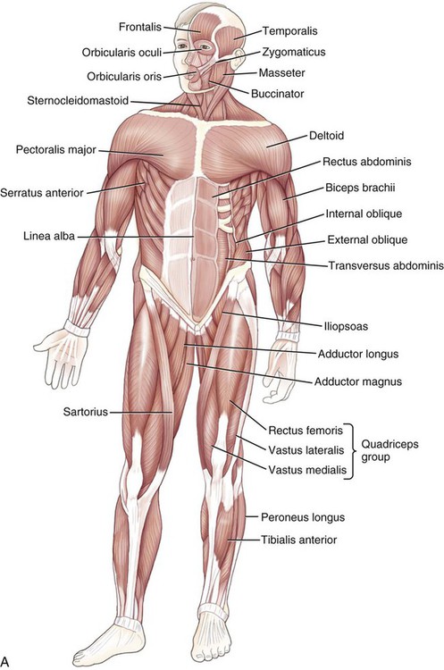
Learning terminology of the musculoskeletal system (MS) (Fig. 3-1) is best done sequentially. Studying the names and locations of the bones and bone markings will help you recognize the names and locations of the joints and ligaments. Learning the names of the bones and muscle actions will be reinforced in the names of the muscles and tendons. Remembering the terms for surface anatomy and directional terms studied in the second chapter will help you with the terms for many of the structures in this body system.

Functions of the Musculoskeletal System and Connective Tissue
The musculoskeletal system is composed of three interrelated parts: the bones, joints (articulations), and muscles. Bones are connected to one another by fibrous bands of tissue called ligaments. Muscles are attached to the bone by bands of tissue called tendons. The tough fibrous covering of the muscles (and some nerves and blood vessels) is called the fascia.1 These structures:
Anatomy and Physiology
Skeletal System
 Note
NoteCartilage
• Elastic cartilage, with its stretchy elastin fibers, forms parts of the ears and nose, along with the temporary bones in the fetal skeleton. This elastic cartilage is later replaced by bone in a process termed endochondral ossification.
• Fibrocartilage, named for the bundles of collagen fibers in it, is found in the discs between the backbones (vertebrae) and pubic bones in the pelvis.
• Hyaline cartilage (also termed glassy cartilage for its appearance), that covers the ends of the long bones and serves to cushion and protect the joints, is called articular cartilage. Hyaline cartilage also forms the attachments of the ribs to the breastbone (costochondral cartilage) and parts of the voice box, windpipe, and bronchi.
The covering of the elastic and hyaline cartilage (with the exception of the articular cartilage) is called the perichondrium, which is instrumental in supplying the tissue with nutrients. Cartilage lacks a nerve supply, which means that wear and tear joint disease is not sensed until pain is registered by the bones. Also, because cartilage is avascular (meaning that it does not have a blood supply), disorders of the cartilage are often slow to heal.
Bone
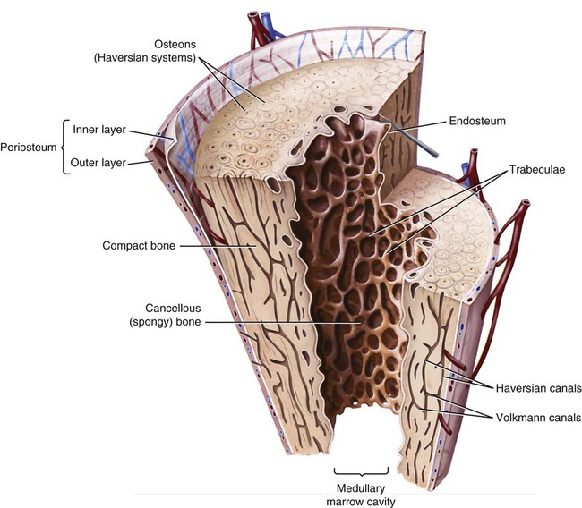
As opposed to cartilage, bones (osseous tissue) are inflexible structures. There are two types of osseous tissue: cortical (also called compact) and cancellous (also called spongy or trabecular) bone (Fig. 3-2). The cortical bone is the dense, stronger, outer segment of bones, whereas cancellous bone is the more open, weaker part.
Skeleton
The skeleton itself can be divided into two parts: the axial skeleton, which is the skull, vertebrae (back bones), and rib cage, and the appendicular skeleton, which is the shoulder and pelvic girdles, and the upper and lower extremities (Fig. 3-1). The shoulder girdle connects the arms to the axial skeleton with the shoulder blades (scapulae) and collarbones (clavicles). The pelvic girdle provides attachment for the legs with its two pelvic (coxal) bones.
Bone Shapes
| Types | Examples |
| long bones | humerus (upper arm bone), femur (thigh bone) |
| short bones | carpal (wrist bone), tarsal (ankle bone) |
| flat bones | sternum (breastbone), scapula (shoulder blade) |
| irregular bones | vertebra (backbone), stapes (a bone in the ear) |
| sesamoid | patella (kneecap) |
 Note
Note
Although the traditional method of explaining the organization of the skeleton is by dividing it into its axial and appendicular components, the ICD-10-PCS Medical Surgical section categorizes the skeleton into three different divisions (Fig. 3-3):
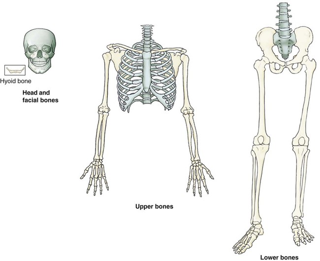
• Head and facial bones: the skull, facial bones, and hyoid bone.
• Upper bones: the left and right upper extremities (arm, hand, and finger bones); left and right glenoid cavity; scapula; clavicle; rib; and the sternum, as well as the cervical and thoracic vertebrae.
• Lower bones: the left and right lower extremities (leg, foot, and toe bones), left and right pelvic bones (ilium, ischium, and pubis), acetabula, and the lumbar vertebrae, sacrum, and coccyx.
Bone Structure
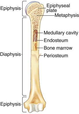
The shaft of a long bone (Fig. 3-4) is termed the diaphysis, whereas the ends are called the epiphyses (sing. epiphysis). The end that is closer to the trunk is termed the proximal epiphysis, whereas the end that is farther away is called the distal epiphysis. The growth plate (also called the epiphyseal plate or the physis) is the site of growth for bone lengthening. This plate is “sealed” when growth stops, typically around the ages of 18-20 in most people.
Bone Markings
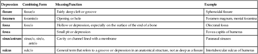
| Depression | Combining Form | Meaning/Function | Example |
| fissure | fissur/o | Fairly deep cleft or groove | Sphenoidal fissure |
| foramen | foramin/o | Opening or hole | Foramen magnum, mental foramina |
| fossa | foss/o | Hollow or depression, especially on the surface of the end of a bone | Olecranal fossa |
| fovea | Small pit or depression | Fovea capitis of humerus | |
| sinus/antrum | sinus/o, sin/o, antr/o | Cavity or channel lined with a membrane | Paranasal sinuses |
| sulcus | sulc/o | General term that refers to a groove or depression in an anatomical structure, not as deep as a fissure | Intertubercular sulcus of humerus |
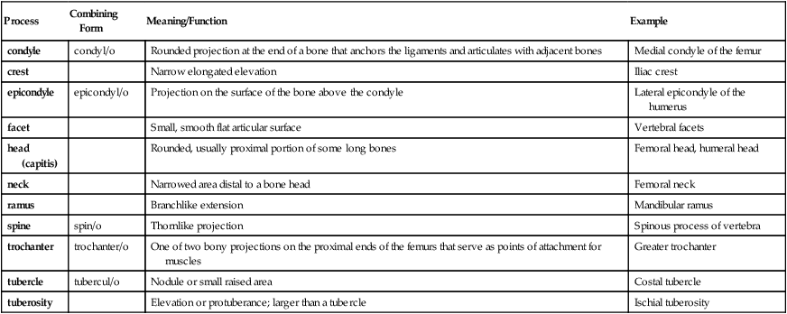
| Process | Combining Form | Meaning/Function | Example |
| condyle | condyl/o | Rounded projection at the end of a bone that anchors the ligaments and articulates with adjacent bones | Medial condyle of the femur |
| crest | Narrow elongated elevation | Iliac crest | |
| epicondyle | epicondyl/o | Projection on the surface of the bone above the condyle | Lateral epicondyle of the humerus |
| facet | Small, smooth flat articular surface | Vertebral facets | |
| head (capitis) | Rounded, usually proximal portion of some long bones | Femoral head, humeral head | |
| neck | Narrowed area distal to a bone head | Femoral neck | |
| ramus | Branchlike extension | Mandibular ramus | |
| spine | spin/o | Thornlike projection | Spinous process of vertebra |
| trochanter | trochanter/o | One of two bony projections on the proximal ends of the femurs that serve as points of attachment for muscles | Greater trochanter |
| tubercle | tubercul/o | Nodule or small raised area | Costal tubercle |
| tuberosity | Elevation or protuberance; larger than a tubercle | Ischial tuberosity |
 Exercise 1:
16. Osteoblasts ______________________ bone, whereas osteoclasts ______________________ bone.
17. The shaft of a long bone is called the ______________________; the ends of a long bone are called ______________________(plural!).
18. The outer covering of bone is the _______________________________________, whereas the inner lining is the ______________________.
19. A foramen, a sinus, and a fossa are examples of bone _________________________________. A condyle, a trochanter, and a tuberosity are examples of bone ______________________________________________.
20. A synonym for a sinus is a/an ___________________________________________________________________.
21. What are the five bone shapes? __________________________________________________________________.
22. The axial skeleton comprises ____________________________________________________________________.
23. The appendicular skeleton comprises _____________________________________________________________.
24. The ICD-10-PCS divides the entire skeleton into three categories. What are they? _______________________________________________________________________________________________.
25. Describe the difference between cortical and cancellous bone. ____________________________________.
 To practice labeling the bones of the musculoskeletal system, click on Label It.
Axial Skeleton
The axial skeleton includes the skull, spine, and rib cage (see Fig. 3-1).
Skull
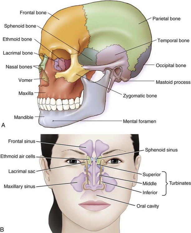
The skull is made up of two parts: the cranium that encloses and protects the brain and the facial bones (Fig. 3-5).
Cranium
Frontal bone: Forms the anterior part of the skull and the forehead. The zygomatic process is the section of the frontal bone that extends toward the cheek.
Parietal bones: Paired bones that form the sides of the cranium.
Occipital bone: Forms the posteroinferior portion of the skull. Notable is a large hole at the ventral surface in this bone, the foramen magnum (magnum means large), which allows brain communication with the spinal cord.
Temporal bones: Paired bones that form the inferior two sides of the cranium. The mastoid process is the small, rounded protruding bone posterior to the ear. The petrous part of the temporal bone is the hard, stonelike portion of the temporal bone that protectively houses the external auditory canal and middle and inner ear. The tympanic portion is the lower area that encircles the eardrum. The zygomatic process is the anterior projection that forms the inferior portion of the cheek. The styloid process is a thin, pointed projection at the base of the temporal bone that serves as a point of attachment for ligaments and muscles.
Ethmoid bone: Single, sievelike bone forming the roof and walls of the nasal cavity. The cribriform plate (also named for its sievelike appearance) is a thin, flat structure perforated by many foramina for the olfactory nerves.
Sphenoid bone: A bat-shaped bone that forms the internal base of the skull, giving shape to the orbits of the eyes. The optic foramina are the openings that allow the optic nerve to pass through. The sella turcica is a saddle-like depression on the body of the sphenoid that holds the pituitary gland. The greater wings (also termed the alisphenoid) are the triangular projections that form the cranial wall adjacent to the petrous part of the temporal bone. The lesser wings (also called orbitosphenoids) are smaller projections that form the back of the orbit and support part of the frontal lobe of the brain.
Paranasal sinuses: Air-filled cavities that are named for the bone where they are located. Each is lined with a mucous membrane (Fig. 3-5, B). The vomer is the bone that forms the inferior/posterior part of the nasal septum.
Hyoid bone: A U-shaped bone located in the neck and attached to the styloid processes of the temporal bone.
Facial Bones
Use Fig. 3-5 to locate the names and locations of the majority of the following facial bones:
Zygoma: Cheekbone. Also called the malar or zygomatic bone.
Lacrimal bones: Paired bones at the corner of each eye that cradle the tear ducts.
Orbit: The bony socket of the eyeball. It is composed of the ethmoid, frontal, lacrimal, maxilla, palatine, sphenoid, and zygomatic bones.
Maxilla: Upper jaw bone. Also called the maxillary bone. The maxillary alveolar processes are the cavities that hold the teeth in.
Mandible: Lower jaw bone. Also called the mandibular bone. The mental foramina are the holes in the central part (body) of the mandible. The rami are posterior vertical projections. The condyloid process is a posterior projection of the ramus that articulates with the temporal bone. The coronoid process is the anterior projection of the ramus. The mandibular notch is the depression between the coronoid and condylar processes.
Palatine bones: Structures that make up part of the roof of the mouth.
Nasal bones: Pair of small bones that make up the bridge of the nose. The vomer is the bone that forms the posterior/inferior part of the nasal septum between the nostrils. The nasal septum is the wall that separates the nostrils.
Rib Cage
The ribs (costa) consist of 12 pairs of thin, flat bones attached to the thoracic vertebrae in the back and to costochondral tissue in the front (Fig. 3-6). The head and neck of the ribs are closest to the vertebrae. The ribs can be categorized as follows:
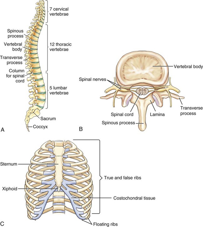
• True ribs: Seven pairs attached directly to the breastbone (sternum) in the front of the body
• False ribs: Five pairs attached to the sternum by cartilage
• Floating ribs: Two pairs of false ribs not attached in the front of the body at all
In addition to ribs, the rib cage includes the sternum, also known as the breastbone. The sharp point at the most inferior aspect of the sternum is called the xiphoid process. The combing form xiph/o is derived from the Greek word for sword, which the xiphoid resembles.
Spine
The spinal or vertebral column is divided into five regions from the neck to the tailbone. It is composed of 26 bones called the vertebrae (Fig. 3-6). The following table lists the bones in the spine.
| Region | Type and Abbreviation |
| cervical | neck bones (C1-C7) |
| thoracic | upper back (T1-T12) |
| lumbar | lower back (L1-L5) |
| sacral | sacrum (S1-S5) (5 bones fused as one) |
| coccygeal (tailbone) | coccyx or tailbone |
C1 is called the atlas for the Greek god who held up the world. C2 is called the axis for its role in permitting rotation of the head. Unique to C2 is the dens (also termed the odontoid process), which is a small, toothlike projection for the first cervical vertebra to rotate around.
 Exercise 2:
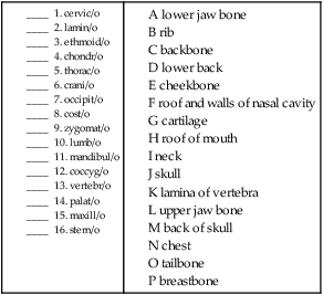
Decode the following terms below using your knowledge of word parts.
17. submandibular _________________________________________________________________________________
18. costochondral __________________________________________________________________________________
19. lumbosacral ____________________________________________________________________________________
20. thoracic ________________________________________________________________________________________
21. substernal ______________________________________________________________________________________
 To practice labeling the axial skeleton, click on Label It.
Appendicular Skeleton
The appendicular skeleton is divided into the upper appendicular and lower appendicular skeletons.
Upper Appendicular Skeleton
The upper appendicular skeleton (Fig. 3-7) includes the shoulder girdle (scapula, clavicle, upper humerus) and the arm bones. Refer to Fig. 3-8A for a closer look at the shoulder.
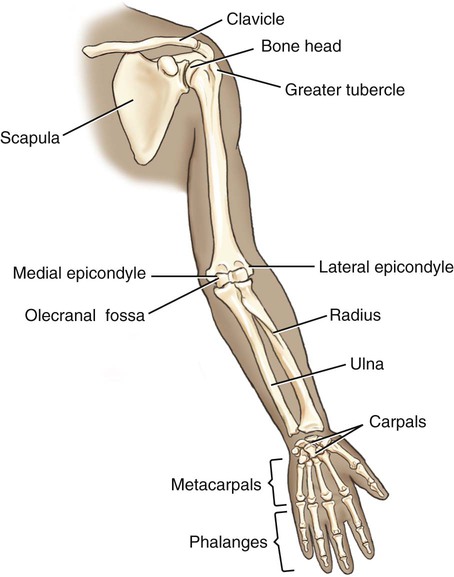
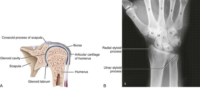
Scapula: The scapulae, or shoulder blades, are flat bones that help to support the arms. The acromion process is the lateral protrusion of the scapula that forms the highest point of the shoulder. The glenoid cavity (the arm socket) is the depression in the scapula that is the seat of the head of the humerus (syn. glenoid fossa). The coracoid process is the beaklike process of the scapula that serves as a point of attachment for muscles and ligaments in the shoulder.
Clavicle: The clavicle, or collarbone, is one of a pair of long, curved horizontal bones that attach to the upper sternum at one end and the acromion process of the scapula at the other. These bones help to stabilize the shoulder anteriorly. A “wishbone” is composed of the fused clavicles of a bird.
Humerus: Upper arm bone. The bone head (caput) is the proximal enlarged round end that articulates with the glenoid cavity. The anatomical neck is immediately below the humeral head, whereas the surgical neck is below the tuberosities distal to the anatomical neck. The greater tubercle is the larger of two protuberances, whereas the adjacent lesser tubercle is obviously the other. Both function as points of attachment for muscles. The long narrow part of the humerus shaft is capped by the medial and lateral epicondyles at the distal end.
Radius: Lower lateral arm bone parallel to the ulna. The distal end articulates with the thumb side of the hand. The ulnar notch is the small cavity that articulates with the ulna at the distal end of the radius. It is also called the sigmoid cavity.
Ulna: Lower medial arm bone. The distal end articulates with the little finger side of the hand. The radial notch is the cavity that articulates with the radius at its proximal end. The radial notch is also termed the lesser sigmoid cavity. The olecranon is a proximal projection of the ulna that forms the tip of the elbow. Commonly known as the funny bone, this structure is actually a process.
Carpus: The eight bones of the carpus (wrist) are each named for a shape: the capitate (head), hamate (hooked), lunate (moon), pisiform (pea), scaphoid (boat), trapezium (table), trapezoid (table), and triquetrum (three corner) (see Fig. 3-8, B for radiograph of the carpus and metacarpus bones).
Metacarpus: One of the five bones that form the middle part of the hand.
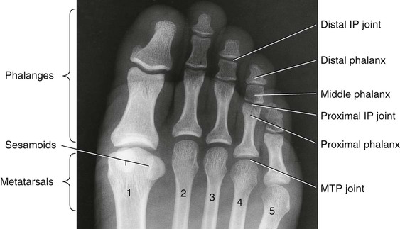
Phalanx: One of the 14 bones that constitute the fingers of the hand, two in the thumb and three in each of the other four fingers (pl. phalanges). The three bones in each of the four fingers are differentiated as proximal, medial, and distal. The joints between these are referred to as proximal and distal interphalangeal (PIP, DIP) joints. When one is referring to a whole finger (or toe), the term digitus is used. The thumb is called the pollex.
 PCS Guideline Alert
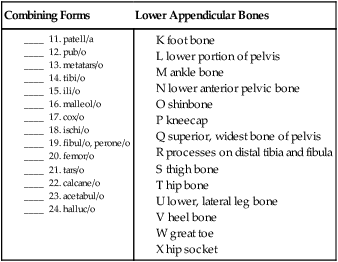
Lower Appendicular Skeleton
The lower half of the appendicular skeleton can be divided into the pelvic girdle (the bony pelvis, sacrum, and coccyx) and the leg bones (Fig. 3-9). The pelvis is composed of two coxal (hip) bones that are connected in the front at the pubic symphysis and in the back at the sacrum. Each coxa is composed of three fused bones that together form the hip socket, the acetabulum, for the head of the thighbone. The three fused bones are the:
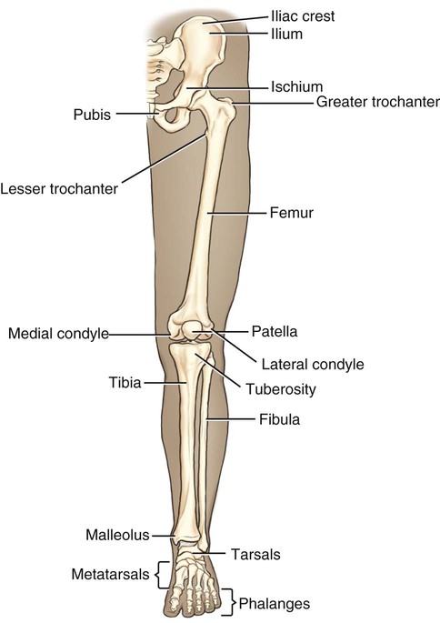
Ilium: The superior and widest bone of the pelvis. The iliac crest is the upper edge of the bone.
Ischium: The lower, posterior portion of the pelvic bone.
Pubis or pubic bone: The lower anterior part of the pelvic bone. The obturator foramina are the two openings between the ischium and pubis on both sides of the pelvic bone. The term obturate means to “stop up” or “block,” and was used to name this opening because on dissection it was found to be filled with muscles and nerves.
The leg is composed of the:
Femur: Thigh bone, upper leg bone. The femoral head articulates with the hipbone at the acetabulum. The femoral neck connects the head to the shaft where two protuberances, the greater and lesser trochanters, serve as sites of muscle attachment. At the distal end of the shaft are medial and lateral condyles that articulate with the condyles of the tibia. Slightly above those are the medial and lateral epicondyles.
Patella: Kneecap, a sesamoid bone that is encased in the quadriceps femoris tendon and helps to protect the knee joint.
Tibia: Shinbone, lower medial leg bone. The proximal end of the tibia has both medial and lateral condyles, whereas the distal end has a medial malleolus, a process that extends inward.
Fibula: Smaller, lower lateral leg bone. The head of the fibula is at its proximal end, where it articulates with the lateral condyle of the tibia. The body (shaft) of the fibula has a diamond-like appearance, with each of the four surfaces named for the direction it faces: anteromedial, anterolateral, posteromedial and posterolateral. The lateral malleolus is the process that extends outward at the distal end of the bone.
Tarsus: One of the seven bones of the ankle including the calcaneus (heel bone), cuboid (box-shaped), cuneiforms (medial, lateral, and intermediate wedge-shaped bones), navicular (boat-shaped), and talus (the second largest tarsal bone). The talus is named from the Latin for its shape like a die (singular of dice), as it was used as a game piece in ancient times.
Metatarsus: One of the five small, long bones in the foot between the tarsals and the phalanges.
Phalanx: One of 14 toe bones, 2 in the great toe and 3 in each of the other four toes. The term for the great toe is the hallux.
 Note
Note Be Careful!
Do not confuse perone/o, meaning fibula, with peritone/o, meaning the lining of the abdomen.
 Exercise 3:
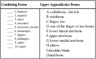
Match the lower appendicular combining forms with their meanings.
| Combining Forms | Lower Appendicular Bones |
25. interphalangeal ________________________________________________________________________________
26. humeroulnar ___________________________________________________________________________________
27. infrapatellar ____________________________________________________________________________________
28. femoral ________________________________________________________________________________________
29. supraclavicular _________________________________________________________________________________
 To practice labeling the appendicular skeleton, click on Label It.
Joints
 PCS Guideline Alert
 PCS Guideline Alert
 PCS Guideline Alert
Joints, or articulations as they are sometimes called, are the parts of the body where two or more bones of the skeleton join. Examples of joints include the knee, which joins the tibia and the femur, and the elbow, which joins the humerus with the radius and ulna. Joints provide range of motion (ROM), the range through which a joint can be extended and flexed. Different joints have different ROMs, ranging from no movement at all to full range of movement. Categorized by ROM, they are as follows:
No ROM: Most synarthroses are immovable joints held together by fibrous cartilaginous tissue. The suture lines of the skull are examples of synarthroses.
Limited ROM: Amphiarthroses are joints joined together by cartilage that are slightly movable, such as the vertebrae of the spine or the pubic bones.
Full ROM: Diarthroses are joints that have free movement. The most commonly known are ball-and-socket joints (such as the hip) and hinge joints (such as the knees). Other examples of diarthroses include the elbows, wrists, shoulders, and ankles. See Fig. 3-10 for an illustration of a knee joint that shows the bones, muscles, tendons, bursae, synovial membrane, and cavity in the knee.
Upper Joints
Upper Spine Joints
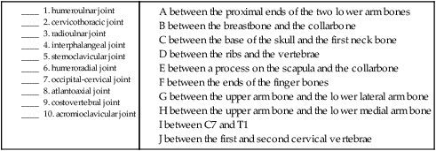
| occipital-cervical joint | The first joint between the base of the skull (occipit/o) and first cervical vertebra (the atlas). |
| cervical vertebral joint | One of the articulations between the seven cervical vertebrae (C1-C7). |
| atlantoaxial joint | Articulation between C1 (atlant/o) and C2 (axi/o). |
| cervical facet joint | Articulation between the adjoining facets of the vertebrae of the neck (cervic/o). |
| cervical vertebral joint | Articulation between two or more of the cervical vertebrae. |
| cervical vertebral disc | The cartilaginous pad between the vertebrae of the neck. |
| cervicothoracic vertebral joint | Articulation between the last cervical vertebra (C7) and the first thoracic (thorac/o) vertebra (T1). |
| cervicothoracic facet joint | Articulation of the facet joints between C7 and T1. |
| cervicothoracic vertebral disc | Intervertebral disk between C7 and T1. |
| thoracic vertebral joint (T2-7) | Any of the articulations between the second (T2) and seventh (T7) thoracic vertebrae. |
| costotransverse joint | Articulation between the posterior end of the six ribs (cost/o) and T2-T7 vertebrae. |
| costovertebral joints | Any of the articulations between the ribs (cost/o) and the thoracic vertebrae, including the floating and false ribs. |
| thoracic facet joints | Any of the articulations between the facet processes of the thoracic vertebrae. |
| thoracic vertebral joints | Any of the articulations between the eighth (T8) and twelfth (T12) vertebrae. |
| thoracic vertebral disc | The intervertebral disc between any of the thoracic vertebral joints. |
| thoracolumbar joint* | Articulation between the final thoracic vertebra (T12) and the first lumbar (lumb/o) vertebra (L1). |
| thoracolumbar facet joints | Articulations between the facet processes of T12 and L1. |
| thoracolumbar disc | Intervertebral disc between T12 and L1. |
Upper Extremity Joints
| sternoclavicular joint | Articulation between the manubrium of the breastbone (stern/o) and the medial end of the collarbone (clavicul/o). |
| acromioclavicular joint | Articulation between the acromion process (acromi/o) of the scapula and the lateral end of the collarbone. |
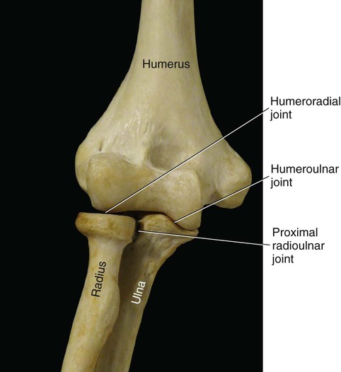
| humeroulnar joint (Fig. 3-10) | Articulation between the upper arm bone (humer/o) and the lower medial arm bone (uln/o). |
| humeroradial joint (Fig. 3-10) | Articulation between the upper arm bone and the lower lateral arm bone (radi/o). |
| proximal radioulnar joint (Fig. 3-10) | Articulation between the lower arm bones nearest to the humerus. |
| carpal joints | Articulations between the individual carpal bones (intercarpal joints) and between the proximal row (scaphoid, lunate, and triquetral) and the distal row (trapezium, trapezoid, capitate, and hamate) termed the midcarpal joint. |
| wrist joints | Articulations between the distal end of the lower lateral arm bone and the carpal bones (radiocarpal joint) and between the distal ends of both lower arm bones (distal radioulnar joint). |
| metacarpal joints | Joints between the distal row of the carpal bones and the five metacarpal bones are metacarpocarpal/carpometacarpal joints. The carpometacarpal joint of the thumb, also known as the trapeziometacarpal joint, connects the trapezium to the first metacarpal bone. |
| metacarpophalangeal joints | Articulations between the metacarpals (metacarp/o) and the proximal finger bones (phalang/o). |
| finger phalangeal joints | Articulations between either the far two bones (distal interphalangeal [DIP]) or near two bones (proximal interphalangeal [PIP]) joints. |
 Exercise 4:
Upper Joints
A between the proximal ends of the two lower arm bones
B between the breastbone and the collarbone
C between the base of the skull and the first neck bone
D between the ribs and the vertebrae
E between a process on the scapula and the collarbone
F between the ends of the finger bones
G between the upper arm bone and the lower lateral arm bone
Lower Joints
Lower Spine Joints
| lumbar vertebral joint | Articulation between two lumbar (lumb/o) vertebrae. |
| lumbar facet joint | Articulation between superior and inferior lumbar facets. |
| lumbar vertebral disc | The cartilaginous pad between the lumbar vertebrae. |
| lumbosacral joint | Articulation between lumbar vertebrae and the sacrum (sacr/o). |
| lumbosacral disc | The cartilaginous pad between the fifth lumbar vertebra and the first sacral vertebra. |
| sacrococcygeal joint | Articulation between the sacrum and the coccyx (coccyg/o). Synonym is the coccygeal joint. |
| sacroiliac joint | Articulation between the sacrum and the ilium (ili/o) of the pelvic bone. |
Lower Extremity Joints
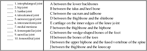
| hip joint | Articulation between the acetabulum of the hip and the femur. |
| knee joint | Articulations between the thighbone and the shinbone, and the thighbone and the kneecap. |
| femoropatellar joint | The articulation between the femur (femor/o) and the patella (patell/a). |
| femorotibial joint | The articulation between the femur and the tibia (tibi/o). |
| lateral meniscus | A crescent-shaped pad of cartilage that cushions the knee joint to the side. |
| medial meniscus | A crescent-shaped pad of cartilage that cushions the knee joint in the middle. |
| inferior tibiofibular joint (ankle joint) | Articulation between the lower end of the tibia and fibula (fibul/o). |
| tarsal joint | One of the many articulations between the seven tarsal bones. |
| calcaneocuboid joint | Articulation between the calcaneus (calcane/o) and the cuboid (cuboid/o). |
| cuboidonavicular joint | Articulation between the cuboid and the navicular (navicul/o). |
| cuneonavicular joint | Articulation between the cuneiform (cune/o) bones and the navicular. |
| intercuneiform joint | Articulation between the three cuneiform bones. |
| subtalar (talocalcaneal) joint | Articulation between the talus (tal/o) and the calcaneus. |
| talocalcaneonavicular joint | Articulation between the talus, calcaneus and navicular bones. |
| metatarsal-tarsal joint | Articulation between a metatarsal and one of the tarsal (tars/o) bones. |
| metatarsal-phalangeal joint | Articulation between one of the metatarsals (metatars/o) and a proximal phalanx (phalang/o). |
| toe phalangeal joint (Fig. 3-11) | One of the articulations between the phalanges of the toes. |
| PIP (proximal interphalangeal joint) | Articulation between the proximal and medial phalanges. |
| DIP (distal interphalangeal joint) | Articulation between the distal and medial phalanges. |
 Exercise 5:
Lower Joints
B between the talus and heel bone
C between the sacrum and tailbone
D between the thighbone and the shinbone
E cartilage on the inner edges of the knee joint
F between the thighbone and the hipbone
G between the wedge-shaped bones of the foot
H between the bones of the toes
I between the upper hipbone and the fused vertebrae of the spine
Ligaments and Bursae
 PCS Guideline Alert
Head and Neck Ligaments
| alar ligament of axis | The winglike (al/i) ligament that connects the skull to the second cervical vertebra. |
| cervical interspinous ligament | The ligaments that connect the spinous processes of the cervical (cervic/o) vertebrae. |
| cervical intertransverse ligament | The ligaments that connect the cervical vertebrae between the transverse processes. |
| cervical ligamentum flavum | The yellow, bandlike ligaments that connect the laminae of the vertebrae of the neck. |
| lateral temporomandibular ligament | The ligament that attaches the temporal (tempor/o) and mandibular (mandibul/o) bones. |
| sphenomandibular ligament | The ligament that attaches the sphenoid bone (sphen/o) to the mandible. |
| stylomandibular ligament | The ligament that connects the styloid (styl/o) process of the temporal bone to the mandible. |
| transverse ligament of atlas | The transverse ligament of the atlas is instrumental in holding the dens of the second cervical vertebrae against the anterior arch for the purpose of stabilizing the neck. |
Shoulder Ligaments and Bursa
| acromioclavicular ligament | The ligament that joins the acromion process (acromi/o) to the collarbones (clavicul/o). |
| coracoacromial ligament | The ligament that connects the coracoid process (corac/o) to the acromion process. |
| coracoclavicular ligament | The ligament that connects the coracoid process to the collarbone. |
| coracohumeral ligament | The ligament that joins the coracoid process with the humerus (humer/o). |
| glenohumeral ligament | The ligament that joins the glenoid cavity (glen/o) to the humerus. |
| glenoid ligament (labrum) | The ligament within the glenoid cavity. Also termed the glenoid labrum. |
| interclavicular ligament | The ligament between the collarbones. |
| sternoclavicular ligament | The ligament joining the sternum (stern/o) to the clavicles. |
| subacromial bursa | The subacromial bursa is located between the head of the humerus and under (sub-) the acromion process. |
| transverse humeral ligament | The transverse humeral ligament is between the greater and lesser tubercles (tuberosities) of the humerus. |
Elbow Ligaments and Bursa
| annular ligament | The annular ligament is a ringlike structure that encircles the head of the radius. |
| olecranon bursa | The olecranon bursa cushions the elbow joint. |
| radial collateral ligament | The radial collateral ligament connects the humerus to the radius. |
| ulnar collateral ligament (UCL) | The ulnar collateral ligament has an anterior and posterior portion, both connecting the humerus to the ulna. |
Wrist Ligaments
| palmar ulnocarpal ligament | Connects the ulnar (uln/o) styloid process to carpal (carp/o) (the lunate, capitate, and triquetral) bones. |
| radial collateral carpal ligament | Connects the styloid process of the radius to the wrist bones. |
| radiocarpal ligament | Connects the radius (radi/o) to the wrist bones. Also termed the volar radiocarpal. Volar means pertaining to the palm of the hand or the sole of the foot. |
| radioulnar ligament | Connects the radius and the ulna. |
| ulnar collateral carpal ligament | Connects the ulna to the wrist bones. |
Hand Ligaments
| carpometacarpal ligament | Connects the wrist bones to the metacarpals (metacarp/o). |
| intercarpal ligament | Connects the carpal bones. |
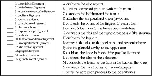
| interphalangeal ligament | Connects the bones of the fingers (phalang/o) to each other. |
| lunotriquetral ligament | Connects the lunate and the triquetral bones. |
| metacarpal ligament | Connects the metacarpals. |
| metacarpophalangeal ligament | Connects the metacarpals to the phalanges. |
| pisohamate ligament | Connects the pisiform and hamate bones. |
| pisometacarpal ligament | Connects the pisiform and metacarpal bones. |
| scapholunate ligament | Connects the scaphoid (scaphoid/o) to the lunate bone. |
| scaphotrapezium ligament | Connects the scaphoid to the trapezium. |
Trunk Ligaments
| iliolumbar ligament | Connects the ilium (ili/o) to the lumbar (lumb/o) vertebrae. |
| interspinous ligament | Connects the spinous processes of the vertebrae. |
| intertransverse ligament | Connects the transverse processes to each other. |
| ligamentum flavum | Broad yellow ligament that connects the laminae of the vertebrae. |
| pubic ligament | Connects the pubic bones. |
| sacrococcygeal ligament | Connects the sacrum (sacr/o) and the coccyx (coccyg/o). |
| sacroiliac ligament | Connects the sacrum (sacr/o) to the ilium. |
| sacrospinous ligament | Connects the sacrum (sacr/o) to the spinous processes of the ischial spine. |
| sacrotuberous ligament | Connects the sacrum (sacr/o) to the tuberosity of the ischium. |
| supraspinous ligament | Connects the tops (supra-) of the spinous processes. |
Thorax Ligaments
| costotransverse ligament | Connects the back of the neck of a rib (cost/o) to the adjacent transverse process of the corresponding vertebra. |
| costoxiphoid ligament | Connects the ribs (cost/o) and the xiphoid process of the sternum. |
| sternocostal ligament | Connects the sternum (stern/o) and the ribs. |
 Note
NoteHip Ligaments and Bursa
| iliofemoral ligament | Connects the ilium and the femur (femor/o). |
| ischiofemoral ligament | Connects the ischium (ischi/o) and the femur. |
| pubofemoral ligament | Connects the pubis (pub/o) and the femur. |
| transverse acetabular ligament | Ligament that is part of the acetabular labrum and serves to keep the femoral head in place. |
| trochanteric bursa | Cushions the hip joint. |
Knee Ligaments and Bursa (Fig. 3-12)
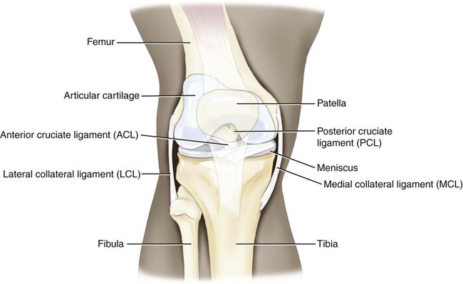
| anterior cruciate ligament (ACL) | Connects the femur and tibia by crossing the front of the knee. |
| lateral collateral ligament (LCL) | Connects the femur and tibia on the outer (lateral) side of the knee. |
| ligament of head of fibula | Connects the fibula to the tibia over the patella (patell/a). |
| medial collateral ligament (MCL) | Connects the femur and tibia at the inner (medial) part of the knee. |
| patellar ligament | Connects the kneecap to the tibia. |
| popliteal ligament | Connects the femur to the tibia in the back of the knee. |
| posterior cruciate ligament (PCL) | Connects the femur to the tibia by crossing in the back of the knee. |
| prepatellar bursa | Largest bursa in knee joint. Cushions the knee in front (pre-) of the patellar ligament. |
Ankle Ligaments
| calcaneofibular ligament | Connects the heel bone (calcane/o) and the fibula (fibul/o). |
| deltoid ligament | Triangular-shaped ligament that connects the medial malleolus of the tibia to the navicular, calcaneus and talus bones. |
| ligament of the lateral malleolus | Connects the lateral malleolus of the fibula to the bones of the foot. May be the calcaneofibular ligament, or the anterior or posterior talofibular ligaments. |
| talofibular ligament | Connects the talus (tal/o) to the fibula with anterior and posterior segments. |
Foot Ligaments
| calcaneocuboid ligament | Connects the heel bone to the cuboid bone. |
| cuneonavicular ligament | Connects the cuneiform (cune/o) bones to the navicular bone. |
| intercuneiform ligament | Connects the cuneiform bones to each other. |
| interphalangeal ligament | Connects the phalanges to each other. |
| metatarsal ligament | Connects the metatarsals to each other. |
| metarsophalangeal ligament | Connects the metatarsals (metatars/o) to the phalanges. |
| subtalar ligament | Connects the talus to the calcaneus. |
| talocalcaneonavicular ligament | Connects the talus to the heel bone and the navicular (navicul/o) bone. |
| tarsometatarsal ligament | Connects the tarsals (tars/o) to the metatarsals. |
 Exercise 6:
Ligaments and Bursae
B joins the coracoid process with the humerus
C connects the ischium and the femur
D attaches the temporal and lower jawbones
E connects the bones of the fingers to each other
F connects the ilium to the lower back vertebrae
G connects the ribs and the xiphoid process of the sternum
I connects the talus to the heel bone and navicular bone
J joins the glenoid cavity to the upper arm
K cushions the knee in front of the patellar ligament
L connects the talus to the calcaneus
M connects the femur to the tibia in the back of the knee
Muscles
• Skeletal muscle that allows the skeleton to move voluntarily
• Smooth muscle that is responsible for involuntary movement of the organs
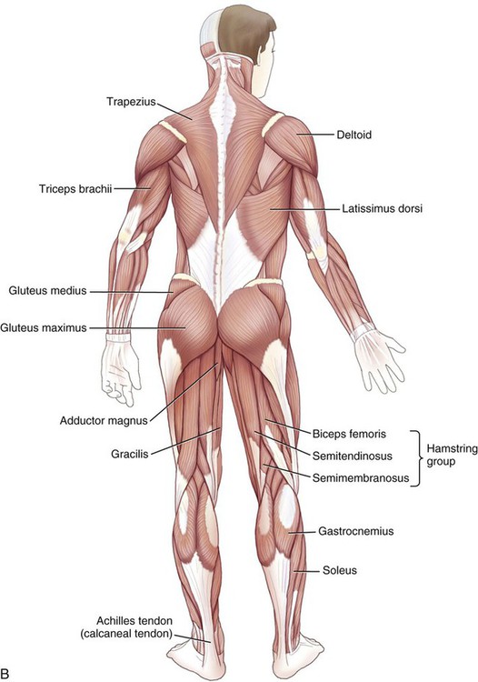
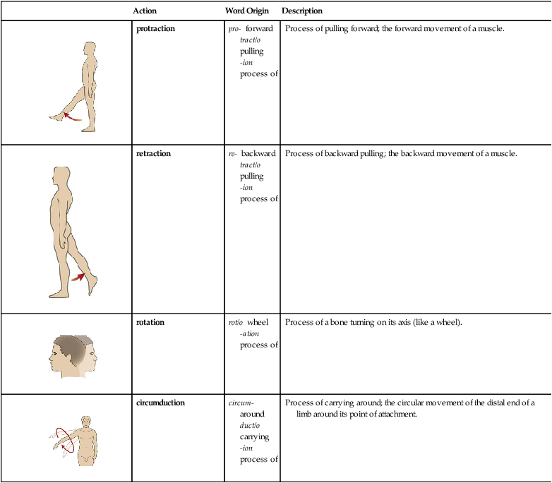
Fig. 3-13 shows posterior and anterior views of the major skeletal muscles of the body. ICD-10-PCS requires knowledge of all of the muscles and knowledge of where they are in the body. The muscles will be presented with their locations and word part meanings, as well as grouped as they are in the classification.
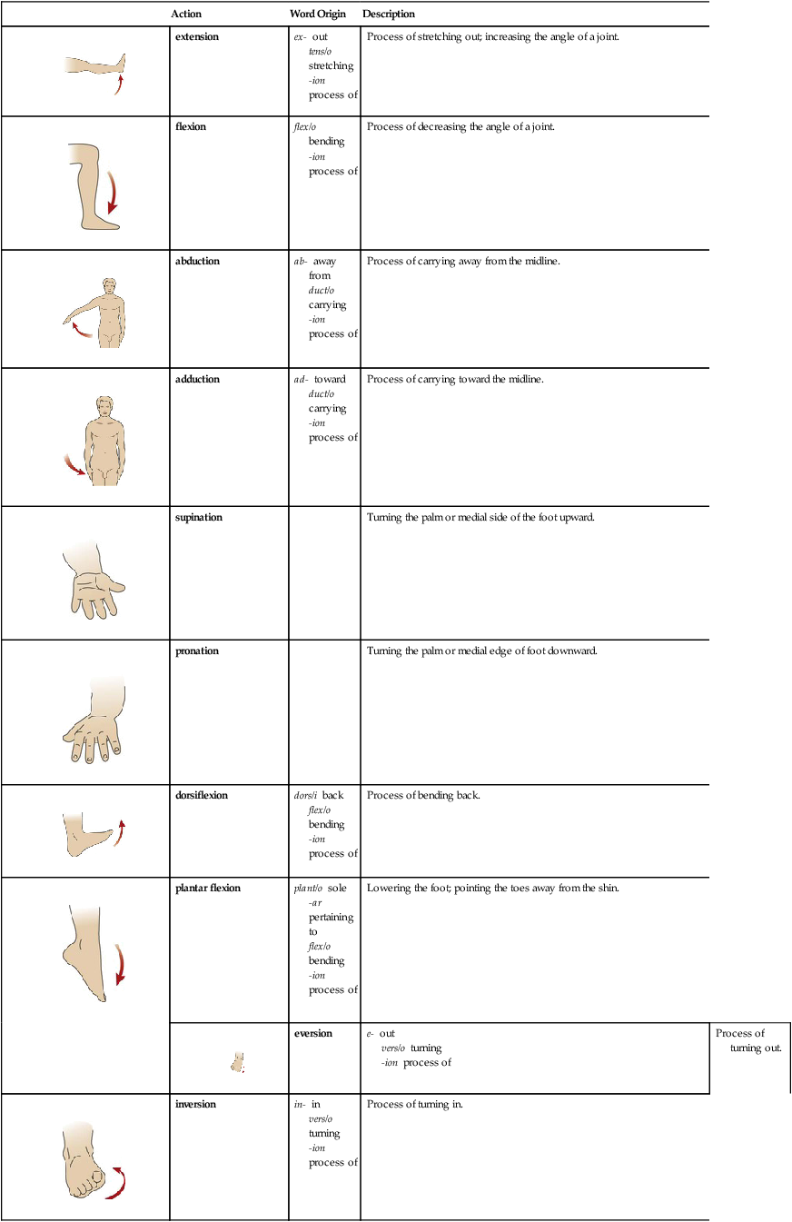
| Action | Word Origin | Description | |
 |
extension | ex- out tens/o stretching -ion process of |
Process of stretching out; increasing the angle of a joint. |
 |
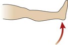
flexion | flex/o bending -ion process of |
Process of decreasing the angle of a joint. |
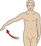 |
abduction | ab- away from duct/o carrying -ion process of |
Process of carrying away from the midline. |
 |
adduction | ad- toward duct/o carrying -ion process of |
Process of carrying toward the midline. |
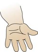 |
supination | Turning the palm or medial side of the foot upward. | |
 |
pronation | Turning the palm or medial edge of foot downward. | |
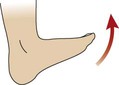 |
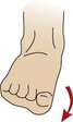
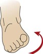
dorsiflexion | dors/i back flex/o bending -ion process of |
Process of bending back. |
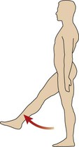
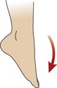 |
plantar flexion | plant/o sole -ar pertaining to flex/o bending -ion process of |
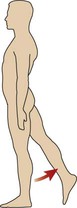
Lowering the foot; pointing the toes away from the shin. |
 |
eversion | e- out vers/o turning -ion process of |
Process of turning out. |
 |
inversion | in- in vers/o turning -ion process of |
Process of turning in. |
 |
protraction | pro- forward tract/o pulling -ion process of |
Process of pulling forward; the forward movement of a muscle. |
 |
retraction | re- backward tract/o pulling -ion process of |
Process of backward pulling; the backward movement of a muscle. |
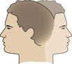 |
rotation | rot/o wheel -ation process of |
Process of a bone turning on its axis (like a wheel). |
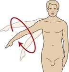 |
circumduction | circum- around duct/o carrying -ion process of |
Process of carrying around; the circular movement of the distal end of a limb around its point of attachment. |
 Exercise 7:
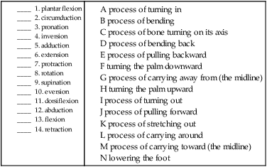
Muscle Naming Conventions
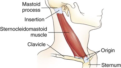
There are some general naming conventions that will help you. Refer to the table below for examples of how muscles are named by their location, number of insertions, size, shape, and muscle action. The final type of naming convention that appears in the table is by its origins and insertion. For example, the sternocleidomastoid muscle (Fig. 3-14) originates in the sternum and collarbone and inserts on the mastoid process.
| Naming Device | Name of Muscle | Word Origin | Definition |
| location | zygomaticus | zygomatic/o zygoma -us noun ending |
Cheek muscle. |
| number of insertions | biceps brachii | bi- two *ceps heads brachi/o arm -i noun ending |
Muscle that flexes upper arm. |
| size | gluteus maximus | glute/o buttock -us noun ending maxim/o large -us noun ending |
Large buttock muscle. |
| shape | deltoid | delt/o triangle -oid like |
Triangular muscle in shoulder. |
| muscle action | adductor longus | ad- toward duct/o carrying -or one who long/o long -us noun ending |
Upper leg muscle that carries the leg back to the midline. |
| origin/insertion | sternocleidomastoid | stern/o breastbone cleid/o collarbone mastoid/o mastoid process |
Muscle that originates in the sternum and collarbone and inserts on the mastoid process (Fig. 3-14). |
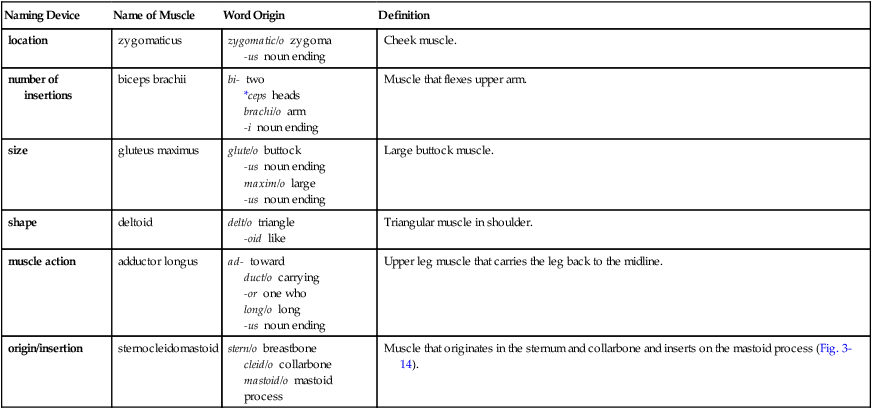
*ceps is a variation of cephal/o, the combining form for head. These word parts are used for “the head” or the beginning of a structure and also are used for bones (bone heads), as well as muscle heads.
Additionally, the naming themes use synonyms and antonyms. Here are some shortcut opposites with their meanings:
• Brevis is short and longus is long.
• Minimus is small and maximus, magnus, and vastus are large.
• Lateralis is to the side and medialis is to the middle.
• Adductors pull toward the midline, whereas abductors pull away from the midline.
Head Muscles
| auricularis muscle | Moves the ears (auricul/o). |
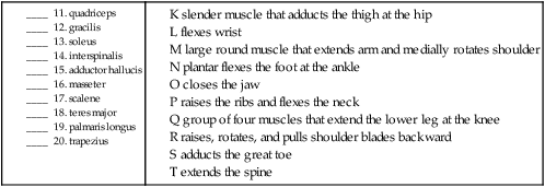
| masseter muscle | Closes the jaw. |
| pterygoid muscle | Winglike muscle that raises, lowers, and allows side-to-side movement of the mandible. |
| splenius capitis muscle | Bandlike muscle that holds the head upright; located in back of neck. |
| temporalis muscle | Raises the lower jaw. |
| temporoparietalis muscle | Paired scalp muscles over the temporal and parietal regions. |
| facial muscles | A group of muscles of the face. |
| buccinator muscles | Cheek (bucc/o) muscles; compress cheek against teeth. |
| corrugator supercilii muscle | Wrinkles forehead (above eyelids). Cili/o refers to the small hairs in the eyelids or eyelashes. The term supercilious, meaning haughty, is derived from one arching his/her eyebrows disdainfully. Corrug/o means wrinkled, as in corrugated cardboard. |
| depressor anguli oris muscle | Lowers corners of mouth (or/o). |
| depressor labii inferioris muscle | Lowers lower lip (labi/o). |
| depressor septi nasi muscle | Lowers wall in nose (nas/o). |
| depressor supercilii muscle | Lowers eyebrows. |
| levator anguli oris muscle | Raises corners of mouth. |
| levator labii superioris alaeque nasi muscle | Raises upper lip and nostrils (wings) of nose. |
| levator labii superioris muscle | Raises upper lip. |
| mentalis muscle | Wrinkles chin (ment/o), protrudes lower lip. |
| nasalis muscle | Raises corners of nostrils. |
| occipitofrontalis muscle | Also called epicranius muscle for its location on top of the skull. Composed of two muscles that are joined by a broad, flat tendon, the galea aponeurotica. |
| orbicularis oris muscle | Encircles mouth and serves to pucker lips. The “kissing” muscle. |
| procerus muscle | Draws eyebrows together and down (as in frowning). |
| risorius muscle | Pulls the angles of the mouth backward. The “smiling” muscle. |
| zygomaticus muscle | Raises angle of mouth up and to the side. |
Neck Muscles
| anterior vertebral muscle | Group of muscles (longi colli and capitis, recti capitis anterior and lateralis) located in the front of the backbones that help the head and neck bend. Note that coll/o means “neck,” like in the word collar. |
| arytenoid muscle | Moves vocal cords together (adduction). |
| cricothyroid muscle | Lengthens and tenses vocal cords. |
| infrahyoid muscle | A group of four muscles (omohyoid, thyrohyoid, sternohyoid, and sternothyroid) under the hyoid bone that move the hyoid and larynx downward while speaking. The thyr/o reference is due to the proximity of these muscles to the thyroid gland. |
| levator scapulae muscle | Raises the shoulder blades. |
| platysma muscle | Broad flat muscle that tenses the lower face and neck. |
| scalene muscle | Raises the ribs and flexes the neck. |
| splenius cervicis muscle | Bandlike muscle that bends and extends the neck. |
| sternocleidomastoid muscle | Rotates and tilts head to opposite side. |
| suprahyoid muscle | Group of muscles (geniohyoid, mylohyoid, digastric and stylohyoid) that raise the tongue and widen the esophagus. |
| thyroarytenoid muscle | Thickens the vocal cords. |
Tongue, Palate, and Pharynx Muscles
| chondroglossus muscle | Lowers the tongue (gloss/o). |
| genioglossus muscle | Lowers and protrudes the tongue. |
| hypoglossus muscle | Lowers and pulls tongue backwards. |
| inferior longitudinal muscle | Located under and extending the length of the tongue; changes shape of tongue for chewing and swallowing. |
| levator veli palatini muscle | Raises the soft palate while swallowing. |
| palatoglossal muscle | Raises tongue. |
| palatopharyngeal muscle | Aids in swallowing. |
| pharyngeal constrictor muscle | Narrows throat (pharyng/o) for swallowing. |
| salpingopharyngeus muscle | Raises nasopharynx (part of the throat behind nasal cavity). |
| styloglossus muscle | Raises and retracts tongue for swallowing. |
| stylopharyngeus muscle | Raises voicebox and nasopharynx; used in swallowing. |
| superior longitudinal muscle | Located on dorsum of tongue; changes shape of tongue for chewing and swallowing. |
| tensor veli palatini muscle | Controls tension of the soft palate. |
Shoulder Muscles
| deltoid muscle | Triangular muscle that flexes, abducts, and extends the arm. |
| infraspinatus muscle | Laterally rotates arm. Named for its origin in the infraspinous fossa of the scapula. |
| subscapularis muscle | Adducts and medially rotates arm. Located under the scapula. |
| supraspinatus muscle | Assists deltoid at the shoulder. Named for its origin in the supraspinous fossa of the scapula. |
| teres major muscle | Large round muscle that extends arm and medially rotates shoulder. |
| teres minor muscle | Smaller round muscle that laterally rotates arm. |
 Note
Note Note
NoteUpper Arm Muscles
| biceps brachii muscle | “Two-headed” arm (brachi/o) muscle that flexes and supinates forearm at elbow. |
| brachialis muscle | Flexes forearm at elbow. |
| coracobrachialis muscle | Flexes and medially rotates arm at shoulder. |
| triceps brachii muscle | “Three-headed” muscle that extends forearm at elbow. |
Lower Arm Muscles
| anatomical snuffbox | Also called the radial fossa, this is a triangular depression on the radial side of the back of the hand. Named for its sometime use in earlier times as a place to put powdered tobacco for sniffing. |
| brachioradialis muscle | Flexes forearm at elbow. |
| extensor carpi radialis muscle | Extends and abducts hand at the wrist. |
| extensor carpi ulnaris muscle | Extends and adducts hand at the wrist. |
| flexor carpi radialis muscle | Flexes and abducts hand at the wrist. |
| flexor carpi ulnaris muscle | Flexes and adducts hand at the wrist. |
| palmaris longus muscle | Flexes wrist. |
| pronator quadratus muscle | Pronates forearm. Named for its four sides (two insertions and two origins). |
| pronator teres muscle | Round muscle that pronates forearm and flexes the elbow. |
Hand Muscles
| hypothenar eminence | Group of muscles (abductor, adductor, and opponens digiti minimi) that move the little finger. |
| palmar interosseous muscle | Also called the interossei volares with volar meaning “pertaining to the palm of the hand or sole of the foot.” Located between the bones of the palm of the hand, these serve to flex the fingers at the metacarpophalangeal joints and extend the interphalangeal joints. |
| thenar muscle | Group of muscles (abductor, flexor, and opponens pollicis brevis with pollic/o referring to the pollex, the thumb) that move the thumb. |
Trunk Muscles
| coccygeus muscle | Pulls the tailbone forward (“tucking the tail”) and supports the organs of the pelvis. |
| erector spinae muscle | Extends and bends the spine and head to the side. |
| interspinalis muscle | Extends the spine. Located between the spinous processes of the backbones. |
| intertransversarius muscle | Located between the transverse processes of the backbones, this muscle serves to flex the trunk sideways. |
| latissimus dorsi muscle | Adducts, extends and medially rotates the humerus. |
| levator ani muscle | Helps to raise the pelvic floor around the anus. |
| quadratus lumborum muscle | Extends and flexes the vertebral column to the side. Named for its insertion on four of the lumbar vertebrae. |
| rhomboid major muscle | Pulls the shoulder blade back and rotates to lower the glenoid cavity. |
| rhomboid minor muscle | A smaller version of rhomboid major, rhomboid minor also pulls the shoulder blade back and rotates to lower the glenoid cavity. |
| serratus posterior muscle | A notched appearance in the back of the body, these muscles raise the ribs. |
| transversospinalis muscle | Located between the transverse and spinous processes of the backbones, they act as rotators of the spine. |
| trapezius muscle | A broad, table-like muscle, this one raises, rotates and pulls the shoulder blades backward. |
Thorax Muscles
| intercostal muscle | Located between (inter-) the ribs (cost/o), they assist in inhalation. |
| levatores costarum muscle | 12 small muscles that attach the ribs to the backbones; help to raise the ribs in respiration. |
| pectoralis major muscle | Large chest (pector/o) muscle that flexes, adducts, and medially rotates the arm at the shoulder. |
| pectoralis minor muscle | Smaller chest muscle that raises and lowers the shoulder blades. |
| serratus anterior muscle | Notched muscle in the front of the body that rotates the shoulder blades upward. |
| subclavius muscle | Muscle located under (sub-) the collarbone (clav/o) that pulls it downward. |
| subcostal muscle | Muscles that pull the ribs (cost/o) down (sub-) in respiration. |
| transverse thoracis muscle | Chest muscles that pull the ribs and costal cartilage downward. Named for their location crossing the chest from the breastbone to the ribs. |
Abdomen Muscles
| external oblique muscle | The outer diagonal muscles that flex and rotate the torso. |
| internal oblique muscle | The inner diagonal muscles that support and compress the abdomen as well as rotate the vertebral column. |
| pyramidalis muscle | Tenses the linea alba, the “white line” that is a tendon located in the midline of the abdominal muscles. |
| rectus abdominis muscle | Straight abdominal muscles that compress the internal abdominal organs and flex the trunk. |
| transverse abdominis muscle | Crosswise abdominal muscles that compress and support internal abdominal organs. |
Perineum Muscles
| bulbospongiosus muscle | Contracts the vagina in females and empties the urethra in males. |
| cremaster muscle | Raises and lowers the scrotum in males. |
| deep transverse perineal muscle | Provides support for the perineum. |
| ischiocavernosus muscle | Assists the bulbospongiosus muscle. |
| superficial transverse perineal muscle | Helps resist increased intrapelvic pressure. |
Hip Muscles
| gemellus muscle | The two forms, superior and inferior, of this muscle laterally rotate and extend the thigh from the hip. Also allows for abduction of the flexed thigh at the hip. Its name is derived from its Latin meaning of “little twin.” |
| gluteus maximus muscle | Extends the thigh at the hip and rotates the thigh laterally. Glute/o is the combining form for buttocks. |
| gluteus medius muscle | Abducts and medially rotates the thigh at the hip. |
| gluteus minimus muscle | Abducts and medially rotates the thigh at the hip. |
| iliacus muscle | Flexes hips and stabilizes the hip joint. Named for its attachment to the ilium. |
| obturator muscles | Rotate and abduct the thigh. Named for their location in the obturator foramen. |
| piriformis muscle | Pear-shaped muscle that rotates the thigh. |
| psoas muscle | One of two muscles (major and minor) that flexes the hip, trunk, and vertebral column. Psoa means “loin.” |
| quadratus femoris muscle | Rotates the thigh laterally at the hip. |
| tensor fasciae latae muscle | Tensors are muscles that tighten a structure; fasciae is the plural form of the bandlike covering of muscles, and latae is the plural form meaning “broad.” These muscles abduct, medially rotate, and flex the thigh at the hip. |
 Be Careful!
Upper Leg Muscles
| adductor brevis muscle | This is the shorter muscle that carries the thigh toward the midline. |
| adductor longus muscle | This is the longer muscle that carries the thigh toward the midline and also rotates the thigh medially at the hip. |
| adductor magnus muscle | Large muscle that adducts the thigh at the hip. |
| biceps femoris muscle | “Two-headed” muscle that flexes the leg at the knee, rotates it laterally, and extends the thigh at the hip. One of the three “hamstrings,” muscles in the back of the thigh. |
| gracilis muscle | Slender muscle that adducts the thigh at the hip. |
| pectineus muscle | Adducts and flexes the thigh at the hip. |
| quadriceps muscle | Also called the quadriceps, this is a “four-headed” group of thigh muscles. The muscles are the three vastus muscles (intermediate, lateral, and medial) and the rectus muscle. They extend the lower leg at the knee joint. |
| rectus femoris muscle | Straight muscle in the thigh. One of the muscles in the quadriceps. |
| sartorius muscle | Sartorius refers to a tailor in the sense that this is the muscle extended when one sits in a cross legged position. |
| semimembranosus muscle | Extends thigh at hip. Flexes and rotates leg medially. One of the three “hamstrings,” muscles in the back of the thigh. |
| semitendinosus muscle | Extends thigh at hip. Flexes and medially rotates leg at knee. One of the three “hamstrings,” muscles in the back of the thigh. |
| vastus intermedius muscle | One of the quadriceps. Extends the lower leg at the knee. |
| vastus lateralis muscle | One of the quadriceps. Extends the lower leg at the knee. |
| vastus medialis muscle | One of the quadriceps. Extends the lower leg at the knee. |
Lower Leg Muscles
| extensor digitorum longus muscle | Extends lateral four digits and dorsiflexes the foot at the ankle. |
| extensor hallucis longus muscle | Extends the great toe and dorsiflexes the foot at the ankle. |
| fibularis brevis muscle | Everts the foot. |
| fibularis longus muscle | Everts the foot. |
| flexor digitorum longus muscle | Flexes the lateral four digits and plantar flexes the foot at the ankle. |
| flexor hallucis longus muscle | Flexes the great toe and plantar flexes the foot at the ankle. |
| gastrocnemius muscle | Plantar flexes the foot at the ankle, raises the heel during walking, and flexes the leg at the knee joint. |
| peroneus brevis muscle | Same as fibularis brevis (perone/o = fibula). |
| peroneus longus muscle | Same as the fibularis longus. |
| popliteus muscle | Flexes the leg at the knee. Named for its location in the space behind the knee. |
| soleus muscle | Plantar flexes the foot at the ankle. (Soleus is named for the shape of the fish of the same name and is another muscle of the calf of the leg.) |
| tibialis anterior muscle | Muscle in the front of the shinbone that dorsiflexes the foot at the ankle and inverts the foot. If you need to remember that it inverts the foot, remember that the tibia is the inner of the two lower leg bones. |
| tibialis posterior muscle | Muscle in the back of the shinbone that plantar flexes the foot at the ankle and inverts the foot. |
Foot Muscles
| abductor hallucis muscle | Abducts and flexes the great toe (halluc/o). |
| adductor hallucis muscle | Adducts the great toe. |
| extensor digitorum brevis muscle | Extends the lateral digits (2-4). Located on the upper surface of the foot. |
| extensor hallucis brevis muscle | Flexes the proximal phalanx of the great toe. |
| flexor digitorum brevis muscle | Flexes the lateral four digits and their interphalangeal joints. |
| flexor hallucis brevis muscle | The short great toe muscle that allows it to bend. |
| quadratus plantae muscle | The four (quadra-) muscles on the sole of the foot (plant/o) that assist in flexing the lateral four digits. |
 Exercise 8:
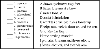
Match the muscle to its definition.
 To practice labeling some of the major muscles, click on Label It.
Tendons
Combining Forms for the Anatomy of the Musculoskeletal System
| Meaning | Combining Form |
| acromion | acromi/o |
| bone marrow | myel/o |
| bone | oste/o, osse/o, oss/i |
| bursa | burs/o |
| calcaneus (heel bone) | calcane/o |
| carpus (wrist) | carp/o |
| cartilage | chondr/o, cartilag/o |
| chin | ment/o, geni/o |
| clavicle (collarbone) | clavicul/o, cleid/o |
| coccyx (tailbone) | coccyg/o |
| condyle | condyl/o |
| elbow (olecranon) | olecran/o |
| epicondyle | epicondyl/o |
| ethmoid | ethmoid/o |
| femur (thigh bone) | femor/o |
| fibula (lower lateral leg bone) | fibul/o, perone/o |
| finger, toe (whole), digitus | dactyl/o, digit/o |
| foramen | foramin/o |
| frontal bone | front/o |
| glenoid | glen/o |
| hallux | halluc/o |
| humerus (upper arm bone) | humer/o |
| ilium | ili/o |
| ischium | ischi/o |
| jaw (entire) | gnath/o |
| joint (articulation) | arthr/o, articul/o |
| lacrima | lacrim/o |
| lamina | lamin/o |
| ligament | ligament/o, syndesm/o |
| lower back | lumb/o |
| mandible (lower jaw bone) | mandibul/o |
| maxilla (upper jaw bone) | maxill/o |
| meniscus | menisc/o |
| metacarpus (hand bone) | metacarp/o |
| metatarsus (foot bone) | metatars/o |
| muscle (heart) | myocardi/o, cardiomy/o |
| muscle (smooth) | leiomy/o |
| muscle (skeletal) | rhabdomy/o |
| muscle | my/o, myos/o, muscul/o |
| neck | cervic/o |
| occiput | occipit/o |
| olecranon | olecran/o |
| palatine bone | palat/o |
| parietal bone | pariet/o |
| patella (kneecap) | patell/o, patell/a |
| pelvis | pelv/i, pelv/o |
| phalanx (one of the bones of the fingers or toes) | phalang/o |
| pubis (pubic bone) | pub/o |
| radius (lower lateral arm bone) | radi/o |
| rib (costa) | cost/o |
| sacrum | sacr/o |
| scapula (shoulder blade) | scapul/o |
| sinus | sin/o, sinus/o, antr/o |
| skeleton | skelet/o |
| skull (cranium) | crani/o |
| sphenoid | sphenoid/o |
| spinal column, spine | spin/o, rachi/o, vertebr/o |
| sternum, breastbone | stern/o |
| tarsus (anklebone) | tars/o |
| temporal bone | tempor/o |
| tendon | tendin/o, tendon/o, ten/o, tend/o |
| thorax (chest) | thorac/o |
| tibia (shinbone) | tibi/o |
| ulna | uln/o |
| vertebra (backbone) | vertebr/o, spondyl/o |
| vomer | vomer/o |
| xiphoid process | xiph/o |
| zygoma (cheekbone) | zygomat/o |
Prefixes for the Anatomy of the Musculoskeletal System
| Prefix | Meaning |
| ab- | away from |
| ad- | toward |
| amphi- | both |
| bi- | two |
| circum- | around |
| dia- | through, complete |
| endo-, end- | within |
| epi- | above, upon |
| ex-, e- | out |
| in- | in |
| inter- | between |
| intra- | within |
| peri- | surrounding, around |
| pro- | forward |
| re- | back |
| syn- | together, joined |
Suffixes for the Anatomy of the Musculoskeletal System
| Suffix | Meaning |
| -ar, -al, -ic, -ous, -eal | pertaining to |
| -blast | embryonic |
| -clast | breaking down |
| -cyte | cell |
| -genesis | production, origin |
| -oid | resembling, like |
| -physis | growth |
| -poiesis | formation |
| -sis | condition |
| -um | structure |
 You can review the anatomy of the musculoskeletal system by clicking on Body Spectrum, then Muscular and Skeletal.
 Exercise 9:
Emergency Room Report
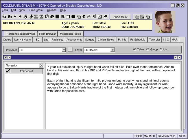
Use the emergency room record above to answer the following questions.
1. Dylan’s injury is to the fleshy area of the palm near the thumb, the “thenar eminence.” Explain the difference between a DIP (distal interphalangeal joint) and a PIP (proximal interphalangeal) joint.
________________________________________________________________________________________________
2. Not being able to “flex” a body part means one is unable to ______________________________________.
3. “First digit, R hand” means _____________________________________________________________________.
4. The first metacarpal is a bone of the ____________________________________________________________.


