Chapter 42 Muscular Dystrophy: How Should It Be Treated?
Improvements in healthcare have resulted in longevity for patients with muscular dystrophies. Until a cure is found, therapeutic and supportive care is essential in preventing complications and improving the affected child’s quality of life (QOL). This chapter reviews the available evidence from a surgeon’s perspective to guide in the management of this condition.
STEROID THERAPY
Currently, corticosteroids are the only class of drug that has been extensively studied in this condition. In 1989, a large, multicenter, randomized, placebocontrolled study in the United States showed an unequivocal improvement in muscle force with prednisone in patients with Duchenne muscular dystrophy (DMD), and that a low-dose (0.75 mg/kg) daily regimen was as effective as the standard 1.5 mg/kg.1 The improvement in muscle force seemed to be fairly rapid, within a matter of weeks, and followed by a plateau. However, the substantial side effects, particularly weight gain, weighed heavily against the potential benefits, and the authors recommended further research regarding use of steroids as a long-term therapy for muscular dystrophy. After the initial trial, another randomized study examined the use of alternate dosing regimens, and duration of therapy was reported.2 Although many studies provided Level III and IV evidence,3–8 they showed that use of a relatively high dose of deflazacort resulted in long-term maintenance of pulmonary function, and the age at which boys became full-time wheelchair users increased by several years over boys who did not take deflazacort. Some debate remains as to the exact dosage regimen (daily vs. alternative schedules) of steroids to compare both efficacy and adverse effects.9,10 Long-term steroid effects on quality and length of life are not yet known. Nevertheless, current Level III and IV evidence suggests that all boys with DMD should be given the opportunity to start these medications early in their disease course, well before they reach the stage of full-time wheelchair use.
NECK HYPEREXTENSION
Neck hyperextension with progressive increase of lordosis associated with a limitation in flexion of the cervical spine forces these patients to assume awkward compensatory postures to maintain balance and level vision, worsening their QOL significantly. Level IV evidence suggests posterior interspinous cervical fusion in seven patients led to a better sitting position, to easier nursing, and better cosmesis.11 The mean angle between C2-C7 decreased from an average of 30 degrees before surgery to an average of 19 degrees at 1-year of follow-up. Clinically, these patients had more control over their head position after surgery.
SHOULDER
Orthopedic intervention for the shoulder is mainly confined to fascioscapulohumeral dystrophy. Surgical indications that have been described include inability to abduct or elevate arms and loss of power with inability to hold arm sustained in abduction and elevation. As the muscles stabilizing the scapula become involved, the scapula starts to wing, leading to issues with cosmesis and problems with garment wear particularly in female patients.12 Surgery should be performed only if the deltoid strength is 4/5 or better. Furthermore, although improvement in appearance and diminution of pain and fatigue can be maintained over the long term, the gains in motion may be lost over time.
Scapulothoracic Fusion
Copeland and colleagues12 reported Level IV evidence with average 16-year follow-up period (range, 14 months to 44 years) of scapulothoracic arthrodesis in patients aged 16 to 35 years. From the 13 initial patients (20 fusions), 10 patients (14 shoulders) were available for long-term follow-up. Complications included pleuritic pain (resolved spontaneously within 1 week), hemopneumothorax (requiring drainage), rib and scapula fractures, screw pullout, and painful nonunion requiring revision surgery. These patients had an average flexion of 123 degrees and an average abduction of 103 degrees. All patients eventually had improved function, activities of daily living, and cosmesis.
Diab and coauthors13 report their Level IV evidence of 11 scapulothoracic arthrodesis in 8 patients with adolescent and infantile forms of fascioscapulohumeral muscular dystrophy. They fused the scapula to the thorax in 25 degrees of abduction using 16-gauge wires, a plate, or washers on the posteromedial scapular surface to prevent wire pullout and iliac crest autograft. In all cases, scapular winging and shoulder fatigue and pain initially were eliminated. Active abduction and forward flexion improved to an average of 145 and 144 degrees, respectively. At 6.3 years after surgery, seven shoulders retained motion, whereas four lost motion because of progressive weakness of deltoid with no evidence of failure of the arthrodesis. No short- or long-term respiratory compromise was reported.
Scapulopexy
Scapulopexy has been proposed as an alternative to theoretically avoid the problems of reduced respiratory function, and the shoulder can be mobilized almost immediately after surgery, reducing the risk for muscle atrophy and complications such as stress fractures and nonunion. Giannini and coworkers14 have reported their case series (Level 4 evidence) with 9 patients (18 shoulders). The scapula was repositioned over the rib cage and fixed to the ribs with metal wires. Arm abduction increased from an average of 68 degrees before surgery to 96 degrees after surgery. Arm flexion increased from an average of 57 degrees before surgery to 116 degrees after surgery. The position of the scapula obtained at the time of surgery was maintained, and the patients had satisfactory cosmetic results at 9.9 years of follow-up. This technique does not require grafts and reduces postoperative complications such as pneumothorax or hemothorax.
SPINE
Steroid Treatment and the Development of Scoliosis
Alman and researchers15 reported Level 2 evidence showing that steroid treatment slows the progression of scoliosis in male patients with DMD. This was a nonrandomized, comparative study with 7- to 10-year-old boys who were able to walk. Thirty patients were treated with deflazacort (treatment group), and 24 were not (control group). Patients were matched for age and pulmonary function at baseline, and were managed for at least 5 years. A curve of 20 degrees or greater developed during the follow-up period in 16 (67%) of the 24 patients in the control group, but in only 5 (17%) of the 30 patients in the treatment group. Fifteen of the 24 patients in the control group underwent spine surgery, at a mean age of 13 years, whereas only 5 of the 30 patients in the treatment group underwent spine surgery, at a mean age of 15 years. Kaplan–Meier analysis demonstrated a significant difference between the two groups with regard to development of scoliosis of 20 degrees or more (P < 0.001). Adverse effects with steroids included cataract (10 patients, none of which interfered with visual acuity or required surgical treatment), stress fractures (3 patients treated with immobilization and/or rest), and weight gain (patients in the treatment group weighed a mean of 3.7 kg more than did those in the control group). Because the families rather than the physician chose the treatment, there may be an inherent selection bias because it is possible that the families who saw more physical deterioration in their child decided against steroid treatment, resulting in the selection of a less severely affected treatment group. These patients were continued on steroid therapy even while the patients were using the wheelchair full time. The future of spines of these patients once skeletal maturity is reached is currently unclear and warrants further study.
Prediction of Spinal Deformity
Several studies have shown that scoliosis develops in almost all patients with DMD, with the exception of those who have a stable, hyperextended spine.16–18 The progression of spinal deformity leads to a deterioration in pulmonary function in patients with DMD. Yamashita and colleagues19 reported a case series (Level IV evidence) showing the correlation of age at and value of the plateau of vital capacity (VC) with the severity of progression of spine deformity. They show in a retrospective study of 36 patients that rapid and severe progression of spinal deformity could be expected in patients whose VC plateau was less than 1900 mL and in those in whom it occurred before age 14 years.
To further refine their criteria, Yamashita and colleagues20 published their results on a cohort of 11 patients (Level IV) using multiple discriminant analysis on the basis of 4 predictors: (1) VC at age 10 years, (2) age at which ambulation ceased, (3) curve pattern of spinal scoliosis, and (4) Cobb angles at age 10 years. In these 11 patients, multiple discriminant analysis correctly predicted the severity of the clinical course of 91.7% of patients with DMD. VC at age 10 was found to be the strongest predictor among the variables. These patients could be identified earlier on as candidates for surgical intervention of spinal deformity.
Surgical Management
Spinal fusion surgery with instrumentation remains the mainstay of treatment for patients with DMD with scoliosis. Cheuk and investigators21 reviewed the available evidence in the literature for spinal surgery in patients with DMD. They evaluated publications from 1861 to January 2006, and because there were no randomized or quasi-randomized clinical trials to evaluate the effectiveness of scoliosis surgery in patients with DMD, they were unable to make any evidence-based recommendation for clinical practice. They recommend that patients should be informed about the uncertainty of benefits and potential risks of surgery for scoliosis.
The potential advantages of spinal surgery described in the literature are to obtain and maintain sitting balance and correction of pelvic obliquity to preserve the ability of the patient to be mobile in a wheelchair for the remainder of his life. In addition, cosmetic improvement, no need for orthopedic braces, easier nursing care, and pain relief are other mentioned factors. Nevertheless, the effects of spinal surgery on respiratory function and life expectancy are still controversial. Velasco and coauthors22 recommend posterior spinal fusion for scoliosis in nonambulatory patients with DMD earlier in the course of progression based on Level III evidence. They found that in 20 patients with a minimum of 2 pulmonary function tests (PFTs) before surgery and 2 after surgery, posterior spinal fusion for scoliosis in DMD slowed the rate of respiratory decline in percentage forced VC from 8% per year before surgery to 3.9% per year after surgery (p < 0.0001). In an expanded group of 56 patients with DMD with variable numbers of PFTs, the rate of decline also slowed after surgery from 4% per year to 1.75% per year as determined by within-subjects mixed-model regression analysis (P < 0.0001).
Quality of Life
Mercado and coworkers23 report a systematic literature review pertaining to QOL in patients with neuromuscular scoliosis who underwent spinal fusion. The need to operate early in the course of the deformity frequently when curves are relatively mild, as well as the change in the course of the disease caused by steroids, makes measuring the impact of scoliosis surgery on QOL in patients with muscular dystrophy more complex. Based on self-report, the surgery may improve sitting ability, posture, simplify nursing care, and improve self-esteem. The effect on the respiratory and cardiac function is unknown.24 The authors conclude that given the large emotional family investment in surgery, and the uncontrolled nature of the surgical series, the evidence in support of improving QOL of these patients is weak (grade C recommendation25).
Intraoperative Blood Loss
Intraoperative blood loss is high in patients with DMD who are undergoing posterior spinal fusion for scoliosis. Shapiro and coworkers26 have reported their results of a cohort of 56 patients divided into 2 groups: 1 group received tranexamic acid (TXA) whereas the other did not (Level III evidence). The groups were comparable in relation to surgical technique, age at surgery, number of levels fused, preoperative deformity, and surgical time. Blood loss with TXA decreased by 42% compared with those not treated with TXA. Accounting for patient weight and estimated blood volume, mean percentage blood loss with and without TXA was 47% ± 28% versus 112% ± 67% (P < 0.001). This physiologic indicator shows that blood loss with TXA decreased by 58% compared with those patients not treated with TXA. TXA was also showed to significantly reduce intraoperative blood loss after accounting for surgical time. Intraoperative homologous blood replacement by whole blood and packed red blood cells and autologous cell saver replacement are also significantly reduced with TXA.
Distal Extent of Fusion
Most authors previously recommended fixation to the sacrum or pelvis for scoliosis in DMD to correct pelvic obliquity. However, this has been questioned because the curve size and pelvic obliquity are small in early cases, and pelvic fixation has a greater complication rate. Mubarak and colleagues27 published Level III evidence comparing pelvic versus lumbar fixation for scoliosis in 22 wheelchair-mobilized patients with DMD. They found that in small curves with minimal preoperative pelvic obliquity (15 degrees or less), instrumentation and fusion to L5 was adequate at the time of a 34-month follow-up. However, in a later study, comparing pelvic and lumbar fixation in 48 cases of DMD, Alman and Kim28 disagreed (Level III evidence). Using Luque instrumentation and following Mubarak and colleagues’ criteria27 (fixation to L5 when Cobb angle < 40 degrees and pelvic obliquity < 10 degrees), they found progression of pelvic obliquity in 32 of 38 cases in their lumbar fixation group (with 2 cases requiring revision), but no progression at all in 10 cases in the sacral fixation group. They recommend sacral fixation in general and especially when the apex of the curve is below L1. They recognized, however, that sublaminar wire instrumentation in the lumbar spine could be responsible for the failure in the lumbar fixation group, and suggested that the use of screws or hooks into the lumbar spine may avoid this problem.
Sengupta and coworkers29 report on their cohort of 50 patients (Level III evidence) with DMD using newer constructs with lumbar pedicle screws. They divided their patients into two groups who were observed for a minimum of 3 years. The first group consisted of 31 patients with an average age of 14 years. This group underwent a spinal fusion that extended to the pelvis. The average preoperative Cobb angle was 48 degrees, and the average preoperative pelvic obliquity was 20 degrees. The second group consisted of 19 patients with an average age at surgery of 11.7 years. The patients in this group were treated with thoracic sublaminar wires and lumbar pedicle screws. The average preoperative Cobb angle was 19.8 degrees, and the average preoperative pelvic obliquity was 9 degrees. The authors document improved correction and maintenance of correction in the group that underwent fusion with pedicle screw fixation to L5. They acknowledge that the preoperative deformity was greater in the patients who had fusion to the pelvis and conclude that lumbar pedicle screw fixation that stops short of the pelvis is adequate in patients who have minimal deformity and pelvic obliquity.
HIP AND KNEE FLEXION CONTRACTURES
Contractures and progressive weakness of the lower extremities render walking increasingly difficult for patients with DMD. Extensive rehabilitation and orthopedic measures, in addition to medical treatment, have been directed toward prolongation of the ability to walk. Manzur and coauthors30 reported a randomized, controlled trial (Level II evidence) in 1992 to evaluate the role of early surgical treatment of contractures in 20 boys with DMD. Their entry criteria were designed to recruit a cohort of boys with a relatively uniform clinical severity, in the age range of 4 to 6 years. These boys were divided into two groups. The 10 boys in the surgical treatment group had the operation within 1 month of randomization by a single surgeon. Surgery was standardized and consisted of open release at the hip of sartorius, superficial head of rectus femoris and tensor fascia lata. A long incision was made along the lateral aspect of the thigh to expose the deep fascia, which was incised anterior to the lateral intermuscular septum, and a strip of fascia approximately 2 cm wide was removed. The tendo-Achilles (TA) was routinely lengthened percutaneously. Hamstring tendons were released percutaneously if there were knee flexion contractures. They were discharged 4 to 6 days after surgery, walking without any orthotic support. No passive stretching or physiotherapy was prescribed. The second control group continued with routine management including regular passive stretching of TA, iliotibial bands (ITBs), and hip flexors. The stretching was performed daily by parents after demonstration by the physiotherapist and was kept under surveillance at each follow-up attendance. Regression analysis showed no significant difference between the two groups in any of the parameters measured. No measurable difference was found between the surgical and control groups. Surgery was effective in reducing the TA contractures in all of the boys who underwent surgery. However, freedom from the need for regular passive stretching was short-lived, and 7 of the 10 boys had recurrence of the TA contractures within 1 to 2 years after surgery. Based on these results, the authors did not recommend surgery as standard treatment.
Bakker and de Groot31 did a structured literature review on the effects of knee-ankle-foot orthosis (KAFO) for patients with DMD. They searched the literature from 1966 to 1997 based on their inclusion criteria. No RCT had been performed to determine the effectiveness of KAFO. Only four nonrandomized, controlled studies were described. They suggest the possibility of a publication bias (only best results published), patient selection bias (only motivated families), and the absence of blinding with measurement bias leading to a positive influence on the results. All reviewed studies described surgical Achilles tenotomy. The scientific strength of the studies reviewed was poor. Results showed that the KAFO can prolong assisted walking, but it is uncertain whether it can prolong functional walking. The boys that benefit most have a relatively low rate of deterioration, are capable of enduring an operation, and are well motivated.
FOOT AND ANKLE
Equinus and equinovarus deformity of the ankle are serious problems associated with DMD. Hyde and colleagues32 reported Level II evidence, a randomized, prospective, multicenter trial, looking at passive stretching techniques with or without the use of below-knee night splints early in the course of the disease on the evolution of ankle equinus. Twenty-seven boys were evaluated and randomized using an incomplete block design using age and muscle strength as variables for stratification. The authors show that boys who wore night splints in addition to performing passive stretching of the TA benefited by an annual delay of 23% in the development of contractures compared with those who did not.
Main et al.33 published Level III evidence regarding serial casting of ankles in DMD to reduce TA contractures and to allow fitting of KAFOs at the end of functional walking. TA range and age at loss of standing with KAFOs were prospectively assessed in 9 patients who underwent serial casting before rehabilitation into KAFOs and compared with a group of 20 patients with DMD who had TA (group II) or TA and ITB surgical resection (group III). Mean age at loss of functional ability to walk and rehabilitation with KAFOs was 10.3 years (8.7–11.7 years) in group I, 9.3 years (7.7–12 years) in group II, and 9.5 years (8.3–11.7 years) in group III. The authors conclude that although further studies will be required to evaluate the effect of all these variables in a larger prospective, randomized cohort, the results of this pilot study suggest that serial casting can be considered in patients with DMD with moderate TA contractures and in whom there is no significant ITB tightness. The results of this study have to be interpreted with caution because the number of patients is too small. Furthermore, this was not a prospective study, and the groups could not be randomized or matched for age at intervention, use of steroids, and other relevant factors that may have contributed to the duration of ambulation in KAFOs.
Scher and Mubarak34 reported Level 3 evidence for the use of posterior tibial tendon transfer in patients with DMD with regard to foot deformity and ambulation. Fifty-seven patients were divided into three treatment groups. Group I contained patients who had surgery to maintain or resume ambulation. Group II included patients who had stopped walking and did not have the potential to resume ambulation or had no desire to do so. They had surgery to correct and maintain foot position. Group III (n = 25) patients did not have any surgery. All 32 patients in groups I and II had posterior tibial tendon transfer and Achilles tendon lengthening as part of the procedure. The mean age at cessation of ambulation for those who had surgery was 11.2 years versus 10.3 years for those who did not have surgery (P < 0.005). Thirty-four available patients (20 in groups I and II, and 10 in group III) were assessed for outcomes and foot position. Of the 24 patients who had lower extremity surgery, 23 (96%) were comfortable in any shoes or sneakers versus 6 (60%) in the group who did not have surgery. The authors indicate a positive role of foot surgery for these patients.
In contrast, Leitch and coworkers35 presented their Level III evidence with 88 full-time wheelchair patients with DMD divided into 2 groups. The first group comprised 30 boys with DMD who had had foot surgery including an open or percutaneous TA lengthening or tenotomy, and 26 of the 30 patients in this group also had a tibialis posterior tendon transfer. The second group consisted of boys who did not have surgery. A self-report questionnaire was used to assess difficulty with shoe wear, foot pain, foot hypersensitivity, and cosmetic concerns with regard to the feet, and a foot examination was performed. No significant differences were found between patients who did and did not receive foot surgery with respect to shoe wear (P > 0.05), pain (P > 0.05), hypersensitivity (P > 0.05), or cosmesis (P > 0.05). Hindfoot motion was significantly better in patients who did not have surgery (P < 0.05). Equinus contractures were significantly worse in patients who did not have surgery (P < 0.05). The authors conclude that surgery changes the position of the feet. However, all patients, regardless of whether they had surgery, had minimal concerns about their feet cosmesis, minimal difficulty with shoe wear, and infrequent pain. They recommend that full-time wheelchair users with DMD do well with or without surgery. Therefore, given the greater risks for surgery in this population, foot surgery should be performed only in children with specific foot problems that surgery can definitely address. The main caveat was that the patients were not randomized, and preoperative data for all patients were not available. Therefore, it is possible that patients who had more severe pain, concerns about the appearance of their feet, or difficulty with shoe wear may have been more likely to receive surgery.
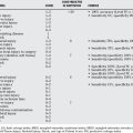
Based on the current level of evidence available, full-time wheelchair sitters can be advised that the frequency of poor outcome is low in patients who do not receive surgery and that surgery can change the shape of their feet, but the outcomes of treatment appear to be roughly similar with or without surgery. Table 42-1 summarizes the current available evidence for surgery in patients with DMD.
| STATEMENT | LEVEL OF EVIDENCE/GRADE OF RECOMMENDATION | REFERENCES |
|---|---|---|
QOL, quality of life; TA, tendo-Achilles.
1 Mendell JR, Moxley RT, Griggs RC, et al. Randomized, double-blind six-month trial of prednisone in Duchenne’s muscular dystrophy. N Engl J Med. 1989;320:1592-1597.
2 Beenakker EA, Fock JM, Van Tol MJ, et al. Intermittent prednisone therapy in Duchenne muscular dystrophy: A randomized controlled trial. Arch Neurol. 2005;62:128-132.
3 Alman BA. Duchenne muscular dystrophy and steroids: Pharmacologic treatment in the absence of effective gene therapy. J Pediatr Orthop. 2005;25:554-556.
4 Biggar WD, Gingras M, Fehlings DL, et al. Deflazacort treatment of Duchenne muscular dystrophy. J Pediatr. 2001;138:45-50.
5 Biggar WD, Harris VA, Eliasoph L, Alman B. Long-term benefits of deflazacort treatment for boys with Duchenne muscular dystrophy in their second decade. Neuromuscul Disord. 2006;16:249-255.
6 Biggar WD, Politano L, Harris VA, et al. Deflazacort in Duchenne muscular dystrophy: A comparison of two different protocols. Neuromuscul Disord. 2004;14(8-9):476-482.
7 Carter GT, McDonald CM. Preserving function in Duchenne dystrophy with long-term pulse prednisone therapy. Am J Phys Med Rehabil. 2000;79:455-458.
8 Fenichel GM, Florence JM, Pestronk A, et al. Long-term benefit from prednisone therapy in Duchenne muscular dystrophy. Neurology. 1991;41:1874-1877.
9 Bushby K, Muntoni F, Urtizberea A, et al. Report on the 124th ENMC International Workshop. Treatment of Duchenne muscular dystrophy; defining the gold standards of management in the use of corticosteroids. 2-4 April 2004, Naarden, The Netherlands. Neuromuscul Disord. 2004;14(8-9):526-534.
10 Dubowitz V. Prednisone for Duchenne muscular dystrophy. Lancet Neurol. 2005;4:264.
11 Giannini S, Faldini C, Pagkrati S, et al. Surgical treatment of neck hyperextension in duchenne muscular dystrophy by posterior interspinous fusion. Spine. 2006;31:1805-1809.
12 Copeland SA, Levy O, Warner GC, Dodenhoff RM. The shoulder in patients with muscular dystrophy. Clin Orthop Relat Res.; 368; 1999; 80-91.
13 Diab M, Darras BT, Shapiro F. Scapulothoracic fusion for facioscapulohumeral muscular dystrophy. J Bone Joint Surg Am. 2005;87:2267-2275.
14 Giannini S, Ceccarelli F, Faldini C, et al. Scapulopexy of winged scapula secondary to facioscapulohumeral muscular dystrophy. Clin Orthop Relat Res. 2006;449:288-294.
15 Alman BA, Raza SN, Biggar WD. Steroid treatment and the development of scoliosis in males with duchenne muscular dystrophy. J Bone Joint Surg Am. 2004;86-A:519-524.
16 Wilkins KE, Gibson DA. The patterns of spinal deformity in Duchenne muscular dystrophy. J Bone Joint Surg Am. 1976;58:24-32.
17 Smith AD, Koreska J, Moseley CF. Progression of scoliosis in Duchenne muscular dystrophy. J Bone Joint Surg Am. 1989;71:1066-1074.
18 Oda T, Shimizu N, Yonenobu K, et al. Longitudinal study of spinal deformity in Duchenne muscular dystrophy. J Pediatr Orthop. 1993;13:478-488.
19 Yamashita T, Kanaya K, Yokogushi K, et al. Correlation between progression of spinal deformity and pulmonary function in Duchenne muscular dystrophy. J Pediatr Orthop. 2001;21:113-116.
20 Yamashita T, Kanaya K, Kawaguchi S, et al. Prediction of progression of spinal deformity in Duchenne muscular dystrophy: A preliminary report. Spine. 2001;26:E223-E226.
21 Cheuk DK, Wong V, Wraige E, et al. Surgery for scoliosis in Duchenne muscular dystrophy. Cochrane Database Syst Rev.; 1; 2007; CD005375.
22 Velasco MV, Colin AA, Zurakowski D, et al. Posterior spinal fusion for scoliosis in Duchenne muscular dystrophy diminishes the rate of respiratory decline. Spine. 2007;32:459-465.
23 Mercado E, Alman B, Wright JG. Does spinal fusion influence quality of life in neuromuscular scoliosis? Spine. 2007;32(19 suppl):S120-S125.
24 Andersson GB, Bridwell KH, Danielsson A, et al. Evidence-based medicine summary statement. Spine. 2007;32(19 suppl):S64-S65.
25 Wright JG, Einhorn TA, Heckman JD. Grades of recommendation. J Bone Joint Surg Am. 2005;87:1909-1910.
26 Shapiro F, Zurakowski D, Sethna NF. Tranexamic acid diminishes intraoperative blood loss and transfusion in spinal fusions for duchenne muscular dystrophy scoliosis. Spine. 2007;32:2278-2283.
27 Mubarak SJ, Morin WD, Leach J. Spinal fusion in Duchenne muscular dystrophy—fixation and fusion to the sacropelvis? J Pediatr Orthop. 1993;13:752-757.
28 Alman BA, Kim HK. Pelvic obliquity after fusion of the spine in Duchenne muscular dystrophy. J Bone Joint Surg Br. 1999;81:821-824.
29 Sengupta DK, Mehdian SH, McConnell JR, et al. Pelvic or lumbar fixation for the surgical management of scoliosis in duchenne muscular dystrophy. Spine. 2002;27:2072-2079.
30 Manzur AY, Hyde SA, Rodillo E, et al. A randomized controlled trial of early surgery in Duchenne muscular dystrophy. Neuromuscul Disord. 1992;2(5-6):379-387.
31 Bakker JP, de Groot IJ, Beckerman H, et al. The effects of knee-ankle-foot orthoses in the treatment of Duchenne muscular dystrophy: Review of the literature. Clin Rehabil. 2000;14:343-359.
32 Hyde SA, Filytrup I, Glent S, et al. A randomized comparative study of two methods for controlling Tendo Achilles contracture in Duchenne muscular dystrophy. Neuromuscul Disord. 2000;10(4-5):257-263.
33 Main M, Mercuri E, Haliloglu G, Baker, et al. Serial casting of the ankles in Duchenne muscular dystrophy: can it be an alternative to surgery? Neuromuscular Disord. 2007;17(33):227-230.
34 Scher DM, Mubarak SJ. Surgical prevention of foot deformity in patients with Duchenne muscular dystrophy. J Pediatr Orthop. 2002;22:384-391.
35 Leitch KK, Raza N, Biggar D, et al. Should foot surgery be performed for children with Duchenne muscular dystrophy? J Pediatr Orthop. 2005;25:95-97.


