Mucinous Cystic Pancreatic Tumor

Published on 19/07/2015 by admin

Filed under Radiology

Last modified 22/04/2025

Print this page

rate 1 star rate 2 star rate 3 star rate 4 star rate 5 star
Your rating: none, Average: 0 (0 votes)

This article have been viewed 2973 times

 “Macrocystic” pattern: Few (< 6) macrocystic locules, which are relatively large (> 2 cm)

image Usually of simple fluid attenuation, but may be mildly hyperdense due to hemorrhage or protein
image Frequent peripheral curvilinear calcifications or calcifications in septations (16% of cases)
image Presence of thick wall, mural nodularity, or thick septations suggests invasive malignancy
image No apparent communication with main pancreatic duct
• MR: Usually simple fluid signal (high T2; low T1), but may be slightly less T2 hyperintense due to mucin content

image Internal septations (which are T2 hypointense) easier to perceive on MRI compared to CT
image MRCP: No communication with main pancreatic duct

PATHOLOGY

• Tumor shares clinical and pathologic characteristics of biliary tumors, ovarian tumors, and IPMN
• Presence of ovarian stroma lining cyst is key feature for diagnosis of mucinous cystic neoplasm (MCN)

CLINICAL ISSUES

• Strong preponderance in middle-aged women (99%)
• Considered premalignant or frankly malignant: Rate of malignancy in different series ranges between 10-40%
• Risk factors for invasive malignancy: Older age, lesion size, mural nodularity, thick wall, patient symptoms (pain, pancreatitis), and ↑ CEA and CA 19-9
• MCNs typically undergo complete surgical resection with adjuvant chemotherapy for malignant lesions
• Malignant MCNs receive adjuvant chemotherapy after surgical resection
image
(Left) Graphic of a mucinous cystic tumor shows a multiseptate, mucin-filled, cystic mass in the pancreatic tail that displaces the pancreatic duct.

image
(Right) Axial CECT in a 35-year-old woman demonstrates a large complex cystic mass image arising from the pancreatic tail with multiple internal cystic locules and septations, some of which are thick image. This mass was found to be a mucinous cystic neoplasm (MCN) with invasive adenocarcinoma at surgery.
image
(Left) Axial CECT in a 46-year-old woman demonstrates a large, simple-appearing cyst image 
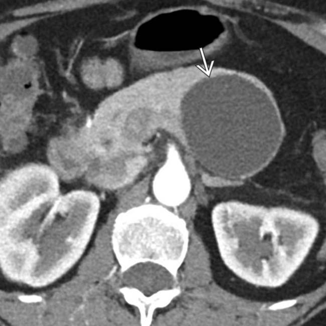
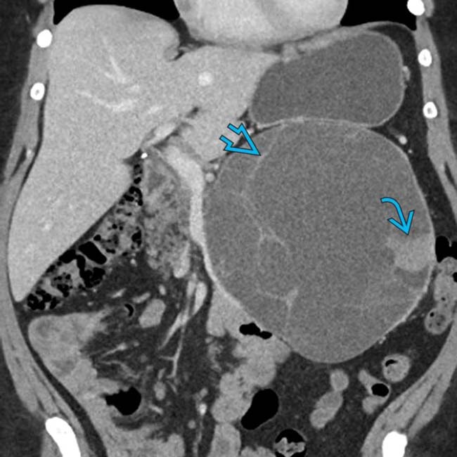
arising from the upstream pancreatic body.

image
(Right) Axial T2 FS MR in the same patient confirms the lack of complexity within the T2-hyperintense cyst image
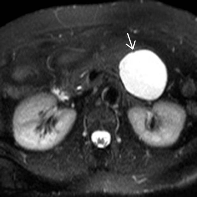
. MR can sometimes show complexity and suspicious features that might be difficult to identify on CT. This was found to be a MCN with low-grade dysplasia at surgery.

TERMINOLOGY

Synonyms

• Mucinous cystic neoplasm (MCN), mucinous macrocystic neoplasm or adenoma, mucinous cystadenoma or cystadenocarcinoma

Definitions

• Thick-walled, unilocular or multilocular pancreatic tumor composed of large, mucin-containing cysts

image Some sources suggest presence of ovarian stroma within lesion is necessary for diagnosis
• MCN and intraductal papillary mucinous neoplasm (IPMN) are together classified as pancreatic mucinous tumors

IMAGING

General Features

• Best diagnostic clue

image Large, unilocular or multilocular encapsulated cystic mass with septations and thick wall in pancreatic tail
• Location

image Tail of pancreas (more common)
image Presents as single lesion (not multifocal like IPMN)
• Size

image 2-12 cm in diameter

– MCNs usually larger than serous cystadenoma or IPMN
image Size is predictor of biologic behavior

– Lesions < 40 mm found to have low risk of malignancy in recent series
• Morphology

image Typically round/ovoid, but may be ill defined/irregular

Radiographic Findings

• ERCP

image Displacement and narrowing of main pancreatic duct adjacent to tumor
image Can differentiate IPMN (which communicate with main pancreatic duct) from MCN (which do not communicate)

CT Findings

• Unilocular or multilocular encapsulated cyst in pancreatic body/tail with frequent internal septations

image “Macrocystic” pattern: Few (< 6) macrocystic locules that are relatively large (> 2 cm)
• Usually of simple fluid attenuation, but may be mildly hyperdense due to hemorrhage or protein
• Frequent peripheral curvilinear calcifications or calcifications in septations (16% of cases)
• Presence of thick, irregular wall, internal mural nodularity, or thick septations suggest invasive malignancy

image Upstream pancreatic ductal dilatation or atrophy suggest invasive malignancy
• No apparent communication with main pancreatic duct

image In rare instances, some series have suggested possible communication with pancreatic duct in very small minority of lesions

MR Findings

• Typically show simple fluid signal (high T2; low T1), but may be slightly less T2 hyperintense due to mucin content

image May show areas of T1 hyperintensity due to internal hemorrhage, proteinaceous content, or mucin
• Internal septations (typically T2 hypointense) easier to perceive on MR compared to CT
• Calcifications often not visible on MR: If visible, are low signal on all pulse sequences
• Thick, enhancing septations, wall thickening, and mural nodularity on T1WI C+ suggest invasive malignancy
• MRCP: No communication with pancreatic duct
• DWI: No role in differentiating mucinous from nonmucinous lesions or benign from malignant

Ultrasonographic Findings

• Grayscale ultrasound

image Multiloculated cystic mass with echogenic internal septa

– Often associated with thick wall ± mural nodularity
image Can also appear as unilocular anechoic mass

Angiographic Findings

• Conventional

image Predominantly avascular mass
image Cyst wall and solid component

– Show small areas of vascular blush and neovascularity
image Displacement of surrounding arteries and veins by cysts

Imaging Recommendations

• CECT or MR

DIFFERENTIAL DIAGNOSIS

Pancreatic Pseudocyst

• Usually known history of pancreatitis or alcoholism ± imaging stigmata of chronic pancreatitis (pancreatic calcifications, ductal beading, etc.)
• Loculated cyst with adjacent peripancreatic fat stranding/inflammation
• Evolves over time from acute peripancreatic fluid collection into loculated pseudocyst
• Communication with pancreatic duct is frequent (70% of cases) and may be visible on ERCP or MRCP
• Lab data: Increased amylase in cyst and serum
• When occurring in pancreatic tail, may simulate unilocular mucinous cystic neoplasm

Pancreatic Serous Cystadenoma

• Well-circumscribed, lobulated cystic mass most often occurring in pancreatic head
• Classic appearance (“microcystic” or “sponge” lesion): Many small cysts separated by thin septa and with central scar demonstrating calcification

image Calcification more common in serous than mucinous pancreatic neoplasms (38% vs. 16%)
• Macrocystic, oligocystic, and unilocular variants of serous cystadenoma difficult to distinguish from MCN

image Thick wall and mural nodularity unusual with serous cystadenoma

Pancreatic IPMN

• Mucin-producing neoplasms which are classified into 3 types with different risks of malignancy

image Side-branch IPMN: Arise in pancreatic duct side branch and carry risk of invasive malignancy of 17%
image Main pancreatic duct (MPD): Arise in main pancreatic duct and carry high risk of malignancy (58%)
image Combined type IPMN: Features of both side branch and main duct IPMN with prognosis similar to main duct IPMN
• Side-branch or combined type IPMN: Cyst (± nodularity, septations, calcifications) that communicates with pancreatic duct

Cystic Pancreatic Neuroendocrine Tumor

• Cystic neuroendocrine tumors more likely to be non-insulin producing and nonsyndromic
• Cystic lesion without pancreatic ductal dilatation or atrophy

image Differentiate from IPMN/MCN by presence of peripheral hyperenhancement on arterial phase CECT or MR

Pancreatic Epithelial (True) Cyst

• Usually small and multiple nonenhancing cysts without pancreatic ductal dilatation
• Rare lesions most commonly seen in patients with von Hippel-Lindau and autosomal dominant polycystic kidney disease

Lymphangioma (Mesenteric Cyst)

• Water density cyst with imperceptible wall and internal thin septations
• May abut pancreas mimicking pancreatic cystic neoplasm

Lymphoepithelial Cyst

• Rare benign cystic lesion that abuts and protrudes into pancreas mimicking pancreatic cystic lesion
• Well-defined water density cyst most commonly seen in pancreatic body or tail
• May be unilocular or multilocular ± internal septations and fat density

PATHOLOGY

General Features

• Etiology

image Uncertain
• Embryology, anatomy

image May be related to germ cell migration during 1st 8 weeks of gestation
• Neoplasm with number of cysts 2-6 cm in diameter seen in 95% of cases
• Tumor shares clinical and histopathologic characteristics of biliary tumors, ovarian tumors, and IPMN

image WHO classification emphasizes ovarian stroma lining cyst to be key feature for diagnosis of MCN
• Hypovascular mass with sparse neovascularity

Gross Pathologic & Surgical Features

• Large mass encapsulated by thick fibrous capsule
• Smooth and round, but may have lobulated surface
• Cut section: Multilocular or unilocular large cysts

image Individual cystic components measure > 2 cm with thin septations < 2 mm
• Cystic cavity may be filled with thick mucoid material and clear, green, or blood-tinged fluid
• Solid papillary projections protruding into interior of tumor are sign of invasive malignancy

Microscopic Features

• Characteristic ovarian-type stroma with spindle cells
• Tall, mucin-producing columnar cells
• Subtended by densely cellular mesenchymal stroma

CLINICAL ISSUES

Presentation

• Most common signs/symptoms

image Many MCNs are asymptomatic and incidentally discovered
image May present with epigastric pain, recurrent pancreatitis (10%), palpable mass (12%), or mass effect on adjacent structures (bowel obstruction, gastric outlet obstruction, jaundice)
image Rarely presents with local invasion/distant metastases
image Very rarely presents with systemic manifestations caused by tumor production of gastrin or other hormones
• Lab data

image Increased levels of serum and cyst fluid CEA
image Increased cyst fluid levels of CA 19-9 (80% of cases) and CA 72-4
image Cyst fluid levels of CA 72-4 (> 4 units/mL)

– 80% sensitivity, 95% specificity for tumor
image Low amylase levels
• Biopsy: Invasive cancer and benign epithelium can coexist in same lesion, making biopsy unreliable; benign biopsy result does not exclude malignancy
• Diagnosis based on a combination of preoperative imaging (CT or MR), endoscopic ultrasound with cyst aspiration/cytology/tumor markers, and post-operative histopathologic analysis

Demographics

• Age

image Mean: 50 years (range: 20-95 years)

– 50% between 40-60 years
• Gender

image M:F = 1:99
image Termed “mother” tumor due to predisposition for middle-aged females
• Epidemiology

image Less common than IPMN and serous cystadenomas
image 25% of all resected pancreatic cysts

Natural History & Prognosis

• Complications

image Due to mass effect

– Bowel obstruction
– Pancreatic duct narrowing or extrinsic obstruction
• Considered premalignant or frankly malignant

image Range from adenomas to invasive carcinoma (depending on degree of atypia)
image Malignant MCNs are lesions containing high-grade dysplasia or invasive carcinoma
image Even benign MCNs carry potential for malignant transformation and are considered premalignant
• Because almost all MCNs are resected, true natural history not completely certain: Rate of malignancy in different series ranges between 10-40%
• Overall 5-year survival of MCNs is 98% for benign lesions and 62% for lesions with high-grade dysplasia or invasive carcinoma

image Recent study suggests MCNs may have better prognosis than previously thought, with 5-year survival of 75% for MCN with invasive carcinoma
• Major risk factors for invasive malignancy

image Older patient age, larger lesion size, mural nodularity, thick wall, peripheral calcification, patient symptoms (pain, pancreatitis, etc.), and elevated CEA/CA 19-9
• Completely excised in patients with benign MCN: Excellent prognosis with extraordinarily low risk of recurrent disease
• Incompletely excised, marsupialized, or drained: Poor prognosis

Treatment

• Traditionally all MCNs have undergone complete surgical excision (usually distal pancreatectomy and splenectomy)

image Lesions in poor surgical candidates may be conservatively managed with close serial imaging follow-up
• Benign MCNs with complete surgical resection do not require further treatment or follow-up
• Malignant MCNs receive adjuvant chemotherapy after surgical resection

DIAGNOSTIC CHECKLIST

Consider

• Differentiation from other cystic pancreatic lesions may not be possible based on CT/MR alone, and may require cyst aspiration with cytology and tumor markers

Image Interpretation Pearls

• Large, unilocular or multiloculated cystic mass with enhancing septa and thick wall in pancreatic body or tail
image
(Left) Axial CECT in a 38-year-old woman demonstrates a large complex cystic mass image extending exophytically downwards from the pancreatic tail with multiple septations and enhancing mural nodularity image.

image
(Right) Coronal CECT in the same patient demonstrates the large size of the mass, as well as the presence of extensive septations image and mural enhancing soft tissue image, both of which raise suspicion for malignancy. This was found to be a MCN with high-grade dysplasia at surgery.
image
(Left) Axial CECT in a 34-year-old woman demonstrates a simple unilocular cyst image 
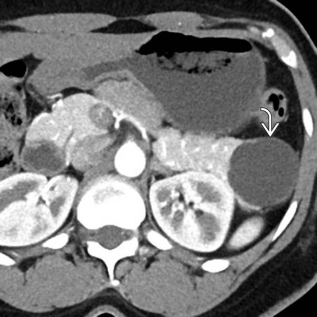
in the pancreatic tail, found to be a MCN with low-grade dysplasia at surgery. There is no mural nodularity or a thick-wall to suggest invasive malignancy.

image
(Right) Axial CECT shows a large cystic mass image 
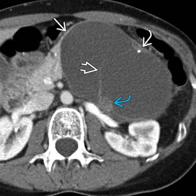
arising from the pancreatic body. The mass has large cystic spaces separated by septa image. Also noted are peripheral calcification image and soft tissue mural nodularity image. This is the classic imaging presentation of MCN.
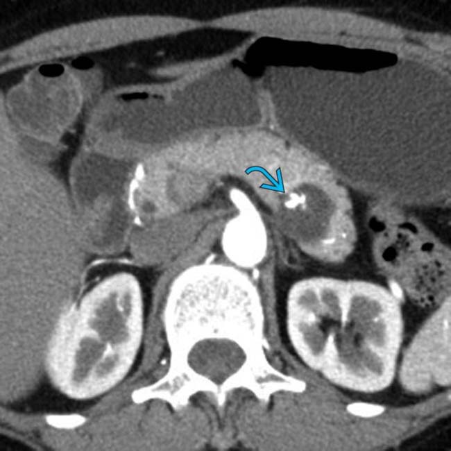
image
(Left) Axial CECT in a 61-year-old woman demonstrates a cystic lesion in the pancreatic tail with coarse mural calcification image.

image
(Right) Axial T2 MR in the same patient better demonstrates the presence of internal septations within the T2-hyperintense cyst image. MCN with low-grade dysplasia was found at surgery.
image
(Left) Axial CECT in a 34-year-old woman demonstrates a simple-appearing cyst image 
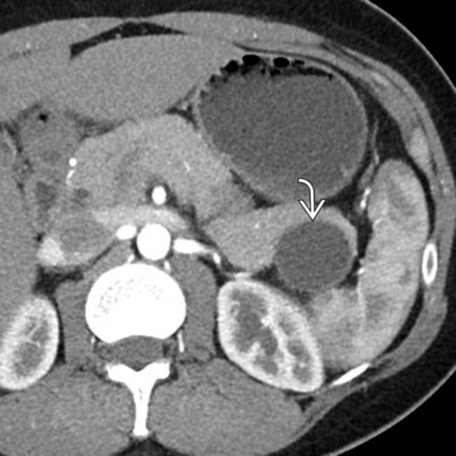
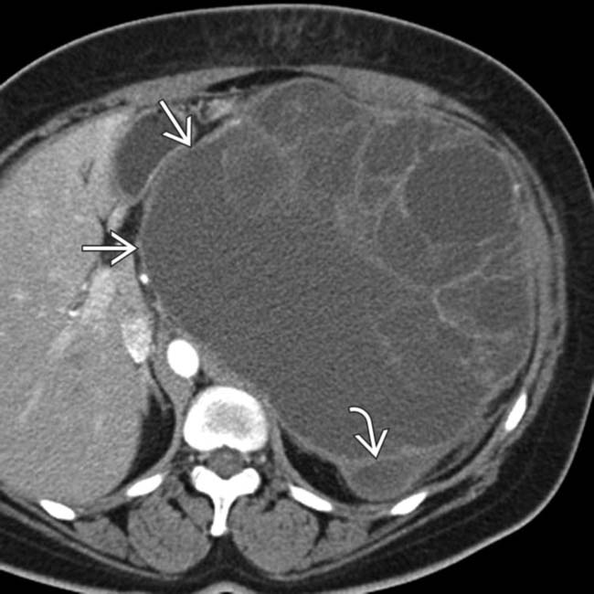
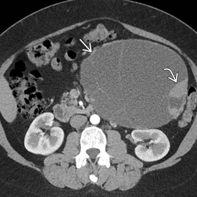
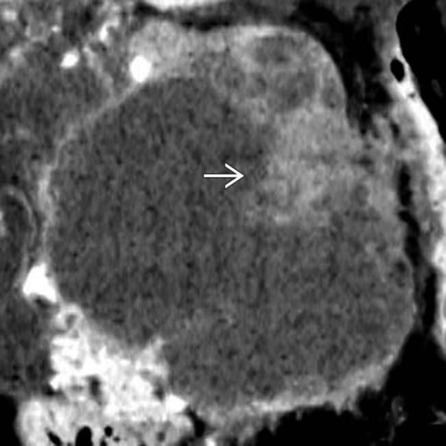
arising from the pancreatic tail. No internal complexity is visualized.

image
(Right) Axial T1 MR in the same patient demonstrates layering T1-hyperintense blood products image 
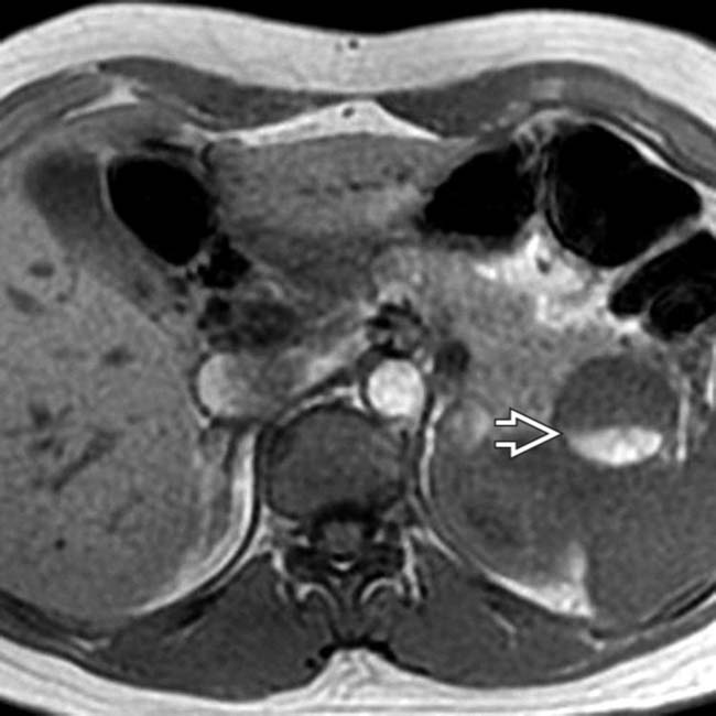
within the cyst, representing a hematocrit level. The presence of hemorrhage within an MCN is quite unusual.
image
(Left) Coronal volume-rendered CECT demonstrates a pancreatic cystic mass with prominent peripheral rim-like calcification image
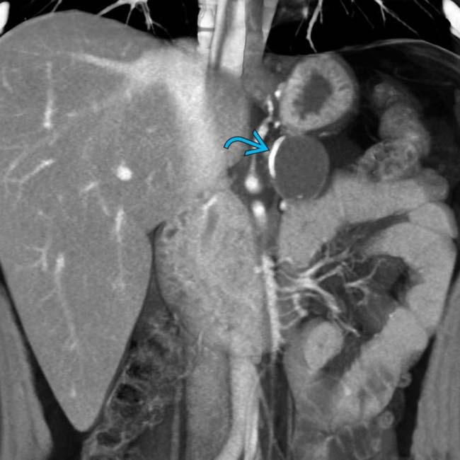
. Calcifications are seen in roughly 16% of MCNs.

image
(Right) Axial CECT demonstrates a large complex cystic mass image 
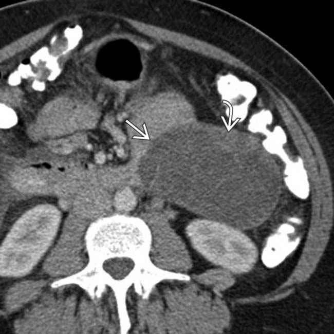
arising from the pancreatic tail with multiple internal septations and solid mural nodularity image. The complexity of the lesion raises suspicion for malignancy, which was confirmed after surgery where an MCN with invasive carcinoma was found.
image
(Left) Axial T2 FS MR demonstrates a large cystic mass image 
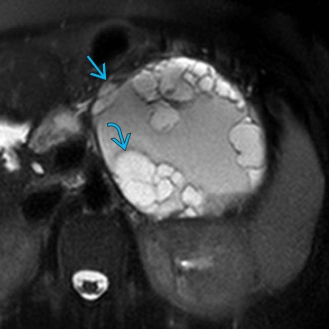
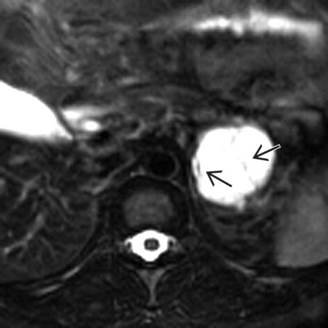
arising from the pancreatic tail with innumerable internal T2-hyperintense cystic locules image.

image
(Right) Axial T2 MR in the same patient demonstrates that the large cystic mass has layering debris with a fluid-fluid level image, probably representing old blood products or proteinaceous debris. This was found to be an MCN with invasive carcinoma at resection.
image
(Left) Axial CECT in a 33-year-old woman shows a cystic mass image 
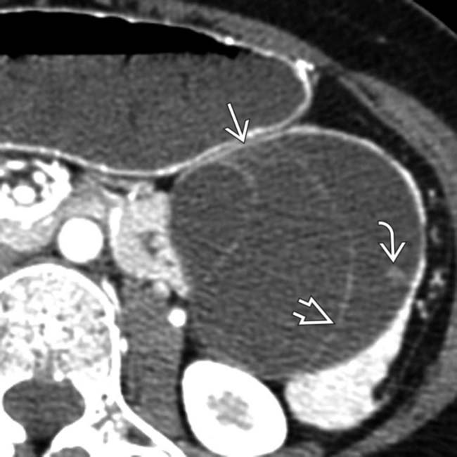
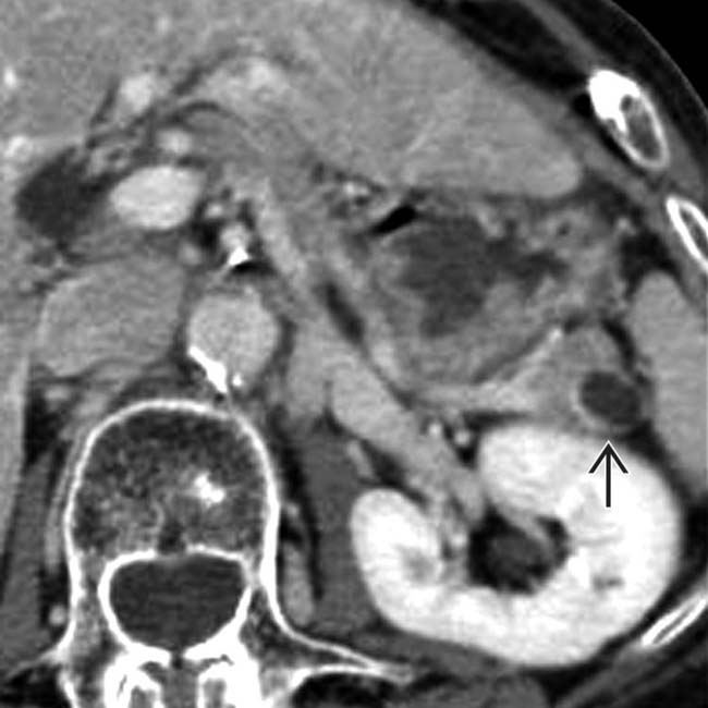
with internal septations image originating from the tail of the pancreas. There is a single mural nodule image. At surgery, a benign MCN was resected with no evidence of adenocarcinoma.

image
(Right) Axial CECT shows a large, complex cystic pancreatic body mass image 
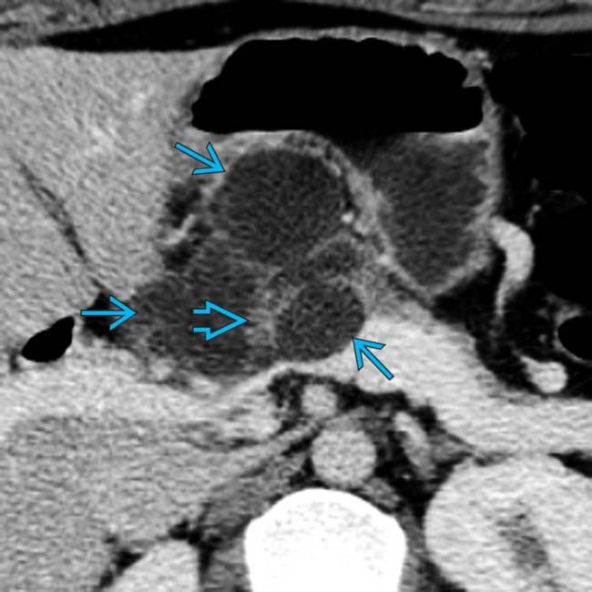
with internally enhancing soft tissue image. EUS-guided biopsy revealed mucinous adenocarcinoma. The presence of enhancing soft tissue within the cyst is strongly suspicious for malignancy.
image
(Left) Axial CECT in a 50-year-old man with vague abdominal pain shows a large complex cystic mass image 
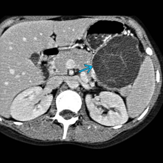
in the body/tail segment of the pancreas. Note the septa and the large cystic spaces within the mass.

image
(Right) Gross pathology from the same case shows that the resected mass image 
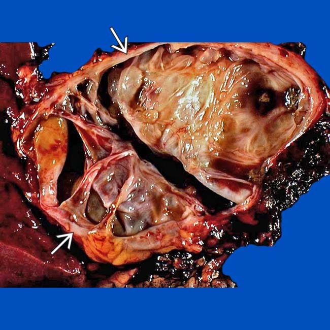
was full of mucinous fluid and had thickened septa. Histologic exam showed cellular atypia, and the lesion was considered a low-grade malignancy.
image
(Left) Axial CECT shows a cystic mucinous tumor in the pancreatic tail, with a few large cystic spaces separated by visible septa, as well as focal calcifications image.

image
(Right) Coronal MRCP shows a cystic mucinous tumor image 
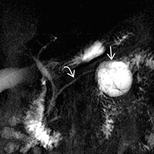
in the pancreatic tail with a few cystic spaces and septa. The pancreatic duct image is deviated but otherwise normal.
image
Axial CECT shows a cystic mucinous tumor image 
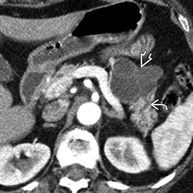
in the pancreatic body containing a few cystic spaces and septa. The tumor compresses the pancreatic duct, causing distal dilatation image.

image
Axial CECT shows a unilocular mucinous cystic tumor image in the pancreatic tail. Note the visible noncalcified wall.
image
Axial CECT in a 20-year-old woman shows large pancreatic cysts with enhancing walls thought to represent pseudocysts. Lack of resolution led to cyst aspiration, which yielded thick mucinous material with low amylase and high CA 19-9 levels. The ultimate diagnosis was mucinous cystic tumor.
image
Axial CECT shows a 1.5 cm cystic mass image 
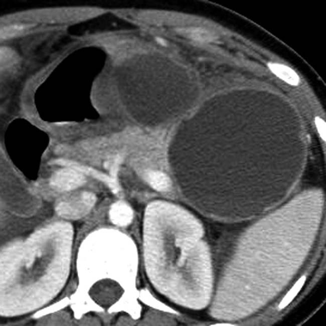
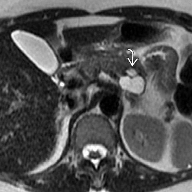
in the pancreatic tail. Mucinous cystic tumor was confirmed at surgical resection.
image
Axial CECT shows a large cystic mass image 
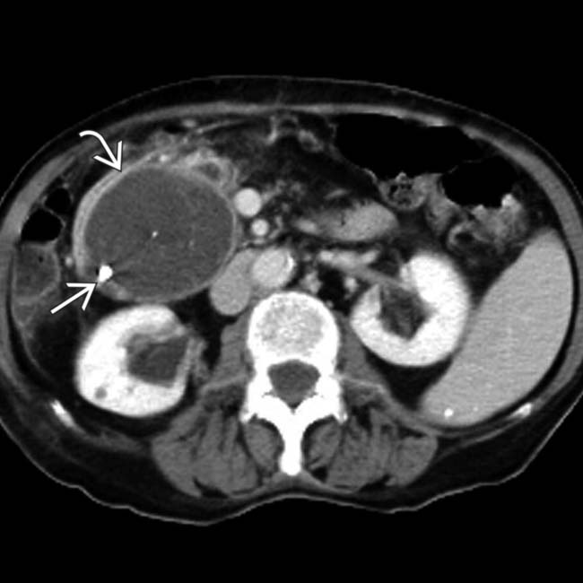
in the head of the pancreas that causes obstruction of the pancreatic duct and common bile duct (not shown). The mass has peripheral wall calcification image and several internal septations. The location of this MCN in the head, and the ductal obstruction are unusual features of this tumor.
image
Axial gadolinium-enhanced MR in a 61-year-old woman shows a well-defined cystic mass image 
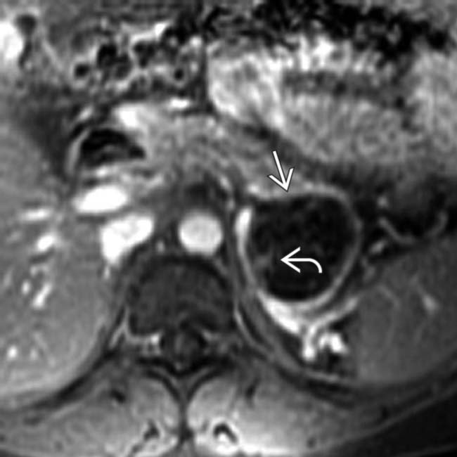
in the pancreatic tail. Note the enhancing wall and septa image.
image
Axial T2WI MR in the same patient reveals thin internal septations image within the cystic mass. Surgical resection was performed and a benign mucinous cystic neoplasm was found.
image
Axial CECT in a 55-year-old woman shows a complex cystic mass image 
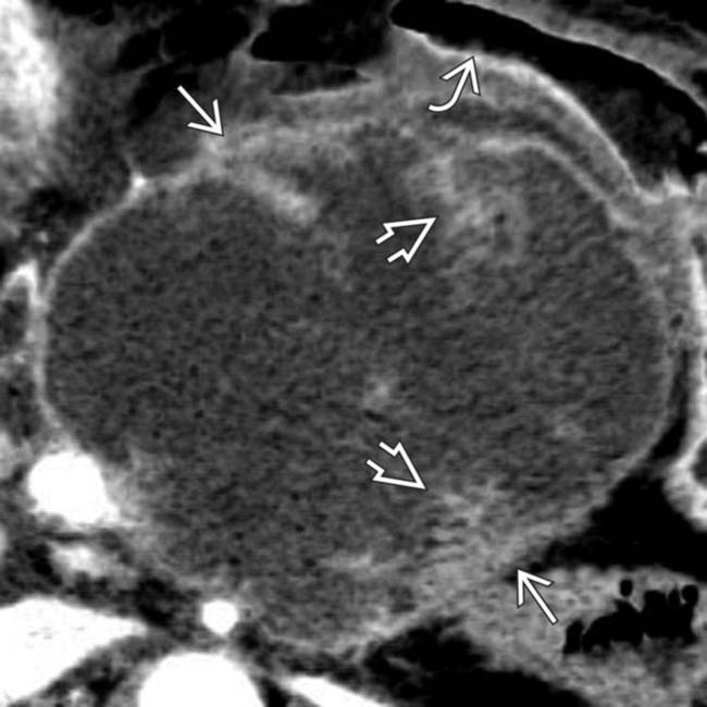
arising from the body of the pancreas. Note the soft tissue mural nodules image, as well as gastric invasion image.
image
A coronal MinIP in the same patient shows the prominent soft tissue mural nodule image strongly favoring a malignant lesion. EUS-guided biopsy revealed mucinous cystadenocarcinoma.
image
Coronal CECT in a 75-year-old woman shows a complex cystic mass image 
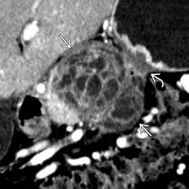
in the body of the pancreas containing both solid and cystic elements. Note that the mass invades into the adjacent serosa of the stomach image causing focal mural thickening.
image
A curved planar reformation of the splenic artery in the same patient shows vascular encasement image 
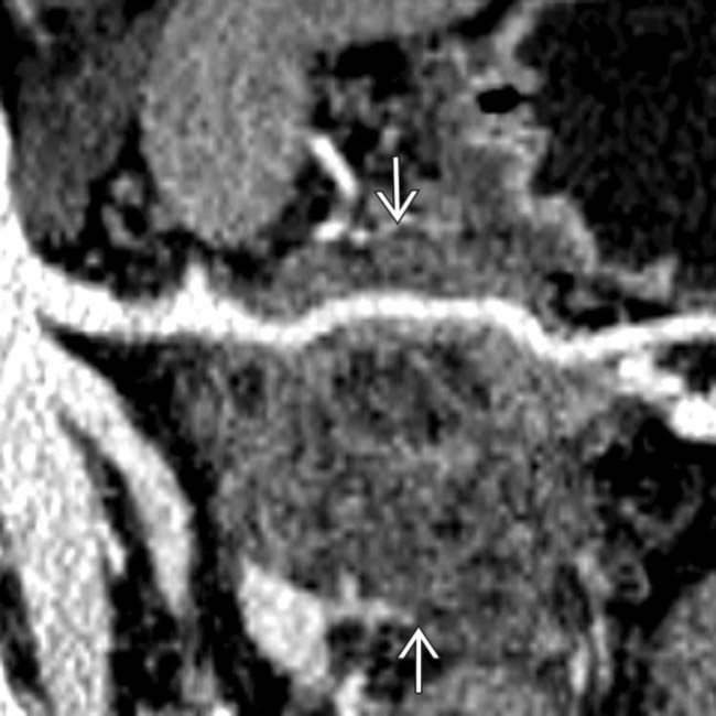
confirming that this is a mucinous cystadenocarcinoma. Gastric invasion and splenic artery encasement make this unresectable for cure.
image
Axial CECT shows a cystic mass image 
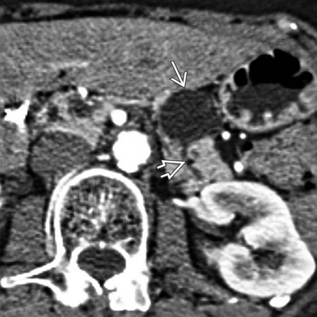
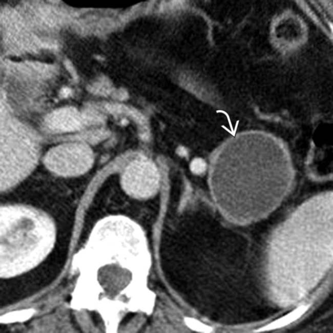
in the body of the pancreas. The pancreatic duct image is only mildly dilated, and there is no glandular atrophy “upstream” from the mass.
image
Axial CECT in the same patient shows septa image 
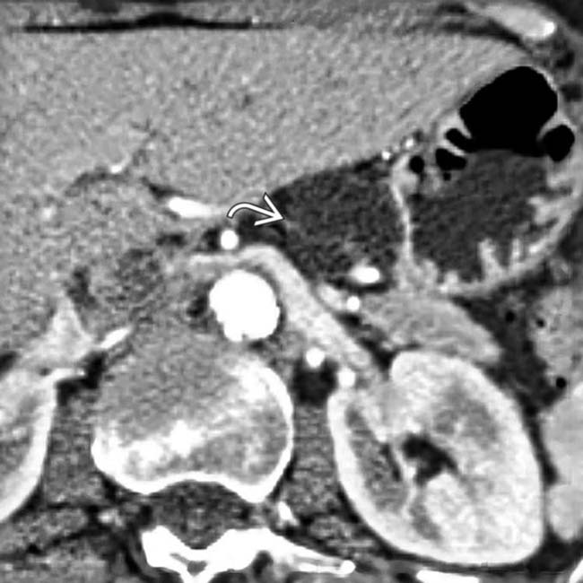
visible within the mass. Endoscopic US confirmed complex cystic features of the mass, and aspiration of cyst contents revealed thick, mucinous fluid with elevated CEA and CA 19-9 markers. Surgical resection confirmed mucinous cystic tumor with dysplastic cells in cyst lining.
image
Axial CECT shows a small, spherical, cystic mass image 
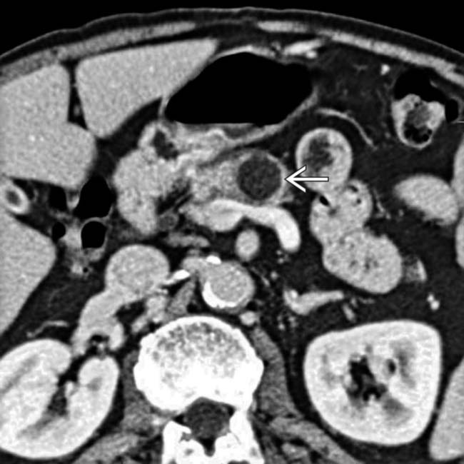
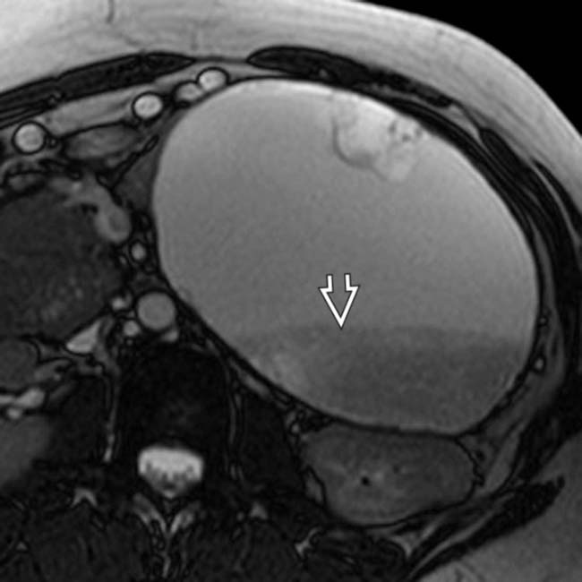
that appears relatively “simple,” although it has a visible wall.
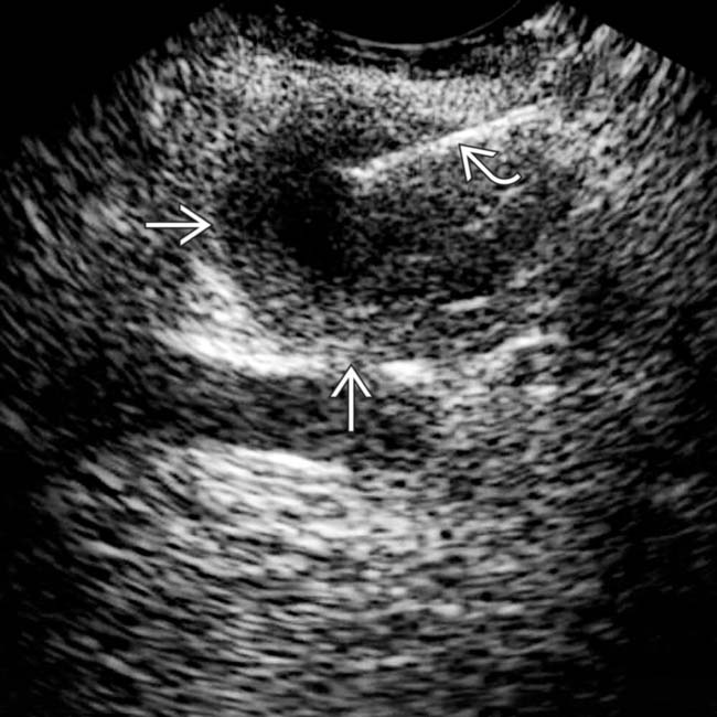
image
Transverse endoscopic ultrasound in the same patient demonstrates more apparent wall thickening image. Needle aspiration image yielded mucinous fluid. Resection showed a mucinous cystic neoplasm with malignant foci in the wall.
image
Axial CECT in a 40-year-old woman with vague abdominal discomfort shows a mucinous cystic tumor image 
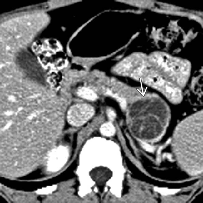
in the pancreatic tail with multiple enhancing septa and displacement of the pancreatic duct.
image
Gross pathologic specimen of a mucinous cystic neoplasm demonstrates a unilocular cyst with a smooth, partially discolored lining image 
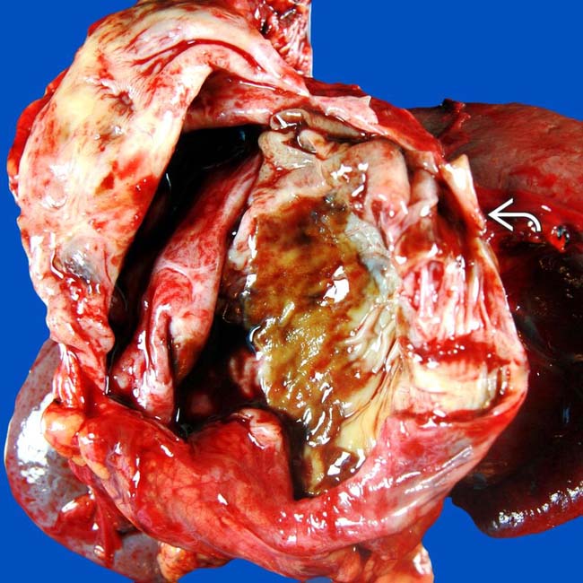
located in the tail of the pancreas. (Courtesy M. Mino-Kenudson, MD.)

SELECTED REFERENCES

1. Park, JW, et al. Mucinous cystic neoplasm of the pancreas: is surgical resection recommended for all surgically fit patients? Pancreatology. 2014; 14(2):131–136.

Pinho, DF, et al. Incidental pancreatic cysts: role of magnetic resonance imaging. Top Magn Reson Imaging. 2014; 23(2):117–128.

Farrell, JJ, et al. Pancreatic cystic neoplasms: management and unanswered questions. Gastroenterology. 2013; 144(6):1303–1315.

Gil, E, et al. Mucinous cystic neoplasms of the pancreas with ovarian stroma. ANZ J Surg. 2013; 83(12):985–990.

Dewhurst, CE, et al. Cystic tumors of the pancreas: imaging and management. Radiol Clin North Am. 2012; 50(3):467–486.

Hong, SK, et al. Targeted cyst wall puncture and aspiration during EUS-FNA increases the diagnostic yield of premalignant and malignant pancreatic cysts. Gastrointest Endosc. 2012; 75(4):775–782.

Kucera, JN, et al. Cystic lesions of the pancreas: radiologic-endosonographic correlation. Radiographics. 2012; 32(7):E283–E301.

Mottola, JC, et al. Diffusion-weighted MRI of focal cystic pancreatic lesions at 3.0-Tesla: preliminary results. Abdom Imaging. 2012; 37(1):110–117.

Valsangkar, NP, et al. 851 resected cystic tumors of the pancreas: a 33-year experience at the Massachusetts General Hospital. Surgery. 2012; 152(3 Suppl 1):S4–12.

Bang, JY, et al. Mucinous cystic neoplasm of the pancreas. Clin Gastroenterol Hepatol. 2011; 9(8):e80–e81.

Le Baleur, Y, et al. Mucinous cystic neoplasms of the pancreas: definition of preoperative imaging criteria for high-risk lesions. Pancreatology. 2011; 11(5):495–499.

Sakorafas, GH, et al. Primary pancreatic cystic neoplasms revisited: part II. Mucinous cystic neoplasms. Surg Oncol. 2011; 20(2):e93–101.

Basturk, O, et al. Pancreatic cysts: pathologic classification, differential diagnosis, and clinical implications. Arch Pathol Lab Med. 2009; 133(3):423–438.

Goh, BK, et al. A review of mucinous cystic neoplasms of the pancreas defined by ovarian-type stroma: clinicopathological features of 344 patients. World J Surg. 2006; 30(12):2236–2245.

Goh, BK, et al. Pancreatic serous oligocystic adenomas: clinicopathologic features and a comparison with serous microcystic adenomas and mucinous cystic neoplasms. World J Surg. 2006; 30(8):1553–1559.

Kim, SY, et al. Macrocystic neoplasms of the pancreas: CT differentiation of serous oligocystic adenoma from mucinous cystadenoma and intraductal papillary mucinous tumor. AJR Am J Roentgenol. 2006; 187(5):1192–1198.

Takeshita, K, et al. Unusual imaging appearances of pancreatic serous cystadenoma: correlation with surgery and pathologic analysis. Abdom Imaging. 2005; 30(5):610–615.

O’Toole, D, et al. Macrocystic pancreatic cystadenoma: The role of EUS and cyst fluid analysis in distinguishing mucinous and serous lesions. Gastrointest Endosc. 2004; 59(7):823–829.

Hara, T, et al. Mucinous cystic tumors of the pancreas. Surg Today. 2002; 32(11):965–969.

Maire, F, et al. Benign inflammatory pancreatic mucinous cystadenomas mimicking locally advanced cystadenocarcinomas. Presentation of 3 cases. Pancreatology. 2002; 2(1):74–78.

Oshikawa, O, et al. Dynamic sonography of pancreatic tumors: comparison with dynamic CT. AJR Am J Roentgenol. 2002; 178(5):1133–1137.

Balci, NC, et al. Radiologic features of cystic, endocrine and other pancreatic neoplasms. Eur J Radiol. 2001; 38(2):113–119.

Friedman, AC, et al. CT of primary cystic pancreatic neoplasms: nihilism may be unwarranted. AJR Am J Roentgenol. 2001; 177(2):469–470.

Grogan, JR, et al. Making sense of mucin-producing pancreatic tumors. AJR Am J Roentgenol. 2001; 176(4):921–929.

Megibow, AJ, et al. Cystic pancreatic masses: cross-sectional imaging observations and serial follow-up. Abdom Imaging. 2001; 26(6):640–647.