Motor System
Reflexes
BACKGROUND
A tendon reflex results from the stimulation of a stretch-sensitive afferent from a neuromuscular spindle which, via a single synapse, stimulates a motor nerve, leading to a muscle contraction. Tendon reflexes are increased in upper motor neurone lesions and decreased in lower motor neurone lesions and muscle abnormalities.
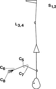
The root values for the reflexes can be recalled by counting from the ankle upwards (Fig. 19.1).
Reflexes can be graded:
WHAT TO DO
Use the whole length of the patella hammer; let the hammer swing. Ensure the patient is relaxed. Avoid telling the patient to relax, as this is guaranteed to produce tension.
Biceps
Place the patient’s hands on his abdomen. Place your index finger on the biceps tendon; swing the hammer on to your finger while watching the biceps muscle (Fig. 19.2).
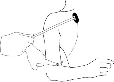
Figure 19.2 Testing the biceps reflex
Supinator
(N.B. Bad name for this reflex; the muscle involved is brachioradialis.)
Place the arm flexed on to the abdomen, place the finger on the radial tuberosity, hit the finger with the hammer and watch the brachioradialis (Fig. 19.3).

Figure 19.3 Testing the supinator reflex
Triceps
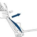
Draw the arm across the chest, holding the wrist with the elbow at 90 degrees. Strike the triceps tendon directly with the patella hammer; watch the muscle (Fig. 19.4).
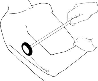
Figure 19.4 Testing the triceps reflex
Finger reflex
Hold the hand in the neutral position, place your hand opposite the fingers and strike the back of your fingers.
Knee reflex
Place the arm under the knee so that the knee is at 90 degrees. Strike the knee below the patella; watch the quadriceps (Fig. 19.5).
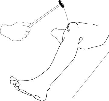
Figure 19.5 Testing the knee reflex
Ankle reflex
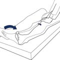
Hold the patient’s foot at 90 degrees with a medial malleolus facing the ceiling. The knee should be flexed and lying to the side. Strike the Achilles tendon directly. Watch the muscles of the calf (Fig. 19.6A).
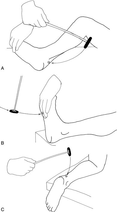
Figure 19.6 The ankle reflex—three ways to get it
Ankle reflex alternatives
1. With the patient’s legs straight, place your hand on the ball of his foot with the ankles at 90 degrees. Strike with your hand and watch the muscles of the calf (Fig. 19.6B).
2. Ask the patient to kneel on a chair so that his ankles are hanging loose over the edge. Strike the Achilles tendon directly (Fig. 19.6C).
Reinforcement
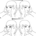
If any reflex is unobtainable directly, ask the patient to perform a reinforcement manœuvre. For the arms, ask the patient to clench his teeth as you swing the hammer. For the legs, ask the patient either to make a fist, or to link hands across his chest and pull one against the other, as you swing the hammer (Fig. 19.7).
 TIP
TIP
An absent reflex sounds dull. It’s worth listening as well as watching.
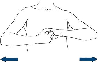
Figure 19.7 Reinforcement
Further manœuvres
Demonstration of clonus
WHAT YOU FIND AND WHAT IT MEANS
ABDOMINAL REFLEXES
What to do
Using an orange stick, lightly scratch the abdominal wall as indicated in Figure 19.8. Watch the abdominal wall; this should contract on the same side.
• Afferents: segmental sensory nerves
• Efferents: segmental motor nerves
• Roots: above the umbilicus, T8–T9; below the umbilicus, T10–T11.
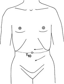
Figure 19.8 Abdominal reflexes
PLANTAR RESPONSE
What to do
Explain to the patient that you are going to stroke the bottom part of his foot. Gently draw an orange stick up a lateral border of the foot and across the foot pad. Watch the big toe and the remainder of the foot (Fig. 19.9).
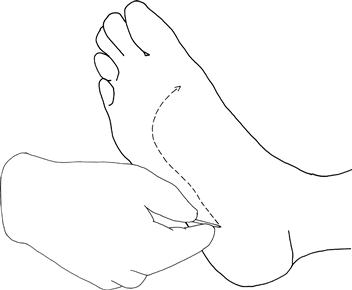
Figure 19.9 Testing the plantar response
What you find
What it means
Alternative stimuli (all trying to elicit the same responses)
• Stimulus on lateral aspect of foot: Chaddock’s reflex.
• Thumb and index finger run down the medial aspect of the tibia: Oppenheim’s reflex.
These alternative stimuli are only useful if present, not if absent.