Chapter 2 Morphology of primary and secondary skin lesions
1. Why do dermatologists use words that no one else understands?
The language of dermatology is unique. It encompasses terms that rarely, if ever, are used in other medical specialties. The use of these correct dermatologic terms is important to accurately describe skin lesions to dermatologists during telephone calls and during rounds and teaching. A good description of a skin lesion enables the listener to formulate a series of differential diagnoses, whereas a poor one does not.
2. But why are the descriptions so long?
Use of appropriate terminology and important clues, such as configuration and skin distribution, effectively paints an accurate picture for the listener. Use of vague terms—spot, bump, rash, and lesion—is not helpful. Such vocabulary is counterproductive to formulating an accurate differential diagnoses. “Grouped vesicles on an erythematous base” immediately suggests herpes simplex, and “brown, friable ‘stuck-on’ papules” accurately describes seborrheic keratoses. “Well-demarcated, erythematous plaques with micaceous, silvery scales located on extensor surfaces” is suggestive of psoriasis. “Violaceous, polygonal papules with Wickham’s striae located on flexural surfaces” is consistent with lichen planus. On the other hand, “red, scaly rash on the foot” describes an enormous, nebulous group of disorders.
3. How can I possibly learn the language of dermatology?
4. What is a primary skin lesion?
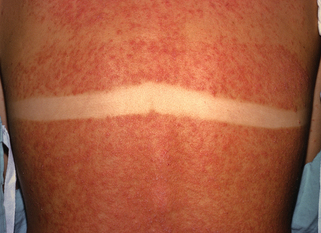
7. How does a primary lesion differ from a secondary lesion?
Secondary skin lesions are created by scratching, scrubbing, or infection. They may also develop normally with time. For example, the primary lesion in a sunburn is a macular erythema (although it could also be a blister), but with resolution, scale and increased pigmentation become prominent. Examples of secondary lesions include:
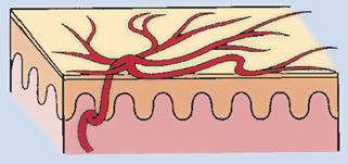
11. What are telangiectasias?
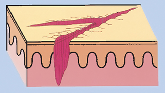
Telangiectasias (Fig. 2-1) are small, dilated, superficial blood vessels (capillaries, arterioles, or venules) that blanch (disappear) with pressure.
12. Are telangiectasias pathognomonic for a certain disease?
No. Telangiectasias may occur in many cutaneous disorders, including collagen vascular diseases such as dermatomyositis, systemic lupus erythematosus, and progressive systemic sclerosis. They are also commonly seen as a consequence of chronic ultraviolet radiation and topical steroid usage. Telangiectasias may also be seen in tumors such as a noduloulcerative basal cell carcinoma, which is classically described as a pearly colored papule with telangiectasias and central ulceration.
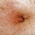
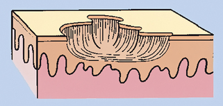
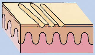
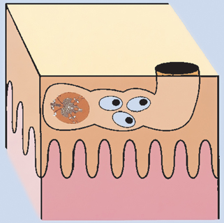
13. What is a burrow?
A burrow (Fig. 2-2) is an elevated channel in the superficial epidermis produced by a parasite, such as the mite Sarcoptes scabiei. Scabies burrows characteristically are located on the wrists and in fingerwebs; the diagnosis is confirmed by demonstrating the mite microscopically in skin scrapings. The human hookworm may also produce a serpiginous burrow; however, demonstrating this organism is much more difficult.
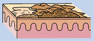
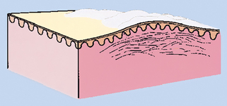
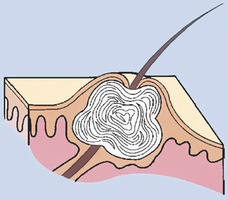
14. What is a comedo?
A comedo (Fig. 2-3) is a folliculocentric collection of sebum and keratin. Comedonal acne characteristically consists of both open (blackheads) and closed comedones (whiteheads). When the contents of a closed comedo are exposed to air, a chemical reaction occurs, imparting the black color of an open comedo.
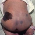
17. List some of the additional descriptive adjectives used in dermatology that refer to color or pigmentation.
• Depigmented: Absence of melanin, a lack of color. Depigmented macules and patches are commonly found in vitiligo.
• Hypopigmented: Lighter than normal skin color; normal number of melanocytes but decreased production of melanin by the melanocytes. The ash-leaf macule of tuberous sclerosis is an example of a hypopigmented macule.
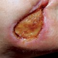
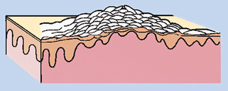
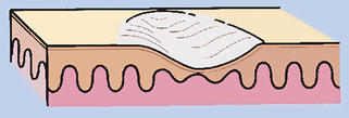
18. How do atrophy and lichenification differ?
Erosion
A partial focal loss of epidermis; heals without scarring
Atrophy (Fig. 2-5) is thinning of the epidermis, dermis, or subcutis (fat). Epidermal atrophy leads to a fine, cigarette-paper wrinkling of the skin surface, whereas dermal and fat atrophy cause a depression in the skin surface.
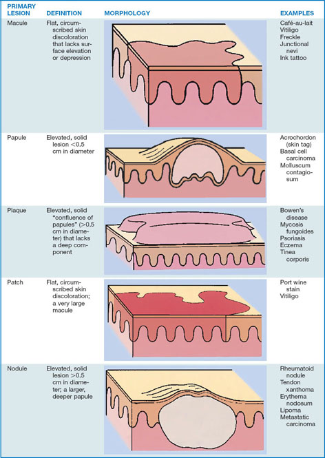
Table 2-2. Secondary Skin Lesions
| SECONDARY LESION | DEFINITION | MORPHOLOGY |
|---|---|---|
| Crust | A collection of cellular debris, dried serum, and blood; a scab Antecedent primary lesion is usually a vesicle, bulla, or pustule |
 |
Scar implies dermoepidermal damage
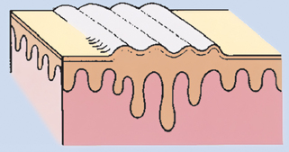
A lichenified lesion (Fig. 2-6) is a focal area of thickened skin produced by chronic scratching or rubbing. The skin lines are accentuated, resembling a washboard (Fig. 2-7).
19. Do skin diseases have characteristic arrangements or configurations?
Some, but not all, cutaneous diseases demonstrate characteristic arrangements or configurations of lesions. Commonly used adjectives include:
• Annular: Used to describe lesions that are ring shaped. Annular plaques are typical findings in granuloma annulare, tinea corporis (ringworm), and erythema marginatum.
• Gyrate: From the Latin gyratus, which means “to turn around in a circle,” gyrate skin lesions are rare presentations. Gyrate erythema that resembles wood grain or topographic maps is seen in erythema gyratum repens, which usually heralds the presence of an internal malignancy.
• Dermatomal: Used to describe lesions that follow neurocutaneous dermatomes. The classic example is herpes zoster (shingles), which demonstrates grouped vesicles on an erythematous base in a dermatomal distribution.
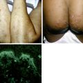
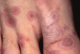
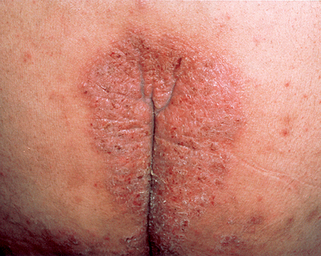
20. What is the Koebner phenomenon?
Traumatizing the epidermis of a patient with a certain preexisting skin disease will cause the same skin disease to form in the traumatized skin. Noticing this skin finding is helpful when creating a differential diagnosis. Only certain diseases are associated with a Koebner phenomenon; lichen planus, lichen nitidus, and psoriasis (Fig. 2-8) are examples.
21. Do skin diseases characteristically occur in certain locations?
Yes. This is the reason that a complete skin examination should be performed on all patients. Seborrheic dermatitis characteristically occurs on the scalp, nasolabial folds, retroauricular areas, eyelids, eyebrows, and presternal areas; it tends to spare the extremities. Psoriasis may resemble seborrheic dermatitis, but it characteristically demonstrates a different distribution, usually involving the extremities (elbows, knees), intergluteal fold, scalp, and nails.
Key Points: Primary and Secondary Lesions
1. The clinical diagnosis of skin disease is accomplished by a complex appreciation of the primary and secondary skin lesions, distribution, color, arrangement, and body site.
1. Cox NH, Coulson IH,. Diagnosis of skin disease. Burns S, Breathnach SM, Cox N, Griffiths C,. Rook’s textbook of dermatology. ed 7. Malden, MA: Blackwell; 2004:5.1-5.10.
2. Rapini R. Clinical and pathologic differential diagnosis. Bolognia JL, Jorizzo JL, Rapini R. Dermatology. London: Mosby; 2003:3-12.