20 Morphologic changes after myeloid hematopoietic growth factors
Cytokine therapies such as erythropoietin, thrombopoietin, and myeloid growth factors such as granulocyte colony-stimulating factor (G-CSF) and granulocyte/macrophage-colony stimulating factor are becoming common. There are characteristic changes in the peripheral blood smear caused by these treatments. Although erythropoietin and thrombopoietin rarely create diagnostic challenges, the morphologic changes in the myeloid cell line may mimic severe infection, acute myeloid leukemia, or myelodysplastic or myeloproliferative neoplasm. Specific changes include transient leukocytosis with immature granulocytic cells, vacuolated and giant neutrophils, toxic granulation, Döhle bodies, hypogranulation, nucleated red blood cells, and as many as 5% blasts in the peripheral blood.*
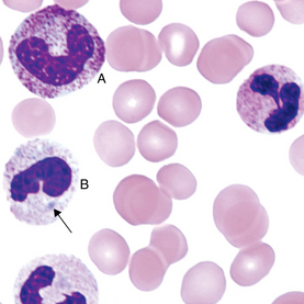
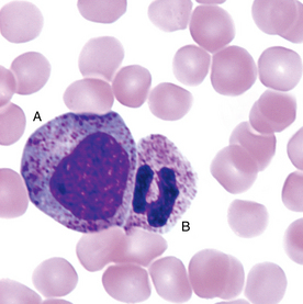
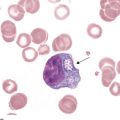
FIGURE 20-3 Immature asynchronous granulocyte (A), and a mature neutrophil with toxic granulation (B).
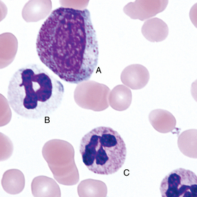
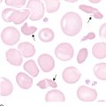
FIGURE 20-4 Giant asynchronous immature granulocyte (A), a hypogranular neutrophil with a Döhle body (B), and a granulocyte with normal granulation (C).