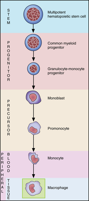
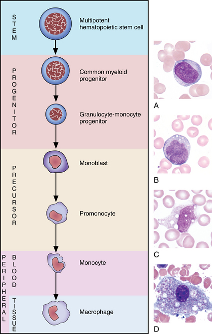
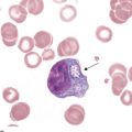
6 Monocyte maturation
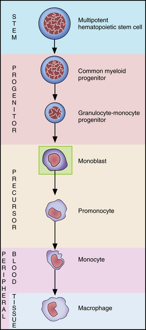
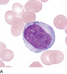
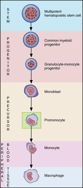
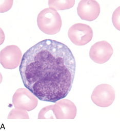
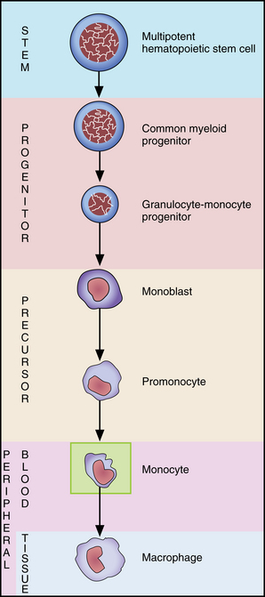
Monocyte
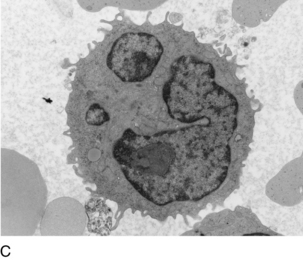
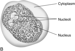
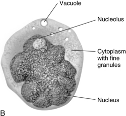
FIGURE 6–6C Electron micrograph of monocyte (×16,500). Specimens for electron microscopy are prepared by embedding tissue in a suitable medium, such as resin. Ultra-thin cross-sections are then prepared. Because this image shows a cross-section, the lobes of the nucleus appear to be separate, but they are not.