Chapter 53 Mohs surgery
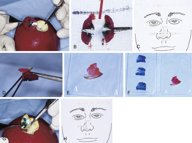
The specimen is then sectioned into manageable pieces, if needed, and these slices are color coded with ink. Using an anatomic cartoon representation of the site, a map is drawn to precisely label the area from which the tumor is taken. The tissue is then submitted to the Mohs histotechnician, who prepares frozen tissue sections under the supervision of the Mohs surgeon. These sections are obtained by cutting the undersurface in a horizontal plane so that the depth and most peripheral skin edge will be available for microscopic examination. Theoretically, 100% of the margin is examined, in contrast to routine histologic processes. If tumor is identified, the map is used as a guide to obtain additional tissue layers only from the areas of positivity until a tumor-free plane is reached. Between stages of Mohs surgery, the wound is covered with a temporary dressing, and the patients wait in a comfortable area. Various steps of the procedure are illustrated in Fig. 53-1 using an apple model.
Davis DA, Pellowski DM, Hanke WC: Preparation of frozen sections, Dermatol Surg 30:1479–1485, 2004.
Key Points: Mohs Surgery
Many dermatologists know the basic techniques and employ them in their practices. However, when patients require more extensive surgery, they usually are referred to a member of the College.