CHAPTER 19 MISCELLANEOUS SURGICAL PATHOLOGY
PATHOLOGY OF PEDIATRIC HAIR SAMPLES
Introduction (Smith et al 2005)


Types of sample and handling
(Garn 1951, Leblond 1951, Eilers 1982, Shelley 1983, Caserio 1987, Crounse 1987, De Berker 1997, Smith et al 2005)


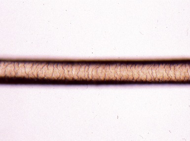
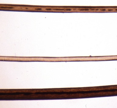
Histopathological features


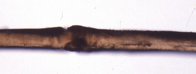
Types of morphological abnormality


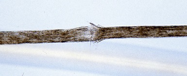
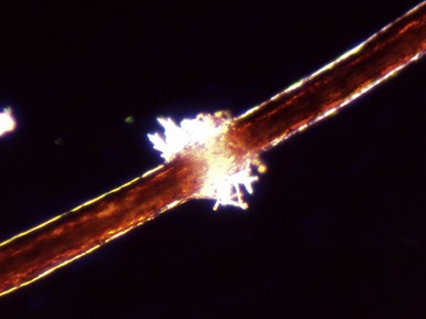
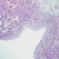
Figs 19.4–19.5 Photomicrographs of hair shafts showing trichorrhexis nodosa, with areas of shaft breakage.
Associations with specific diagnoses


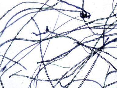
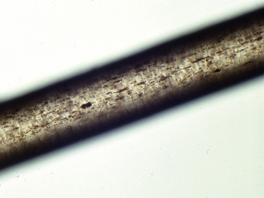
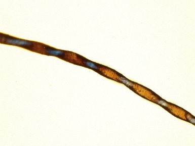
Fig 19.9 Photomicrograph of hair shafts showing abnormal pigment clumping in Chediak–Higashi syndrome.
BLOOD FILM EXAMINATION FOR VACUOLATED LYMPHOCYTES
Introduction (Anderson et al 2005)


Sample preparation and examination


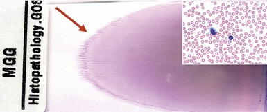
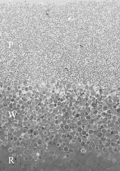
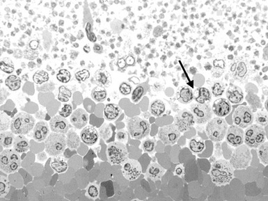
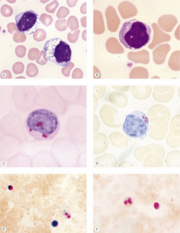
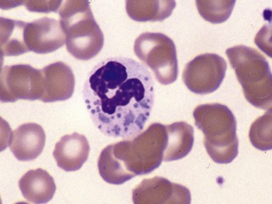
Fig 19.15 Photomicrograph of a blood film showing a range of lymphocyte vacuolation. (A) Routine May-Grunwald–Giemsa stained blood film showing two lymphocytes with many large bold vacuoles, such as are seen in GM1 gangliosidosis, and juvenile Batten’s disease. (B) Routine May-Grunwald–Giemsa stained blood film showing a lymphocyte with discrete small vacuoles seen in Pompe’s disease and adult acid maltase deficiency. (C) Periodic acid–Schiff (PAS) stained blood film showing a lymphocyte with PAS positive inclusions in Pompe’s disease and adult acid maltase deficiency. (D) Toluidine blue stained blood film showing metachromatic cytoplasmic inclusions in a lymphocyte. (E) Enzyme histochemical demonstration of β-galactosidase in a blood film showing normal enzyme activity in a lymphocyte and eosinophil but negative activity in a neutrophil. (F) Enzyme histochemical demonstration of β-galactosidase in a blood film showing absent enzyme activity in a lymphocyte and a granulocyte seen in GM1 gangliosidosis and galactosialidosis.
Histopathological features


All figures in this chapter are from Anderson et al 2005 and Smith et al 2005.
Anderson G, Smith VV, Malone M, Sebire NJ. Blood film examination for vacuolated lymphocytes in the diagnosis of metabolic disorders; retrospective experience of more than 2500 cases from a single center. J Clin Pathol. 2005;58:1305-1310.
Aula P, Autio S, Raivio KO, et al. ‘Salla disease’: a new lysosomal storage disorder. Arch Neurol. 1979;36:88-94.
Caserio RJ. Diagnostic techniques for hair disorders. Part I: Microscopic examination of the hair shaft. Cutis. 1987;40:265-270.
Caserio RJ. Diagnostic techniques for hair disorders. Part II: Microscopic examination of hair bulbs, tips, and casts. Cutis. 1987;40:321-325.
Celik HH, Tore H, Tunali S, Tatar I, Aldur MM. Light and scanning electron microscopic examination of hair in Griscelli syndrome. Saudi Med J. 2007;28:1275-1277.
Crounse RG. The diagnostic value of microscopic examination of human hair. Arch Pathol Lab Med. 1987;111:700-702.
De Berker D. ‘Tiger tail’ pattern on polarized hair microscopic examination is found in healthy infants. Arch Dermatol. 1997;133:1313-1314.
Descargues P, Deraison C, Bonnart C, et al. Spink5-deficient mice mimic Netherton syndrome through degradation of desmoglein 1 by epidermal protease hyperactivity. Nat Genet. 2005;37:56-65.
Eilers RJ. Pathology – epitomes of progress: hair analysis: a potential diagnostic tool. West J Med. 1982;136:423-424.
Garn SM. The examination of hair under the polarizing microscope. Ann. NY Acad Sci. 1951;53:649-652.
Giugliani R, Dutra JC, Pereira ML, et al. GM1 gangliosidosis: clinical and laboratory findings in eight families. Hum Genet. 1985;70:347-354.
Hamm H, Traupe H. Loose anagen hair of childhood: the phenomenon of easily pluckable hair. J Am Acad Dermatol. 1989;20:242-248.
Hansen LK, Wulff K, Brandrup F. Trichothiodystrophy. Hair examination as a diagnostic tool. Ugeskr Laeger. 1993;155:1949-1952.
Hicks J, Metry DW, Barrish J, et al. Uncombable hair (cheveux incoiffables, pili trianguli et canaliculi) syndrome: brief review and role of scanning electron microscopy in diagnosis. Ultrastruct Pathol. 2001;25:99-103.
Ito M, Ho K, Hashimoto K. Pathogenesis in trichorrhexis invaginata (bamboo hair). J Invest Dermatol. 1984;83:1-6.
Kimura S, Goebel HH. Light and electron microscopic study of juvenile neuronal ceroid-lipofuscinosis lymphocytes. Pediatr Neurol. 1988;4:148-152.
Lake BD, Cavanagh NP. Early-juvenile Batten’s disease – a recognisable subgroup distinct from other forms of Batten’s disease. Analysis of 5 patients. J Neurol Sci. 1978;36:265-271.
Leblond CP. Histological structure of hair, with a brief comparison to other epidermal appendages and epidermis itself. Ann. NY Acad Sci. 1951;53:464-475.
Liang C, Kraemer KH, Morris A, et al. Characterization of tiger tail banding and hair shaft abnormalities in trichothiodystrophy. J Am Acad Dermatol. 2005;52:224-232.
Martin JJ, Flament-Durand J, Farriaux JP, Buyssens N, Ketelbant-Balasse P, Jansen C. Menkes kinky-hair disease. A report on its pathology. Acta Neuropathol. 1978;42:25-32.
Menasche G, Ho CH, Sanal O, et al. Griscelli syndrome restricted to hypopigmentation results from a melanophilin defect (GS3) or a MYO5A F-exon deletion (GS1). J Clin Invest. 2003;112:450-456.
Pastural E, Ersoy F, Yalman N, et al. Two genes are responsible for Griscelli syndrome at the same 15q21 locus. Genomics. 2000;63:299-306.
Rossier A, Caldera R, Sarrut S. A pathognomonic sign of Niemann–Pick disease: vacuolated lymphocytes. Arch Fr Pediatr. 1959;16:1358-1361.
Shelley WB. Hair examination using double-stick tape. J Am Acad Dermatol. 1983;8:430-431.
Smith VV, Anderson G, Malone M, Sebire NJ. Light microscopic examination of scalp hair samples as an aid in the diagnosis of pediatric disorders: retrospective review of more than 300 cases from a single center. J Clin Pathol. 2005;58:1294-1298.
Terashima Y, Tsuda K, Isomura S, et al. I-cell disease. Report of three cases. Am J Dis Child. 1975;129:1083-1090.
Williams RS, Marshall PC, Lott IT, Caviness VSJr. The cellular pathology of Menkes steely hair syndrome. Neurology. 1978;28:575-583.