21 Microorganisms
Plasmodium species
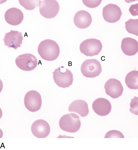
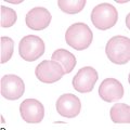
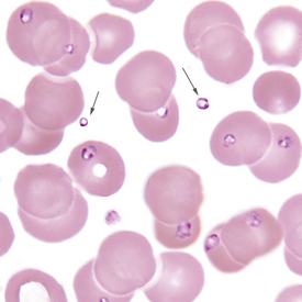
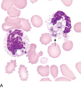
FIGURE 21–1A Plasmodium falciparum rings (A), including applique form (B), and platelet on RBC (C) (PB ×1000).
(Courtesy Indiana Pathology Images).
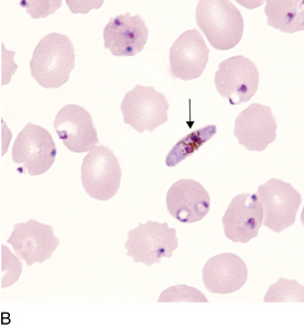
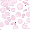
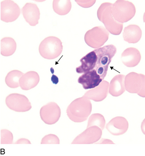
FIGURE 21–1B Plasmodium falciparum rings and crescent (banana-shaped) gametocyte (arrow) (PB ×1000).
(Courtesy Indiana Pathology Images).
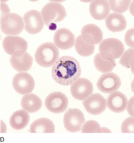
Babesia species
Babesia species may be confused morphologically with Plasmodium falciparum, but lack of pigment and absence of life cycle stages help differentiate Babesia spp. from P. falciparum. Another differentiating factor is the presence of extracellular organisms (Figure 21-2, arrows) that may be seen in Babesia spp. but not in P. falciparum.
Loa loa
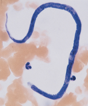
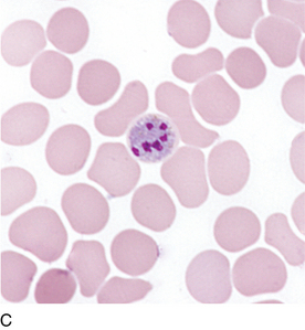
Loa loa is a microfilaria (Figure 21-3). Other microfilariae rarely may be seen in the peripheral blood.
Trypanosomes
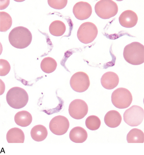

FIGURE 21–4B Trypanosoma cruzi (Giemsa stain, PB ×1000).
(From Marler LM, Siders JA, Simpson A et al: Parasitology image atlas CD-ROM, Indianapolis, IN, 2003, Indiana Pathology Images.)
Trypanosomes are examples of hemoflagellates that may occasionally be encountered in the peripheral blood (Figure 21-4, A and B). Differentiating features may be found in a parasitology text.
Fungi

Bacteria
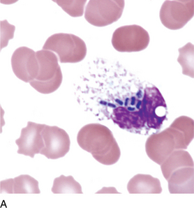
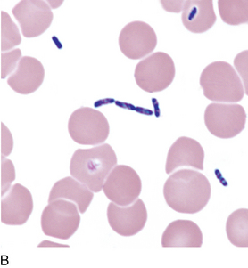
FIGURE 21–6B Extracellular bacteria from same specimen as Figure 21-6A. Extracellular bacteria alone may indicate contamination. Presence of intracellular bacteria may rule out contamination (PB ×1000).