[level-membership-for-dermatology-category]
Microanatomy of the skin
Introduction
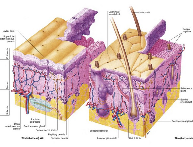
Skin is composed of three layers: the epidermis, the dermis and the subcutis (Fig. 1).
Epidermis
The epidermis is a stratified squamous epithelium that is about 0.1 mm thick, although the thickness is greater (0.8–1.4 mm) on the palms and soles. Its prime function is to act as a protective barrier. The main cells of the epidermis are keratinocytes, which produce the protein keratin. Keratinocytes are squamous cells functionally similar to all other structural epithelial cells as found in the airways and gastrointestinal tract. The four layers of the epidermis (Fig. 2) represent the stages of maturation of keratin by keratinocytes (p. 6).
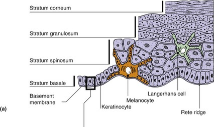
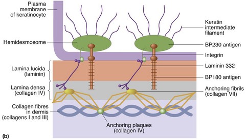
Fig. 2 Cross-sectional anatomy of the epidermis.
(a) Layers of the epidermis and other structures. (b) Detailed view of the basement membrane zone at the dermoepidermal junction. Components are arranged in three layers. The lamina lucida is traversed by filaments connecting the basal cells with the lamina densa, from which anchoring fibrils extend into the papillary dermis. These laminae are the sites of cleavage in certain bullous disorders (p. 84).
Basal cell layer (stratum basale)
The basal cell layer of the epidermis is composed mostly of keratinocytes, which are either dividing or non-dividing. The cells contain keratin tonofibrils (p. 6) and are secured to the basement membrane (see Fig. 2) by hemidesmosomes. Melanocytes make up 5–10% of the basal cell population. These cells synthesize melanin (p. 8) and transfer it via dendritic processes to neighbouring keratinocytes.
Prickle cell layer (stratum spinosum)
Daughter basal cells migrate upwards to form this layer of polyhedral cells, which are interconnected by desmosomes (the ‘prickles’ seen at light microscope level). Keratin tonofibrils form a supportive mesh in the cytoplasm of these cells. Langerhans cells are mostly found in this layer; these dendritic, immunologically active cells are described fully on page 10.
Granular cell layer (stratum granulosum)
Cells become flattened and lose their nuclei in the granular cell layer. Keratohyalin granules are seen in the cytoplasm together with membrane-coating granules (which expel their lipid contents into the intercellular spaces).
Dermis
Collagen fibres make up 70% of the dermis and impart a toughness and strength to the structure. Elastin fibres are loosely arranged in all directions in the dermis and provide elasticity to the skin. They are numerous near hair follicles and sweat glands, and less so in the papillary dermis. The ground substance of the dermis is a semisolid matrix of glycosaminoglycans (GAGs), which allows dermal structures some movement (p. 9).
Subcutaneous layer
The subcutis consists of loose connective tissue and fat (up to 3 cm thick on the abdomen).


[/level-membership-for-dermatology-category][not-level-membership-for-dermatology-category]
Microanatomy of the skin
Introduction
Skin is composed of three layers: the epidermis, the dermis and the subcutis (Fig. 1).
Epidermis
The epidermis is a stratified squamous epithelium that is about 0.1 mm thick, although the thickness is greater (0.8–1.4 mm) on the palms and soles. Its prime function is to act as a protective barrier. The main cells of the epidermis are keratinocytes, which produce the protein keratin. Keratinocytes are squamous cells functionally similar to all other structural epithelial cells as found in the airways and gastrointestinal tract. The four layers of the epidermis (Fig. 2) represent the stages of maturation of keratin by keratinocytes (p. 6).
Fig. 2 Cross-sectional anatomy of the epidermis.
(a) Layers of the epidermis and other structures. (b) Detailed view of the basement membrane zone at the dermoepidermal junction. Components are arranged in three layers. The lamina lucida is traversed by filaments connecting the basal cells with the lamina densa, from which anchoring fibrils extend into the papillary dermis. These laminae are the sites of cleavage in certain bullous disorders (p. 84).
Basal cell layer (stratum basale)
The basal cell layer of the epidermis is composed mostly of keratinocytes, which are either dividing or non-dividing. The cells contain keratin tonofibrils (p. 6) and are secured to the basement membrane (see Fig. 2) by hemidesmosomes. Melanocytes make up 5–10% of the basal cell population. These cells synthesize melanin (p. 8) and transfer it via dendritic processes to neighbouring keratinocytes.
[/not-level-membership-for-dermatology-category]
