[level-membership-for-opthalmology-category]23.1
Metastatic Choroidal Tumor
Clinical Features:
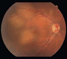
These lesions are typically creamy, yellow, and elevated. They tend to be bilateral and can also be multifocal (Fig. 23.1.1). Associated serous retinal detachments can cause decreased visual acuity when involving the macula. A history of primary malignancy is helpful in confirming the diagnosis.
OCT Features:
There is a localized elevation of the choroid in the location of the tumor, which can have overlying subretinal fluid (Fig. 23.1.2). Cystoid intraretinal fluid overlying the tumor can also be present (Figs 23.1.3 and 23.1.4).
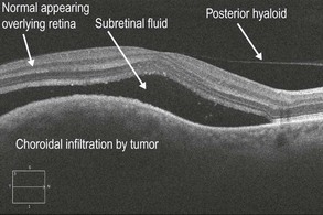
Figure 23.1.2 OCT (corresponding to Figure 23.1.1) shows a hill-like elevation of the choroid due to infiltration by the tumor with mild obscuration of the choriocapillaris. There is overlying subretinal fluid.
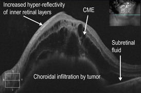
Figure 23.1.3 OCT of a large metastatic choroidal tumor involving the optic nerve and macula shows a hyporeflective elevation of the choroid that is infiltrated by tumor. Overlying this is subretinal fluid and extensive CME. The inner retinal layers are somewhat more hyper-reflective than normal.
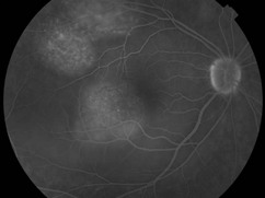
Figure 23.1.4 Fluorescein angiography (corresponding to Figure 23.1.1) shows multiple areas of pinpoint hyperfluorescence overlying each area of choroidal infiltration.
[/level-membership-for-opthalmology-category][not-level-membership-for-opthalmology-category]23.1
Metastatic Choroidal Tumor
Clinical Features:
These lesions are typically creamy, yellow, and elevated. They tend to be bilateral and can also be multifocal (Fig. 23.1.1). Associated serous retinal detachments can cause decreased visual acuity when involving the macula. A history of primary malignancy is helpful in confirming the diagnosis.
