Meninges, ventricular system and blood supply
Meninges
Basic anatomy
Dura mater
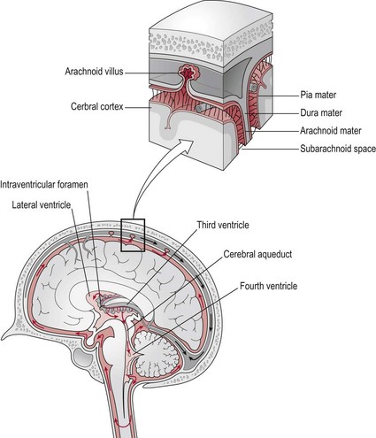
This outer layer is conventionally described as two layers, however they are closely united except along certain lines where they part to surround and support large venous channels termed ‘the dural venous sinuses’ (Fig. 8.1). The two layers comprise:
The ventricular system
Basic anatomy
Ventricles
The ventricular system incorporates a series of interconnected spaces in the core of the brain. The largest spaces are the lateral ventricles, which are bilateral and are the site of the choroid plexus, which produce cerebrospinal fluid. The 3rd ventricle is linked to the lateral ventricles by the interventricular foramina and to the 4th ventricle by the cerebral aqueduct. The 3rd and 4th ventricles lie in midline (Fig. 8.1).
Cerebrospinal fluid
Cerebrospinal fluid (CSF) is a clear fluid produced by the choroid plexuses within the lateral ventricles. The CSF circulates through the ventricular system and ultimately enters the subarachnoid space which surrounds the cerebral cortex, cerebellum, brain stem and spinal cord down to the level of the second sacral vertebra. The CSF is recycled into the dural venous sinuses via the arachnoid villi (Fig. 8.1) and returned to the venous circulation.
Circulatory systems of the brain
Arterial supply
Basic anatomy
Anterior circulation
The carotid arteries and their branches are referred to as the ‘anterior circulation’. The left common carotid artery arises from the aortic arch (Fig. 8.2). The right common carotid arises from the innominate artery. From each common carotid artery there are two branches:
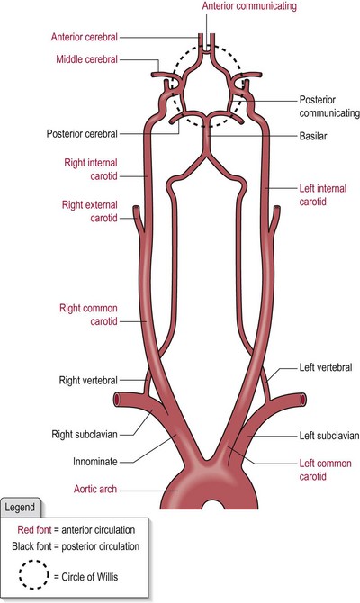
The left and right internal carotid arteries pierce the dura mater at the base of the brain and each bifurcates to form the anterior cerebral artery and the middle cerebral artery (Fig. 8.2).
Posterior circulation
The vertebrobasilar system is referred to as the ‘posterior circulation’ (Fig. 8.2).
The two vertebral arteries join at the junction of the pons and medulla oblongata (S2.10) to form the basilar artery, which then divides at the junction of the pons and mid-brain to form the two posterior cerebral arteries (Fig. 8.2). It also gives off branches to the cerebellum.
The circle of Willis
The circle of Willis (Fig. 8.2) is a polygon shaped network of blood vessels beneath the cerebral hemispheres. The two anterior cerebral arteries are connected by the anterior communicating artery and the posterior cerebral arteries are connected to the ipsilateral internal carotid arteries by the posterior communicating arteries. The six cerebral arteries, bilaterally the anterior, posterior and middle are termed end arteries.
The blood–brain barrier
In other areas of the body, free movement of ions and molecules back and forth across the tissue boundary is permitted. However, in the brain, the endothelial cells of the capillary wall overlap and form tight junctions termed ‘end feet’ with astrocyte cells (S2.7) surrounding them. The astrocytes also provide them with biochemical support. Thus, there is little free movement from blood into the interstitial environment of the neural tissue. There are however, specific transporters across the barrier for critical ions and molecules such as glucose and specific amino acids.
Venous drainage
Basic anatomy
The venous drainage of the brain can be separated into two sub-divisions:
Superficial
The superficial system is composed of dural venous sinuses (Fig. 8.1) located on the surface of the cerebrum. The dural venous sinuses are situated between the two layers of the dura mater and lined with endothelium. They differ from other vessels in that their walls lack the characteristic layering, muscle and valves seen in other veins. Their function is to receive blood from the deep and superficial cerebral veins and cerebrospinal fluid from the subarachnoid space via the arachnoid villi and empty these components into the internal jugular vein to be returned to other organs for recycling.