Melomental folds

Published on 16/03/2015 by admin

Filed under Dermatology

Last modified 22/04/2025

Print this page

rate 1 star rate 2 star rate 3 star rate 4 star rate 5 star
Your rating: none, Average: 3 (1 votes)

This article have been viewed 6395 times

21 Melomental folds

Introduction

The melomental folds (MMFs) are defined as the curvilinear folds running inferiorly from the oral commissure towards the mandible. They form the anatomical division between the lower lip and cheek, as well as the chin and cheek cosmetic units. A number of factors contribute to the formation of MMFs – chiefly loss of soft tissue volume and elasticity in the mid-face, bony atrophy of the mandible, and gravitational pull. The end result is downward descent of mid-facial tissue with festooning of the upper lip over the oral commissure and of the lower medial cheek over the lateral portion of the lower lip / chin subunit, thus forming the marionette-like appearance of the melomental crease.

The MMFs give a downturned appearance to the mouth, which may falsely convey the impression of anger or ‘sourness’. Impressions made within the first few fractions of a second of viewing a face have been found to have a lasting effect on interpersonal relationships. Similarly, subjects’ self-perception can be modified by their own facial expression. As such, many patients feel, correctly, that their MMFs cause them tangible life difficulty. Understandably, treatment of this troublesome area has long been desired. Historically, alternatives such as face or lip lifts yielded disappointingly high MMF recurrence rates, as have numerous resurfacing techniques including dermabrasion, chemical peels, and laser resurfacing. Botulinum toxin injection of the depressor anguli oris does provide some attenuation of the MMF, but oftentimes is not sufficient to fully efface these rhytides. It will be discussed in more detail later in the chapter as an excellent adjunct for MMF correction.

The advent of soft tissue fillers has revolutionized the treatment of this troublesome area. Fillers offer a minimally invasive, reliable treatment modality with an excellent safety profile and minimal post-procedure downtime. Additionally they allow for the possibility of easy titration and stepwise correction, if needed, to achieve the desired cosmetic effect. The ever-expanding library of soft tissue fillers allows for improved tailoring of the product to your patient’s needs.

Anesthesia

Minimizing the pain associated with any procedure is key to insuring patient satisfaction. As injections in the perioral area are frequently more painful than mid-facial areas owing to increased sensory innervation, anesthetic measures are more often necessary. While lidocaine-containing hyaluronic acid (HA) fillers (Juvéderm XC, Restylane-L, Prevelle Silk) or the mixture of calcium hydroxylapatite (Radiesse) with lidocaine significantly decrease the pain of injection, some patients may still benefit from additional pain control measures. In many, a measure as simple as pre-injection cryoanesthesia with ice may be sufficient. If more profound analgesia is judged necessary, the workhorse anesthetic methods for MMF augmentation are topical anesthetic agents and regional blockade of the mental nerve.

Topical anesthetic agents such as 5% lidocaine creams (LMX-5) or lidocaine / prilocaine mixtures (EMLA or Betacaine), while providing adequate anesthesia, require occlusion and application periods in excess of 30 minutes, which may limit their use in a busy clinical practice. A eutectic lidocaine / tetracaine peel (S-Caine peel) has been developed that can achieve anesthesia in as little as 20 minutes and has been found to be superior to EMLA applied under occlusion for 30 minutes. In the authors’ experience, Betacaine Plus ointment can provide adequate anesthesia within 10 minutes without the need for occlusion.

Bilateral mental nerve blocks may be used to achieve anesthesia of the lower facial unit. The mental foramen may be reliably located in the mid-pupillary line approximately 1 cm inferior to the gumline. An intraoral approach just below the second premolar provides a reliable way of executing the block without the pain of a percutaneous approach.

Basic principles for melomental fold correction

The correction of the MMF with soft tissue fillers requires proper selection of filler product in concert with appropriate injection technique and depth of placement. Only with the proper intersection of these three parameters may optimal results be achieved. Generally speaking, the MMFs are more difficult to correct than the melolabial folds, given the role of bony atrophy and significant lower facial volume loss in the pathogenesis of the former. As the deficit in question may be more profound than can be corrected by simply ‘filling a wrinkle’, linear threading or serial puncture techniques may prove inadequate to attenuate more advanced folds. Rebuilding structural support for the oral commissure and deeply set MMFs is best accomplished with techniques that place a larger amount of filler across a broader area, such as cross-hatching or fanning.

MMFs are best addressed with injection into the deep dermis. This in turn demands the use of sturdier filler such as a highly cross-linked HA product (Juvéderm Ultra Plus, Prevelle) or calcium hydroxylapatite (Radiesse). The fold should be injected from inferior to superior, attenuating the rhytide and building a buttress of support for the oral commissure. Additionally, injections should be oriented from medial to lateral as this will minimize the risk of traumatizing the facial vein, which runs just lateral to the MMF. For the most deeply set MMFs, vertical layering of fillers may be necessary to achieve the desired result. For example, if a deeply injected, cross-hatched, heavier weight filler is insufficient to attenuate the MMF, the addition of a more superficially placed, lighter weight filler can efface etched components to the fold.

Injection strategies for melomental fold correction

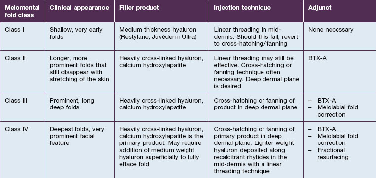
While ultimately a treatment plan must be individualized for a given patient, there are some broad generalizations that may be made. The Validated Melomental Fold Scoring System allows for division of MMFs into four classes based on severity, as summarized in Table 21.1. An excellent and thorough discussion of this classification may be found in the article by Carruthers et al in the Further reading section. This scoring system will be used to more concisely discuss treatment strategies for patients based on the severity of their MMFs.

Class I MMFs are the earliest and shallowest presentation of this rhytide. They unsurprisingly are the easiest to correct and may be treated easily with mid-dermal injection of an intermediate viscosity HA product (Restylane or Juvéderm Ultra) using a linear threading technique.

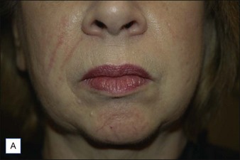
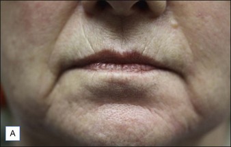
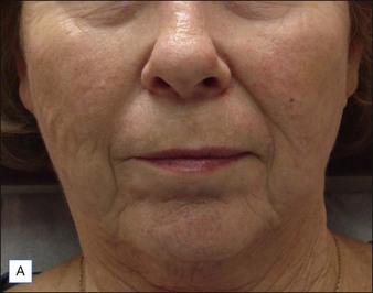
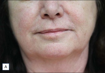
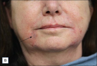
Class II folds are slightly deeper than their class I counterparts, but still will disappear upon stretching of the skin. These folds may require the use of a thicker product that consequently must be injected in the deep dermal plane. Should linear deposition of filler fail to efface these folds, a cross-hatching pattern of filler placement should be adopted. Figure 21.1 demonstrates the use of Radiesse in this area.

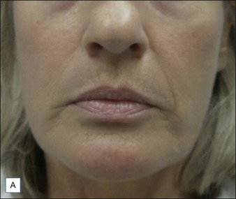
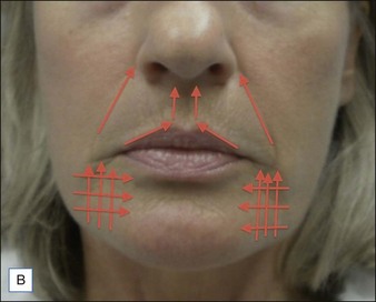
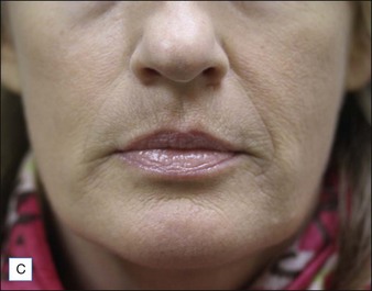
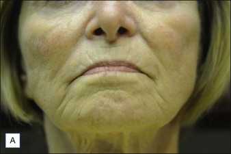
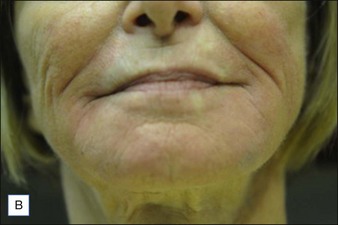
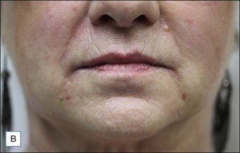
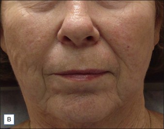
Class III MMFs are prominent, long, and deep. Deep dermal placement in a cross-hatched pattern is the rule. Figure 21.2 demonstrates a patient with class III folds injected with Juvéderm Ultra Plus. Class IV folds are the deepest and the most difficult to correct. They often require vertical layering of multiple filler products to achieve full correction. Even with meticulous technique, deep-plane injection of a thicker filler product may leave remnants of the MMF. This can be dealt with by the addition of a less viscous filler, normally a lower cross-linkage HA product (Restylane, Juvéderm Ultra). This lighter weight filler is injected in the mid-dermis after deep dermal injection of the thicker product to efface the MMF remnant. Normally, the linear threading technique is ideal for this superficially placed filler. Figures 21.3 and 21.4 depict patients with class IV folds who were treated with differing modalities.

Blunt-tipped cannulas offer an opportunity to provide excellent outcomes for filler patients while minimizing the risk of bruising. The authors frequently use these to enhance results in the lower face. A 25-gauge needle is used to make a puncture site on the mandibular margin inferior to the oral commissure. A 27-gauge, 1.5-inch (3.8 cm) cannula is then introduced into this puncture site and used to inject the deep-filling agent. From this single access point, the cannula can atraumatically access the entire area to be filled (Fig. 21.5). The video supplement shows an example of this technique.

image

Choosing the proper filler

As with any location, selection of the proper filler product is nearly as crucial as the technique used to inject it. Dermatologic surgeons are blessed with a broad and ever-expanding armamentarium of filler products to meet the needs of our patients. Fillers may be subdivided into three general categories based upon their absorption characteristics: absorbable, semipermanent, and permanent. The MMFs benefit from the use of sturdier filler, which can be found in any of the above absorption groups.

The first absorbable filler class released was the collagens. These remain the standard for comparison in many labeling trials, but with the advent of the longer lasting HA filler class, collagens have increasingly fallen out of favor and are now less widely available. Human and bovine collagens are generally absorbed within 3 months and are consequently less than ideal for this location. Porcine collagen products do provide the potential of longer lasting effect, but are not widely available at this time.

HA fillers represent the current mainstay in absorbable fillers. These products are non-reactive and, by modifying their degree of cross-linking, their viscosity and time of absorption may be modified. In general, more cross-linked fillers are more viscous (often referred to as ‘thicker’ or ‘heavier’), take longer to be absorbed, and should be injected more deeply in the dermis. As discussed earlier, MMF correction demands deep dermal placement of a sturdy, heavily cross-linked HA such as Juvéderm Ultra Plus or Prevelle.

For very fine etched remnants of the MMF that remain after deposition of the deep filler, mixing a lower cross-linkage HA (Restylane or Juvérderm Ultra) with 1% lidocaine in a 1 : 1 ratio provides an improvised fine line filler that can effectively efface fine lines while minimizing the risk of nodule formation.

Semipermanent fillers such as calcium hydroxylapatite (Radiesse) and poly-l-lactic acid likewise demand deep dermal placement to avoid untoward adverse effects. Calcium hydroxylapatite in particular has shown a very low rate of adverse effects when used for this indication.

Polymethylmethacrylate-containing products are touted as permanent filling products. They have shown a higher rate of complications than their counterparts and should be used only cautiously by an expert injector.

Complications

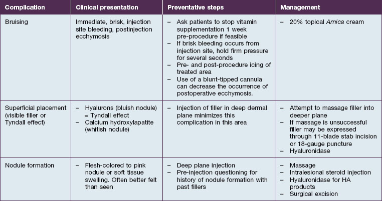
One of the most attractive features of fillers as a cosmetic option is their low rate of adverse effects. Table 21.2 summarizes common complications, and their management. As with any injection, pain is something that must be considered. Strategies for dealing with intraoperative pain management have been addressed earlier in the chapter, and the rate of postoperative pain has consistently been found to be quite low. Bruising is a commonly observed postoperative complication of filler injection (Fig. 21.6). During filler placement it may be prevented by holding firm, direct pressure over any entry points that exhibit brisk bleeding. Postoperative application of ice to the treated areas may also decrease the extent of ecchymosis. Application of 20% topical Arnica has been shown to decrease the duration of post-laser bruising, and has been touted by some experts as a useful agent for managing post-filler bruising as well. Additionally, some injectors find that the use of blunt-tipped cannulas decreases the rate of postoperative bruising (see Fig. 21.5).

Overly superficial placement of filler can lead to visibility of the implant through the skin surface. Owing to the diffraction of light, HA deposits will appear bluish (Tyndall effect). A similar phenomenon can be observed with calcium hydroxylapatite, but this opaque white filler will appear chalky white through the skin surface. Should superficial placement occur, an attempt can be made to massage these malleable products into the proper plane. Failing this, the filler can normally be easily expressed through a needle puncture site or small 11-blade incision.

True immediate hypersensitivity to filler products is rare, but patients should be questioned on history of allergic reaction to past injections. A more common adverse effect is nodule formation. Nodules may be quite disturbing to patients in terms of both causing contour irregularity and tactile sensation. Nodule formation occurs most commonly with calcium hydroxylapatite and poly-l-lactic acid products when they are placed too superficially. As the dominant plane of injection in the MMFs is deep, nodule formation has been found to be rare in this location. Should nodules occur, they may be managed in the following ways. First, massage and watchful waiting may correct small nodules. Intralesional injection of corticosteroid may prove helpful. For persistent nodules that do not resolve with injection and massage, excision may be necessary to provide relief.

Adjunctive treatments

Multiple aging processes affect the perioral cosmetic unit and, as such, filler injection of the MMFs may not be entirely sufficient to restore the area to the desired standard. There are several useful adjuncts that should be considered and these are summarized in Table 21.3.

Table 21.3 Adjunctive measures

Adjunctive measure When to consider Why it works
Botulinum toxin injection of depressor anguli oris

Melolabial fold correction / mid-facial soft tissue augmentation Resurfacing
(e.g. chemical peels, fractional photothermolysis)

Significant synergy has been demonstrated with the combined use of fillers and toxins in the lower face. The depressor anguli oris (DAO), through its connections to the modiolus and dermal connection through SMAS, pulls down on the corner of the mouth as well as contributing directly to the MMF. Thus, with botulinum toxin-mediated relaxation of this muscle, downturning of the oral commissure and improvement of the MMF may be accomplished in one fell swoop. Removal of the inward pull of the DAO’s SMAS connections allows for increased filler effect and duration. Low doses of botulinum toxin are injected at a point 1 cm lateral and 2 cm inferior to the oral commissure. Specifics of toxin injection in this area are outside the scope of this chapter.

An important contributing factor for MMF formation is mid-facial descent with festooning of the cheek and upper lip over the MMF. Mid-facial injection of filler, such as melolabial fold correction, may provide an upward vector for the mid-face, thus attenuating its descent and hence the MMF as in Figure 21.2. Advanced MMF almost universally corresponds with significant skin quality and pigmentation issues. As patients desire not only correction of the fold itself, but correction of their problem, addressing pigment issues with topical products such as retinoids, hydroquinone, or kojic acid can enhance results and has minimal potential morbidity. If a more aggressive approach is needed then resurfacing techniques such as fractional laser resurfacing or chemical peels can address texture and pigmentation issues, as well as fine rhytides, which a filler cannot.

Case Study 1

A 65-year-old female presents to your office requesting ‘Botox’ for her frown lines and melolabial folds. She has never had any cosmetic procedures performed before, but has a high-school reunion the next month and wants to look her best. She has advanced dermatoheliosis with Glogau classification of III, and prominent, class IV, melomental folds. You notice that she has significant festooning of the cheek that accentuates the melomental fold. After explaining your therapeutic plan, you begin by injecting calcium hydroxylapatite to the melolabial folds. The melolabial folds are easily effaced.

After this is complete, calcium hydroxylapatite is injected in a cross-hatched pattern at the dermal–subcutaneous junction beneath the melomental fold. As you had suspected, fine etched remnants of the MMF persist after calcium hydroxylapatite injection. You then, using a linear threading technique, inject the residual rhytides in the mid-dermal plane with a less cross-linked hyaluronic acid product. After massage of the fillers is complete, you palpate the depressor anguli oris muscles bilaterally at the mandible before injecting them with a lower dose of neurotoxin (2 U onabotulinum toxin or incobotulinum toxin, or 5 sU of abobotulinum toxin). At the 2-week follow-up, both you and the patient are pleased with the result.

For advanced melomental folds a multi-pronged approach must be taken to correction. Vertical layering of filler is often necessary, and studies have shown botulinum toxin to have a synergistic effect on filler injection. Patients should be counseled on the likely course of action to improve their awareness and satisfaction with the procedure.

Further reading

Carruthers A, Carruthers J, Hardas B, et al. A validated grading scale for marionette lines. Dermatologic Surgery. 2008;34(suppl 2):S167–S172.

Carruthers A, Carruthers J, Monheit GD, et al. Multicenter, randomized, parallel-group study of onabotulinum toxin A and hyaluronic acid dermal fillers (24-mg/ml smooth, cohesive gel) alone and in combination for lower facial rejuvenation: satisfaction and patient-reported outcomes. Dermatologic Surgery. 2010;36(suppl 4):2121–2134.

Dayan SH, Lieberman ED, Thakkar NN, et al. Botulinum toxin A can positively impact first impression. Dermatologic Surgery. 2008;34:S40–S47.

Don Parsa F, Parsa N, Murariu D. Surgical correction of the frowning mouth. Plastic and Reconstructive Surgery. 2010;2:667–676.

Finzi E, Wasserman E. Treatment of depression with botulinum toxin A: a case series. Dermatologic Surgery. 2006;32:645–649.

Graivier MH, Bass L, et al. Calcium hydroxylapatite for correction of the mid- and lower face: consensus recommendations. Plastic and Reconstructive Surgery. 2007;120(suppl 6):55S–66S.

Park TH, Seo SW, Kim JK, et al. Clinical experience with hyaluronic acid filler complications. Journal of Plastic, Reconstructive and Aesthetic Surgery. 2011;64(7):892–896.

Requena L, Requena C, Christensen L, et al. Adverse reactions to injectable fillers. Journal of the American Academy of Dermatology. 2011;64(1):1–34.

Solish NJ. Assessment of recovery time for the collagen products Dermicol-P35 27G and 30G. Journal of the American Academy of Dermatology. 2010;62(5):824–830.

Tzikas TA. 52-month summary of results using calcium hydroxylapatite for soft tissue augmentation. Dermatologic Surgery. 2008;34:S9–S15.

Van Eijk T, Braun MI. A novel technique to inject hyaluronic acid: the fern technique. Journal of Drugs in Dermatology. 2007;6:806–808.

Weinkle S. Injection techniques for revolumization of the perioral region with hyaluronic acid. Journal of Drugs in Dermatology. 2010;9:367–371.