[level-membership-for-internal-medicine-category]ASTHMA
Asthma is commonly encountered in the long case, and a thorough grasp of the principles of asthma management is essential.
Definition
Asthma: reversible, inflammatory airways disease. Inflammation could be mediated by eosinophils or other cells (lymphocytes and neutrophils).
Case vignette
A 28-year-old female patient has been admitted with fever, chills and rigors. She also has a productive cough and pleuritic chest pains. She has been recently diagnosed with asthma.
She smokes 5–10 cigarettes a day. She works in a bakery and describes symptoms of rhinorrhoea and wheezing while at work and after work. She has been prescribed an inhaler by her GP, but has not been compliant. On examination her temperature is 38°C and respiratory rate 20. Her oxygen saturation is 88% on room air. There are diffuse polyphonic wheezes in the lung fields, with bronchial breath sounds in the left mid to lower zone. Her sputum mug shows rusty purulent sputum.
Approach to the patient
History
Symptoms of chronic cough, especially nocturnal cough, wheezing and complaints of chest tightness, can be clues to consider asthma in the list of differential diagnoses in the dyspnoeic patient. In the known asthmatic, there are some questions that should invariably be asked.
Ask about:
• the current asthma management regimen, and frequency of bronchodilator use. Check whether the patient is using a bronchodilator at an unusually high frequency.
• what the known precipitants of asthma attacks are and how often the patient experiences exacerbations
• whether the patient has ever been hospitalised or treated in the intensive care unit for exacerbation of asthma
• whether the patient has a nocturnal cough
• whether the patient monitors their airway function with a peak flow meter at home. If they do monitor the peak flow, ask how often it is performed and the usual and most recent readings.
• the variability of the peak flow meter readings before and after bronchodilator therapy. Persistent variability is indicative of poor disease control.
• seasonal variation of symptoms and association with exercise
• whether an allergist has been consulted or special tests for allergy (skin prick test and radioallergosorbent (RAST) test) have been performed
• corticosteroid use—how often the patient is prescribed oral steroids, the maximum dose and the minimum dose ever, and the side-effect profile the patient has experienced
• how this chronic condition has affected the patient’s day-to-day life and occupational activities.
Examination
The patient who has had poorly managed asthma since childhood may show evidence of stunted growth. Observe for evidence of dyspnoea and tachypnoea. Notice whether the patient is using accessory muscles for breathing. Check whether the patient has the fine tremor induced by beta agonist therapy. Feel for tracheal tug. Look for evidence of cyanosis. Do not forget to look for evidence of chronic systemic steroid use, such as easy bruising, ecchymoses, cushingoid body habitus and cutaneous striae. Listen to the lung fields for polyphonic wheezes. Perform forced expiratory timing.
Management
The candidate should formulate an ideal ‘asthma management plan’ for every poorly controlled or newly diagnosed asthmatic patient. Two of the most common causes of poor asthma control are non-compliance with medications and poor inhaler technique. Therefore it is important to ascertain the patient’s level of drug compliance and to ask about the inhaler devices used. Elements of a good asthma management plan are as follows:
2. If control is very poor, with frequent exacerbations and frequent bronchodilator use (daily or several times a day), a course of oral corticosteroids together with high-dose inhaled steroids should be commenced. Oral steroids should be tapered and stopped as soon as disease control is achieved.
3. When the level of disease control is suboptimal despite maximum inhaled corticosteroid therapy, an inhaled long-acting beta2 adrenergic receptor agonist such as salmeterol or oxymeterol should be commenced. Combined preparations of inhaled steroids and long-acting bronchodilators are becoming increasingly popular due to their convenience of use, thus improving compliance.
4. Short-acting bronchodilators should be used only in paroxysmal exacerbations of the disease.
5. In special situations, when the level of control is still poor, leukotriene inhibitors and theophylline should be considered as possible additions to the regimen. Leukotriene inhibitors have shown particular benefit in exercise-induced asthma and aspirin-sensitive asthma.
The patient should be given a good insight into his/her disease condition and taught the proper techniques for using an inhaler device. Referral to an asthma educator would be a wise step. Particularly in young and relatively young, active patients, it is important to make an assessment of how the disease affects their day-to-day lives as well as occupational, educational and social activities.
6. Provide the patient with an asthma self-management plan (see box). Such plans have shown benefit to the adult patient with asthma. The plan should include instructions to the patient on how to self-adjust medications according to the symptoms.
7. All asthmatics should be immunised against seasonal influenza and pneumococcal pneumonia.
Asthma self-management plan
One objective of the asthma action plan is to educate and empower the patient to assume some control over the management of this chronic condition. The plan should be able to educate the patient on how to recognise worsening of symptoms and signs of impending danger. The plan should be in writing and individualised to the patient. It should carry the following information:
• the patient’s peak flow readings when stable, when symptomatic, during exacerbations and also when there is a danger of very severe exacerbations
• preventer and reliever medication dose requirements during the abovementioned stages
• instructions on self-adjusted dose increments during exacerbations (up to a maximum dose) and dose decrements when feeling better
• clear instructions on seeking urgent medical help when in danger
• contact details of the doctor and the pharmacist in case the patient needs further assistance.
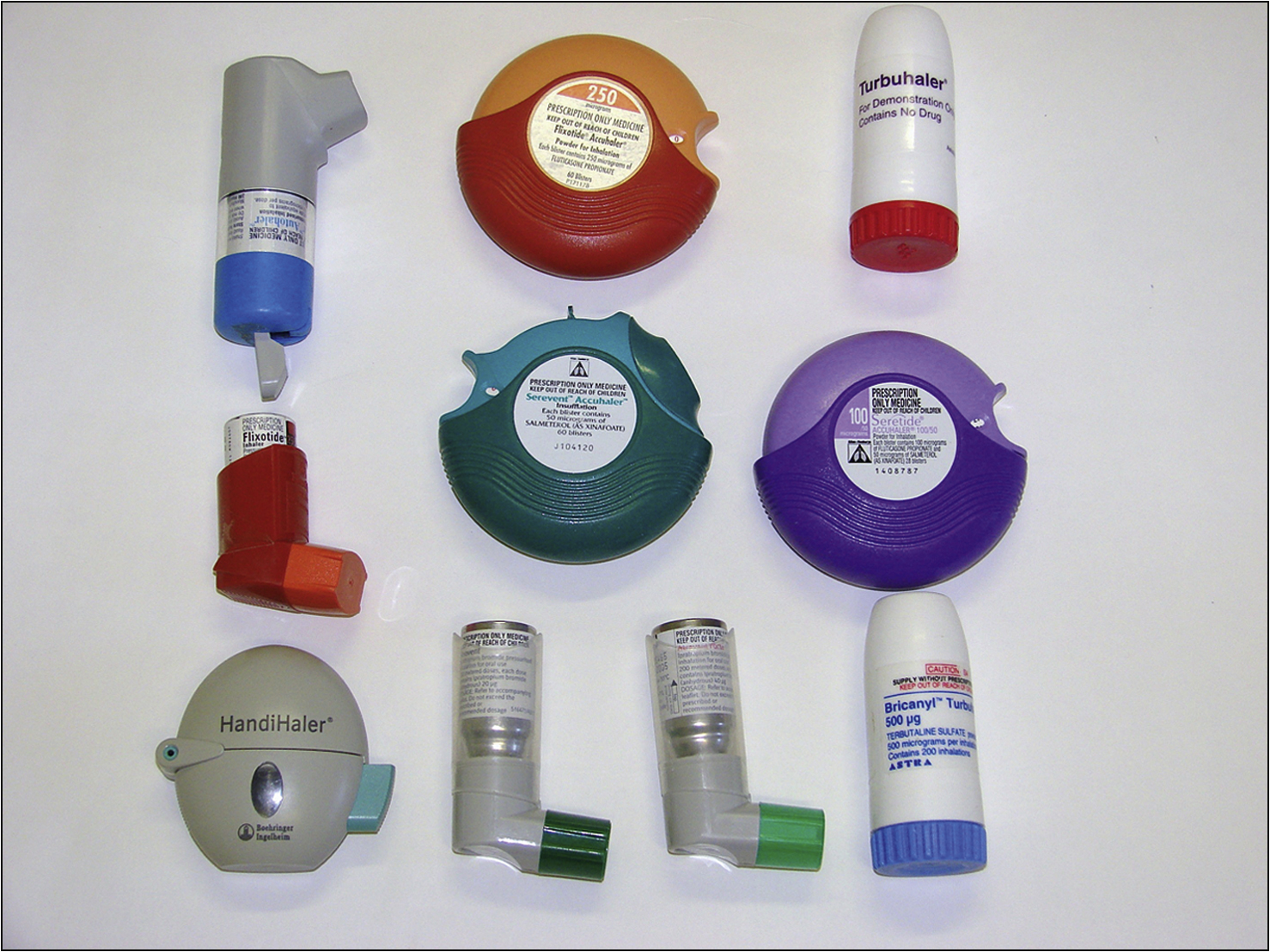
Candidates should be familiar with the different inhaler devices, as some patients know their inhaler device or the medication in it only by its colour and shape (Fig 4.1). Therefore, during the preparation period the candidate should consult the hospital asthma educator or the asthma nurse consultant for further information on the various inhaler devices and become familiar with the proper techniques for use. Candidates should be able to interpret formal lung function study reports quickly and accurately.
Drugs used in asthma
Asthma medications are broadly classified into two categories based on their clinical effects. The first category is the group of medications that improve symptoms (relievers) and the second category prevent exacerbations (preventers).
• Relievers—are short-acting beta2 agonists such as salbutamol, terbutaline, and long-acting beta2 agonists such as efemetorol. Tiotropium and ipratropium bromide are inhaled anticholinergic bronchodilator agents with a slower onset of action. Theophylline, which is capable of relaxing bronchial smooth muscle, is also used to treat severe and acute exacerbations of asthma. However, due to its wide adverse effects profile (nausea, diarrhoea, arrhythmias) it is rarely used these days.
• Preventers—include inhaled corticosteroids such as beclomethasone, budesonide, fluticasone and ciclesonide. Other preventers are leukotriene receptor blockers (montelukast) and cromoglycates (mast cell stabilisers).
– Inhaled corticosteroids have proven benefits in reducing exacerbations, reducing mortality and recurrent hospital admissions. These agents are known to improve overall quality of life in chronic asthmatics. However, long-term high-dose therapy with topical corticosteroids can bring about systemic adverse effects such as cataracts, osteoporosis, glaucoma and cutaneous fragility.
– Cromones such as nedocromil sodium and sodium cromoglycate are capable of preventing early and late bronchoconstrictor reactions to allergen exposure and therefore have particular use in seasonal allergic asthma. They have shown benefit in the prevention of exercise-induced asthma. Nedocromil is useful in the treatment of asthma-associated cough.
Occupational asthma
Occupational asthma is a common occupational morbidity and is quite likely to be encountered in the long case setting. It is a diagnosis in cases of adult-onset asthma.
Approach to the patient
Ask about the patient’s occupation, precise onset of symptoms, diurnal patterns of symptoms and occupational exposure related rhinitis or rhinoconjunctivitis in the past. The patient may report improvement in symptoms outside the workplace. Ask about cigarette smoking, which is known to exacerbate the condition. Most cases of occupational asthma are due to immunoglobulin E (IgE)-mediated immunological response. This form of occupational asthma has a characteristic latency prior to the onset of symptoms after exposure.
Investigations
Occupational asthma is usually investigated by performing serial lung function measurements before and after exposure (at work and away from work on repeated occasions). Serial measurement of peak expiratory flow rate (PEFR) may provide useful information but lack diagnostic accuracy. Referral to an immunologist for blood or skin prick testing for specific IgE may enhance definitive diagnosis.
Management
Early and adequate management of occupational asthma is of prime importance, because failure to control the disease early can lead to a very poor prognosis. The management plan should involve an occupational health physician. Respiratory protective gear, when used properly, helps reduce the risk of occupational asthma but does not prevent its onset. Complete avoidance of allergen exposure is an important first step. Medical management is similar to that of standard asthma management. Remember to discuss the patient’s job and financial issues and also possible worker’s compensation claims (if relevant in the jurisdiction).
Chronic severe asthma
A minority of patients may have recalcitrant disease with hallmark features of frequent severe exacerbations requiring hospitalisation, significant associated morbidity, resistance to commonly used anti-asthma agents and significant steroid dependency. In addition to high mortality rates this patient group suffers from significant drug adverse effects and places a major (disproportionate) burden on the healthcare budget. It is important to ensure that these patients are properly worked up and investigated to exclude non-compliance or missed other diagnoses that could be contributing to the situation.
The management objectives in the patient group are reduction in the number of hospitalisations, steroid weaning and restoration of productivity. Some may respond to very high-dose inhaled steroids such as fluticasone or very high-dose long-acting beta agonists. Other agents that could be used in this setting include cyclosporine, gold and methotrexate. The efficacy of the latter is variable and fraught with significant adverse effects.
CHRONIC OBSTRUCTIVE PULMONARY DISEASE (COPD)
Chronic obstructive pulmonary disease (COPD) or chronic airflow limitation (CAL) is an extremely common long case pathology.
Definition
Chronic obstructive pulmonary disease: irreversible airways disease that incorporates chronic bronchitis, emphysema and chronic asthma with fixed airflow obstruction.
Approach to the patient
History
In the history of patients with known or suspected chronic airflow limitation, enquire about current or previous smoking, occupational exposure to fumes, dust and gases, environmental exposure to such agents and any family history of lung disease. The smoking history (including marijuana) has to be comprehensive and detailed. Also ask about chronic sputum production, wheezing, dyspnoea and the level of effort tolerance. Cardiac disease is common in this patient cohort, and therefore it is important to enquire extensively into this and obtain details.
Examination
Look for tar-stained fingernails, cyanosis, pursed-lip breathing, barrel-shaped chest, subcostal retraction, decreased breath sounds and wheezing on unforced expiration. Particular attention should be focused on excluding a fixed wheeze, which could suggest the presence of a bronchial tumour. Look for evidence of cor pulmonale: elevated JVP, peripheral oedema, parasternal heave and a loud P2.
Smoking-associated comorbidities in patients with smoking-related lung damage (COPD)
• Ischaemic heart disease
• Peripheral vascular disease
• Recurrent respiratory sepsis
• Lung carcinoma
• Carcinoma of head and neck
• Carcinoma of the bladder
• Carcinoma of the oesophagus
• Carcinoma of the colon
• Renal carcinoma
• Peptic ulcer disease
• Sexual dysfunction in men
• Osteoporosis in women
• Secondary polycythaemia
• Depression/anxiety
• Tobacco–alcohol amblyopia
Investigations
Ask for the chest X-ray, looking for evidence of hyperinflation, flattened diaphragmatic shadows, decreased peripheral lung markings and the absence or presence of other lung pathology (lung malignancy in smokers). It should be remembered that only severe emphysema can reliably be diagnosed in a plain chest X-ray.
Other investigations of value include:
1. Spirometry or formal lung function studies, with readings before and after bronchodilator therapy—looking for reversibility of the obstructive airway picture. The total lung capacity would be increased and the vital capacity and carbon dioxide diffusion (DLCO) would be decreased. Patient develops dyspnoea on minimal exertion when forced expiratory volume in 1 second (FEV1) drops to 30% of predicted. Forced expiratory time (FET) is a simple bedside test that can be used to assess lung function. FET of over 6 seconds indicates severe airflow limitation.
3. Haemoglobin level—looking for elevated levels, particularly if arterial partial pressure of oxygen is less than 55 mmHg.
4. Full blood count—looking for erythrocytosis/polycythaemia and elevation of the white cell count if an infection is present. A haematocrit of > 52% in males and > 45% in females is diagnostic of erythrocytosis. A packed cell volume (PCV) of > 55% is very significant and an indication for long-term oxygen therapy.
5. Formal lung function tests—including carbon monoxide diffusion capacity (particularly if the severity of the dyspnoea is out of proportion to the FEV1)and lung volumes. Most patients benefit from a trial of steroids to assess steroid responsiveness with FEV1/forced vital capacity (FVC) measured before and after.
6. High-resolution CT of the lung—to look for dilated terminal airways typical of emphysema and to exclude other parenchymal lung pathology.
7. A sleep study—warranted if obstructive sleep apnoea is suspected (this should be considered if there is polycythaemia or cor pulmonale despite daytime arterial oxygen partial pressure being maintained above 60 mmHg).
8. Alpha1-antitrypsin levels—especially in patients under 40 years of age with a positive family history of emphysema.
9. Sputum microscopy—in infective exacerbations, sputum may contain neutrophils and pathogenic bacteria. Most frequently associated organisms are Moraxella catarrhalis, Haemophilus influenzae and Streptococcus pneumoniae.
10. A trial of steroids is indicated, to assess the patient’s steroid responsiveness. FEV1/FVC is measured before and after the challenge, looking for an improvement of significance.
Management of chronic airflow limitation
Candidates should formulate a suitable plan for the optimal management of the patient’s condition. A sound and practical plan of action would be very useful. The main objective of the optimal management plan is to improve the patient’s activity levels and overall quality of life. This is achieved by treating symptoms, preventing exacerbations and preserving lung functions. Recruit the patient into a pulmonary rehabilitation program and encourage them to undertake light exercise. This helps improve morbidity, quality of life and mortality.
Formulate a collaborative management plan with the participation of the patient’s general practitioner, community nursing sister and other community resources, with the main objective of preventing recurrent hospital admissions due to exacerbations. Physical rehabilitation and progressive exercising should be a major part of the long-term management plan.
The following are the integral components of the plan:
1. Instructions on the different medications and how to use them. Don’t forget to stress the need for good compliance.
2. If the patient is suffering from frequent severe exacerbations, they should be commenced on oral corticosteroids. Start treatment with prednisolone 30 mg and plan to decrease the dose according to the clinical improvement. (IV hydrocortisone is indicated in very severe exacerbation of chronic airway limitation, and the decision to use this should be guided by the clinical findings.)
4. Twice-daily inhaled long-acting bronchodilator therapy should be considered if there is only suboptimal response to inhaled steroids alone. Combined formulations of inhaled steroids and a long-acting beta2 receptor agonist are more appealing to patients due to the convenience of their use.
5. Ipratropium bromide or tiotropium (Spiriva®) via a metered dose inhaler and a spacer device four times a day has also been shown to be beneficial to these patients. Some patients may benefit from theophylline therapy, an agent rarely used these days.
6. Short-acting bronchodilator via a metered dose inhaler should be prescribed, to be taken only as needed.
7. Phosphodiesterase 4 inhibitors such as cilomilast and roflumilast given systemically are also known to control the inflammatory process. There is emerging evidence of its clinical benefits to patients with COPD.
8. Acute exacerbation may warrant antibiotic therapy. One example is the combination of IV ceftriaxone 1 g once daily with oral roxithromycin 150 mg twice daily. Other agents to consider are penicillin, ampicillin, azithromycin and clarythromycin.
On recovery, the patient should be given oral antibiotics (for example, roxithromycin or amoxycillin) at discharge, with instructions to take prophylactically on identification of the earliest signs of an infective exacerbation (patient should be instructed to be on the alert for such symptoms as any unusual cough, sputum production, fever, dyspnoea or malaise). Also instruct the patient to see their general practitioner with a view to recommencing oral corticosteroid therapy in such circumstances.
Rotating different agents may be useful in preventing antibiotic resistance.
9. Advice on and help in stopping smoking (topical nicotine patches or effective anti-craving agents such as bupropion hydrochloride) and avoiding airborne hazards. Varenicline is a novel agent that has shown promise in assisting smoking cessation.
10. Patient education should be provided on the condition and its current severity, contributory lifestyle factors that need modification, and how to slow progression of the disease and prevent complications.
11. Assess the patient’s need for oxygen supplementation at home (see box overleaf).
12. If the patient remains significantly dyspnoeic and incapacitated despite all the above measures, or if the patient has giant pulmonary bullae, consider lung volume reduction surgery (bullectomy).
13. If the patient has cor pulmonale, referral to a cardiologist and further investigation (echo/right heart catheterisation) is indicated. Therapy includes loop diuretics, oxygen, optimising airway therapy and rehabilitation.
14. In resistant patients younger than 55 years, consideration should be given to lung transplantation. This process should be triggered with the patient being referred to a centre that has a lung transplantation program and expertise for screening and work-up thereof.
15. All patients should be advised on appropriate nutrition and regular vaccination against Pneumococcus and influenza virus.
Criteria for home oxygen supplementation
The presence of any one of the following criteria qualifies the patient for home oxygen. Prescribe domiciliary oxygen for at least 19 hours a day.
• PaO2 of < 55 mmHg or arterial oxygen saturations of < 88% at rest
• Resting PaO2 of 56–59 mmHg with cor pulmonale
• PaO2 of < 55 mmHg or arterial oxygen saturations of < 88% on exertion or while asleep
• P-pulmonale (of > 3 mm) and evidence of right ventricular hypertrophy on ECG
• Echocardiographic evidence of right ventricular hypertrophy/strain together with pulmonary hypertension
• PCV of > 0.55
BRONCHIECTASIS
Bronchiectasis is defined as abnormal and permanent dilatation of bronchi with associated pooling of secretions, often leading to recurrent or persistent sepsis.
Approach to the patient
History
In the history of a patient with known or suspected bronchiectasis, ask about symptoms of recurrent cough, purulent sputum, dyspnoea, wheeze, haemoptysis and pleuritic chest pain. Check how often the patient experiences exacerbations and how such exacerbations present (usually there is an increase in the volume of sputum and its degree of purulence, with associated fevers and worsening dyspnoea). Ask how the episodes of exacerbation are managed (usually with multiple oral and parenteral antibiotics together with vigorous chest physiotherapy) and enquire about any chronic prophylactic antibiotic use (e.g. oral fluoroquinolones and inhaled aminoglycosides). Record the date of the most recent exacerbation. Ask about recurrent hospital admissions—frequency and average duration of stay on each occasion. Some respiratory physicians admit patients with bronchiectasis regularly for a prophylactic course of IV antibiotics to keep pathogenic bacterial colonies under adequate control.
Enquire about regular chest physiotherapy (self or by partner), forced expiratory techniques (huffing) and postural drainage. Gain insight into the volume of sputum production.
Ask about complications such as massive haemoptysis (due to bronchoarterial fistulae that need management with embolisation of the relevant segment of the bronchial artery). Significant weight loss is a bad prognostic sign in these patients. Ask about any recent weight loss and about general nutrition and appetite.
Ask how the disease is affecting the patient’s social, occupational and family life. Enquiry into housing, social and economic problems is of great importance. Check about domestic and housing conditions. Ask about any depression associated with the chronic illness and assess the adequacy of the patient’s coping skills and supportive resources. Patient motivation and supportive social or family networks are essential factors in the management of patients with bronchiectasis.
Examiners may be interested in the aetiology of the patient’s condition. Ask about childhood illnesses such as measles and whooping cough, any past history of severe viral (adenovirus/influenza virus) or bacterial (Staphylococcus aureus, Klebsiella sp., anaerobic organisms and tuberculosis) respiratory tract sepsis that could be associated with the onset of symptoms.
The family history may give clues to the aetiology of the disease. Ask about cystic fibrosis and immunodeficiency, including HIV infection. Complaints of recurrent sinusitis and cutaneous sepsis should alert the candidate to the possibility of hypogammaglobulinaemia. Ask about any past history of foreign body aspiration, toxic gas inhalation and aspiration of caustic material, including acidic gastric content.
Check for features of primary ciliary dyskinesia such as recurrent upper respiratory tract infections, otitis media and infertility. Significant asthmatic symptoms should alert candidates to the possibility of allergic bronchopulmonary aspergillosis as a causative factor.
Obtain a detailed history of alcohol consumption, looking for clues of possible recurrent aspiration. A history of pulmonary fibrosis and interstitial lung disease may suggest traction bronchiectasis.
Examination
In the physical examination, look for signs of weight loss, wasting and cachexia. Examine the sputum mug and the temperature chart. Observe for a productive cough, tachypnoea and reduced chest expansion. Look for central or peripheral cyanosis and finger and/or toe clubbing. Some may even have hypertrophic pulmonary osteoarthropathy (HPOA) with tenderness in the wrists and ankles. Percussion of the thorax may show areas of dullness due to consolidation or severe atelectasis. Auscultate for coarse crepitations and wheezing. Check for features of right heart failure due to cor pulmonale and for signs of pulmonary hypertension. Check for hepatosplenomegaly and peripheral oedema. Do not miss situs inversus and dextrocardia with a right-sided apex beat, if present (Kartagener’s syndrome—a form of primary ciliary dyskinesis). Look at the sputum mug and estimate sputum volumes, and check the smell.
Investigations
Investigations of bronchiectasis include:
1. Chest X-ray—looking for cystic air spaces, presence of air-fluid levels in the dilated bronchi, thickened bronchial walls with peribronchial cuffing, with the appearance of ‘tramlines’ and ‘ring shadows’.
2. High-resolution CT scan of the chest—to confirm and better define the above features.
3. Formal lung function tests—looking for a reversible obstructive, restrictive or mixed picture.
4. Sputum microscopy and culture, including prolonged cultures in special media for fungi and tubercle bacilli. Organisms that commonly colonise these patients include Pseudomonas aeruginosa, Burkholderia cepacia (in cystic fibrosis), Haemophilus influenzae, Escherichia coli and Staphylococcus aureus. Bronchial washings too may be of use.
6. Renal function indices—presence of renal failure should signal the diagnosis of possible secondary amyloidosis. Amyloidosis secondary to chronic inflammation usually presents with renal failure and/or hepatosplenomegaly. Cardiac involvement is a rarity.
Where the aetiology is not clear, the following tests may be considered:
7. Fibreoptic bronchoscopy—looking for obstructive lesions
8. Sweat chloride levels—looking for evidence of cystic fibrosis
9. Serum immunoglobulin assay—looking for hypogammaglobulinaemia
10. Sperm assay or respiratory mucosal biopsy—looking for abnormalities of ciliary motility
11. Skin tests and serology for aspergillosis.
Management
The main objectives in the management of bronchiectasis are:
Management consists of the following:
1. Regular twice-daily chest physiotherapy (teach the patient how to self-administer physiotherapy) and training on techniques of postural drainage and forced expiration (huffing).
2. Aerosolised recombinant DNAse to further facilitate clearance of secretions (only in cystic fibrosis). Nebulised NaCl or mucolytics such as N-acetyl cysteine may be of use.
3. Chronic or periodic prophylactic/maintenance antibiotics (see box), depending on the culprit organisms. Patients are managed with single or combination antibiotic therapy. Some patients infected with Pseudomonas sp. benefit from nebulised aminoglycosides.
4. Patients with bronchoconstriction benefit from regular bronchodilator therapy. Some patients may benefit from inhaled steroids.
5. Localised disease can be treated successfully with lung resection surgery.
6. Associated right heart failure can be treated with diuretics and, where there is persistent hypoxia, with chronic oxygen supplementation. Severe right heart failure is managed with heart–lung transplantation in suitable candidates.
7. Allergic bronchopulmonary aspergillosis is treated with high-dose steroids, and hypogammaglobulinaemia is treated with regular infusion of normal immunoglobulins.
8. Nutritional dietary supplements, availability of community-based healthcare resources and psychological counselling where necessary.
CYSTIC FIBROSIS
Cystic fibrosis is the most common cause of bronchiectasis in many parts of the Western world. Therefore, most facts discussed under ‘bronchiectasis’ apply to cystic fibrosis too. This is an autosomal recessive disorder with an incidence of 1 in 2000 and a carrier frequency of 1 in 25 among Caucasians. In most cases, causative mutation is localised to the cystic fibrosis transmembrane conductance regulator (CFTR) gene in the long arm of chromosome 7, but many more genetic mutations associated with cystic fibrosis have been described, making regular genetic testing difficult and inaccurate. In the past, this disease was associated with childhood fatality. With novel management modalities, patients seem to survive well into adulthood and are now commonly encountered in the practice of internal medicine.
Case vignette
A 25-year-old male with known cystic fibrosis presents with sudden-onset severe pleuritic chest pain of the left side. He also complains of severe dyspnoea. He denies any exacerbation of cough, fever or haemoptysis. On examination he is tachypnoeic and is in evident distress. Breath sounds are significantly decreased in the left upper and mid zones of the lung fields. There are coarse crepitations audible elsewhere.
Approach to the patient
History
Ask about:
• details of the initial diagnosis, such as age, first presenting symptoms and diagnostic investigations
• current respiratory symptoms of productive cough, wheeze, fever, dyspnoea and haemoptysis. The patient may have had episodes of spontaneous pneumothorax and epistaxis due to nasal polyps.
• details about recurrent exacerbations of respiratory tract infections, multiple hospital admissions and the various forms of antibiotic therapy
• regular physiotherapy, techniques of forced expiration and postural drainage
• sino-nasal symptoms such as nasal obstruction, worsening nasal discharge, facial pain and cough
• gastrointestinal features of steatorrhoea, intestinal obstruction due to meconium ileus equivalent syndrome, and right upper quadrant pain and/or jaundice due to cholesterol gallstones
• nutrition, appetite and weight loss—malnutrition and weight loss are bad prognostic signs of the disease
• regular pancreatic enzyme supplementation and any side effects of this therapy
• heat intolerance and heat exhaustion in hot weather.
A minority of patients suffer from diabetes mellitus, of which the details should be obtained.
It is important to get a detailed family history.
Women suffering from cystic fibrosis are able to conceive but male patients are usually infertile. Ask about any plans for reproduction, genetic counselling and screening, and the coping strategies of the male patient with infertility. Be ready to discuss the ethical issues surrounding facilitated reproduction in cystic fibrosis.
Examination
Look for wasting, cachexia, stunted growth and features of malnutrition. Check the patient’s weight. Some patients may have permanent vascular access devices for long-term antibiotic therapy. Observe the temperature chart and the sputum mug. Look for tachypnoea, cyanosis, finger and toe clubbing and hypertrophic pulmonary osteoarthropathy. Perform a detailed examination of the respiratory system as described under ‘bronchiectasis’. Perform a detailed abdominal examination, looking for tenderness (particularly in the right upper or lower quadrant) and features of intestinal obstruction (distension, high-pitched bowel sounds or absent bowel sounds). Look for signs of right heart failure due to cor pulmonale.
Investigations
Investigations in cystic fibrosis include initial diagnostic tests and tests to diagnose and monitor the recurrent and persistent complications:
1. Sweat test—a sweat Cl level of > 70 mmol/L is consistent with cystic fibrosis in adults
2. Genetic tests for the most common mutations
4. Formal lung function tests or spirometry
5. Sputum microscopy and culture. Blood cultures in the febrile patient. Discovery of Aspergillus sp. or Pseudomonas sp. in the sputum often suggests colonisation rather than infection.
6. Supine and erect abdominal X-rays—looking for air-fluid levels and dilated loops of small intestine suggesting intestinal obstruction
7. Abdominal ultrasonography—to exclude cholelithiasis, where relevant
8. Full blood count—looking for anaemia of chronic disease or megaloblastic anaemia of malabsorption
9. Liver function tests—rarely these patients develop cirrhosis
10. Renal function indices—looking for nephrotoxic effects of aminoglycosides.
Management
1. Regular chest physiotherapy, forced expiration and postural drainage
2. Regular oral or inhaled aerosolised antibiotics for maintenance and the treatment of minor exacerbations. Intravenous antibiotics for severe exacerbations as guided by the results of the microbiological tests.
3. Recombinant DNAse to facilitate the clearance of respiratory secretions
4. Bronchodilator therapy
5. Oral replacement of pancreatic enzyme supplements and lipid-soluble vitamins
6. Dietary advice and monitoring to ensure adequate nutrition and prevent weight loss
7. Meconium ileus equivalent can be treated with enemas of hypertonic radiocontrast agents.
8. Consideration should be given to lung transplantation in end-stage lung disease with respiratory failure. Patients with severe cor pulmonale may need combined heart–lung transplantation.
9. Psychological and social support as necessary. The young patient with a chronic disorder and relatively short life expectancy may suffer from significant emotional distress.
PNEUMONIA
Approach to the patient
History
Ask the patient about:
• presenting symptoms such as fevers, rigors, chills, cough, dyspnoea and pleurisy
• the appearance of the sputum—if available, ask to have a look at the patient’s sputum mug. Mucopurulent sputum is associated with bacterial pneumonia.
• other associated symptoms such as fatigue, nausea, vomiting, diarrhoea (sometimes may be associated with the antibiotic therapy)
• comorbidities such as COPD, asthma, immunocompromised status (HIV, splenectomy, chemotherapy, corticosteroid use)
• risk factors such as occupational exposure, diabetes, alcoholism (aspiration) and smoking.
Examination
Check the patient’s temperature and also request the temperature chart to ascertain the nature and periodicity of the temperature spikes. Check respiratory rate and pulse rate. Tachypnoea and tachycardia should not be missed. On auscultation note crackles (rales and rhonchi) and bronchial breath sounds. Stony dullness with absent breath sounds may indicate pleural effusion.
Investigations
1. Arterial blood gases, full blood count (looking for leucocytosis and the differential count) and the electrolyte profile, together with renal function indices
2. AP and lateral chest X-ray—looking for consolidation, infiltrates, cavitations, effusion. Distinguish between lobar pneumonia (confined to a lung lobe) and bronchopneumonia (with a diffuse patchy appearance)
3. Sputum microscopy (Gram stain) and culture
4. Atypical serology/viral serology (polymerase chain reaction (PCR) could be useful for atypical bacteria and viruses)
5. Blood culture results
6. Urine results for Legionella and pneumococcal antigen
7. Viral cultures and enzyme immunoassay (EIA) or immunofluorescence for viruses
Pathogens associated with pneumonia
• Streptococcus pneumoniae is the most commonly identified pathogen in most community-acquired pneumonia cases. Chlamydophila pneumoniae and Mycoplasma pneumoniae are common atypical pathogens of community-acquired pneumonia.
• Haemophilus should be considered in patients with chronic lung disease, alcoholic patients and elderly patients.
• If the pneumonia was preceded by influenza virus infection, Staphylococcus aureus should be considered a likely pathogen. The latter has a more aggressive clinical course, with potential pulmonary necrosis and toxic shock.
• Diagnosis of influenza pneumonia and Legionella pneumonia is of significance due to the potential for epidemic outbreaks.
• If aspiration is a possibility, Klebsiella and anaerobic agents should be considered high on the list of pathogenic agents.
• Common pathogens associated with nosocomial pneumonia include Staphylococcus, Pseudomonas and Gram-negative bacteria.
Management
It is important to identify the features that place a patient with pneumonia at high risk, such as advanced age, confusion, hypotension, tachypnoea and elevated serum urea levels (see box). Such features warrant more aggressive therapy and supportive management (i.e. hospitalisation).
1. Antibiotics remain the cornerstone of pneumonia therapy. Bacterial community-acquired pneumonia can be treated with an oral macrolide such as clarithromycin or azithromycin or with a fluoroquinolone if the patient is young and stable. Mild to moderate pneumonia requiring hospitalisation can be treated with an IV third-generation cephalosporin such as ceftriaxone.
2. Patients with severe community-acquired bacterial pneumonia requiring hospitalisation may be treated with an extended-spectrum penicillin such as piperacillin/tazobactam together with an aminoglycoside.
3. Atypical pneumonias respond to macrolides or quinolones.
4. Gram-negative agents need treatment with more potent agents such as fourth-generation cephalosporins such as cefepime, or carbapenems such as meropenem.
5. In addition to antibiotic therapy, patients require antipyrexial agents, oxygen therapy (if hypoxic), analgesics and chest therapy.
6. Pneumonia prevention should be discussed in detail. Older individuals (over 50), patients with comorbidities, immunocompromised patients and healthcare workers should have pneumococcal vaccine and the annual influenza immunisation. Haemophilus vaccine is indicated for patients with sickle cell disease, those who have had splenectomy and those with leukaemias or HIV.
[/level-membership-for-internal-medicine-category][not-level-membership-for-internal-medicine-category]ASTHMA
Asthma is commonly encountered in the long case, and a thorough grasp of the principles of asthma management is essential.
Definition
Asthma: reversible, inflammatory airways disease. Inflammation could be mediated by eosinophils or other cells (lymphocytes and neutrophils).
Case vignette
A 28-year-old female patient has been admitted with fever, chills and rigors. She also has a productive cough and pleuritic chest pains. She has been recently diagnosed with asthma.
She smokes 5–10 cigarettes a day. She works in a bakery and describes symptoms of rhinorrhoea and wheezing while at work and after work. She has been prescribed an inhaler by her GP, but has not been compliant. On examination her temperature is 38°C and respiratory rate 20. Her oxygen saturation is 88% on room air. There are diffuse polyphonic wheezes in the lung fields, with bronchial breath sounds in the left mid to lower zone. Her sputum mug shows rusty purulent sputum.
Approach to the patient
History
Symptoms of chronic cough, especially nocturnal cough, wheezing and complaints of chest tightness, can be clues to consider asthma in the list of differential diagnoses in the dyspnoeic patient. In the known asthmatic, there are some questions that should invariably be asked.
Ask about:
• the current asthma management regimen, and frequency of bronchodilator use. Check whether the patient is using a bronchodilator at an unusually high frequency.
• what the known precipitants of asthma attacks are and how often the patient experiences exacerbations
• whether the patient has ever been hospitalised or treated in the intensive care unit for exacerbation of asthma
• whether the patient has a nocturnal cough
• whether the patient monitors their airway function with a peak flow meter at home. If they do monitor the peak flow, ask how often it is performed and the usual and most recent readings.
• the variability of the peak flow meter readings before and after bronchodilator therapy. Persistent variability is indicative of poor disease control.
• seasonal variation of symptoms and association with exercise
• whether an allergist has been consulted or special tests for allergy (skin prick test and radioallergosorbent (RAST) test) have been performed
• corticosteroid use—how often the patient is prescribed oral steroids, the maximum dose and the minimum dose ever, and the side-effect profile the patient has experienced
• how this chronic condition has affected the patient’s day-to-day life and occupational activities.
Examination
The patient who has had poorly managed asthma since childhood may show evidence of stunted growth. Observe for evidence of dyspnoea and tachypnoea. Notice whether the patient is using accessory muscles for breathing. Check whether the patient has the fine tremor induced by beta agonist therapy. Feel for tracheal tug. Look for evidence of cyanosis. Do not forget to look for evidence of chronic systemic steroid use, such as easy bruising, ecchymoses, cushingoid body habitus and cutaneous striae. Listen to the lung fields for polyphonic wheezes. Perform forced expiratory timing.
Management
The candidate should formulate an ideal ‘asthma management plan’ for every poorly controlled or newly diagnosed asthmatic patient. Two of the most common causes of poor asthma control are non-compliance with medications and poor inhaler technique. Therefore it is important to ascertain the patient’s level of drug compliance and to ask about the inhaler devices used. Elements of a good asthma management plan are as follows:
2. If control is very poor, with frequent exacerbations and frequent bronchodilator use (daily or several times a day), a course of oral corticosteroids together with high-dose inhaled steroids should be commenced. Oral steroids should be tapered and stopped as soon as disease control is achieved.
3. When the level of disease control is suboptimal despite maximum inhaled corticosteroid therapy, an inhaled long-acting beta2 adrenergic receptor agonist such as salmeterol or oxymeterol should be commenced. Combined preparations of inhaled steroids and long-acting bronchodilators are becoming increasingly popular due to their convenience of use, thus improving compliance.
4. Short-acting bronchodilators should be used only in paroxysmal exacerbations of the disease.
5. In special situations, when the level of control is still poor, leukotriene inhibitors and theophylline should be considered as possible additions to the regimen. Leukotriene inhibitors have shown particular benefit in exercise-induced asthma and aspirin-sensitive asthma.
The patient should be given a good insight into his/her disease condition and taught the proper techniques for using an inhaler device. Referral to an asthma educator would be a wise step. Particularly in young and relatively young, active patients, it is important to make an assessment of how the disease affects their day-to-day lives as well as occupational, educational and social activities.
6. Provide the patient with an asthma self-management plan (see box). Such plans have shown benefit to the adult patient with asthma. The plan should include instructions to the patient on how to self-adjust medications according to the symptoms.
7. All asthmatics should be immunised against seasonal influenza and pneumococcal pneumonia.
Asthma self-management plan
One objective of the asthma action plan is to educate and empower the patient to assume some control over the management of this chronic condition. The plan should be able to educate the patient on how to recognise worsening of symptoms and signs of impending danger. The plan should be in writing and individualised to the patient. It should carry the following information:
• the patient’s peak flow readings when stable, when symptomatic, during exacerbations and also when there is a danger of very severe exacerbations
• preventer and reliever medication dose requirements during the abovementioned stages
• instructions on self-adjusted dose increments during exacerbations (up to a maximum dose) and dose decrements when feeling better
• clear instructions on seeking urgent medical help when in danger
• contact details of the doctor and the pharmacist in case the patient needs further assistance.
Candidates should be familiar with the different inhaler devices, as some patients know their inhaler device or the medication in it only by its colour and shape (Fig 4.1). Therefore, during the preparation period the candidate should consult the hospital asthma educator or the asthma nurse consultant for further information on the various inhaler devices and become familiar with the proper techniques for use. Candidates should be able to interpret formal lung function study reports quickly and accurately.
Drugs used in asthma
Asthma medications are broadly classified into two categories based on their clinical effects. The first category is the group of medications that improve symptoms (relievers) and the second category prevent exacerbations (preventers).
• Relievers—are short-acting beta2 agonists such as salbutamol, terbutaline, and long-acting beta2 agonists such as efemetorol. Tiotropium and ipratropium bromide are inhaled anticholinergic bronchodilator agents with a slower onset of action. Theophylline, which is capable of relaxing bronchial smooth muscle, is also used to treat severe and acute exacerbations of asthma. However, due to its wide adverse effects profile (nausea, diarrhoea, arrhythmias) it is rarely used these days.
• Preventers—include inhaled corticosteroids such as beclomethasone, budesonide, fluticasone and ciclesonide. Other preventers are leukotriene receptor blockers (montelukast) and cromoglycates (mast cell stabilisers).
– Inhaled corticosteroids have proven benefits in reducing exacerbations, reducing mortality and recurrent hospital admissions. These agents are known to improve overall quality of life in chronic asthmatics. However, long-term high-dose therapy with topical corticosteroids can bring about systemic adverse effects such as cataracts, osteoporosis, glaucoma and cutaneous fragility.
– Cromones such as nedocromil sodium and sodium cromoglycate are capable of preventing early and late bronchoconstrictor reactions to allergen exposure and therefore have particular use in seasonal allergic asthma. They have shown benefit in the prevention of exercise-induced asthma. Nedocromil is useful in the treatment of asthma-associated cough.
Occupational asthma
Occupational asthma is a common occupational morbidity and is quite likely to be encountered in the long case setting. It is a diagnosis in cases of adult-onset asthma.
Approach to the patient
Ask about the patient’s occupation, precise onset of symptoms, diurnal patterns of symptoms and occupational exposure related rhinitis or rhinoconjunctivitis in the past. The patient may report improvement in symptoms outside the workplace. Ask about cigarette smoking, which is known to exacerbate the condition. Most cases of occupational asthma are due to immunoglobulin E (IgE)-mediated immunological response. This form of occupational asthma has a characteristic latency prior to the onset of symptoms after exposure.
Investigations
Occupational asthma is usually investigated by performing serial lung function measurements before and after exposure (at work and away from work on repeated occasions). Serial measurement of peak expiratory flow rate (PEFR) may provide useful information but lack diagnostic accuracy. Referral to an immunologist for blood or skin prick testing for specific IgE may enhance definitive diagnosis.
Management
Early and adequate management of occupational asthma is of prime importance, because failure to control the disease early can lead to a very poor prognosis. The management plan should involve an occupational health physician. Respiratory protective gear, when used properly, helps reduce the risk of occupational asthma but does not prevent its onset. Complete avoidance of allergen exposure is an important first step. Medical management is similar to that of standard asthma management. Remember to discuss the patient’s job and financial issues and also possible worker’s compensation claims (if relevant in the jurisdiction).
Chronic severe asthma
A minority of patients may have recalcitrant disease with hallmark features of frequent severe exacerbations requiring hospitalisation, significant associated morbidity, resistance to commonly used anti-asthma agents and significant steroid dependency. In addition to high mortality rates this patient group suffers from significant drug adverse effects and places a major (disproportionate) burden on the healthcare budget. It is important to ensure that these patients are properly worked up and investigated to exclude non-compliance or missed other diagnoses that could be contributing to the situation.
The management objectives in the patient group are reduction in the number of hospitalisations, steroid weaning and restoration of productivity. Some may respond to very high-dose inhaled steroids such as fluticasone or very high-dose long-acting beta agonists. Other agents that could be used in this setting include cyclosporine, gold and methotrexate. The efficacy of the latter is variable and fraught with significant adverse effects.
CHRONIC OBSTRUCTIVE PULMONARY DISEASE (COPD)
Chronic obstructive pulmonary disease (COPD) or chronic airflow limitation (CAL) is an extremely common long case pathology.
Definition
Chronic obstructive pulmonary disease: irreversible airways disease that incorporates chronic bronchitis, emphysema and chronic asthma with fixed airflow obstruction.
Approach to the patient
History
In the history of patients with known or suspected chronic airflow limitation, enquire about current or previous smoking, occupational exposure to fumes, dust and gases, environmental exposure to such agents and any family history of lung disease. The smoking history (including marijuana) has to be comprehensive and detailed. Also ask about chronic sputum production, wheezing, dyspnoea and the level of effort tolerance. Cardiac disease is common in this patient cohort, and therefore it is important to enquire extensively into this and obtain details.
Examination
Look for tar-stained fingernails, cyanosis, pursed-lip breathing, barrel-shaped chest, subcostal retraction, decreased breath sounds and wheezing on unforced expiration. Particular attention should be focused on excluding a fixed wheeze, which could suggest the presence of a bronchial tumour. Look for evidence of cor pulmonale: elevated JVP, peripheral oedema, parasternal heave and a loud P2.
Smoking-associated comorbidities in patients with smoking-related lung damage (COPD)
• Ischaemic heart disease
• Peripheral vascular disease
• Recurrent respiratory sepsis
• Lung carcinoma
• Carcinoma of head and neck
• Carcinoma of the bladder
• Carcinoma of the oesophagus
• Carcinoma of the colon
• Renal carcinoma
• Peptic ulcer disease
• Sexual dysfunction in men
• Osteoporosis in women
• Secondary polycythaemia
Buy Membership for Internal Medicine Category to continue reading. Learn more here
[/not-level-membership-for-internal-medicine-category]
