Meckel’s Diverticulum
Meckel’s diverticulum is a common congenital anomaly affecting the small bowel. Typically, the diverticulum is located within 2 feet of the ileocecal valve and arises from the antimesenteric border of the ileum. The protrusion is about 2 inches long and occurs in 2% of the population (Figs. 96–1 through 96–3). The diverticulum may contain stomach, pancreas, and biliary and colonic tissue, which may create symptoms (e.g., peptic ulcer). Significant intestinal hemorrhage, which manifests itself by rectal bleeding, may emanate from Meckel’s diverticulum. Other complications include inflammation, obstruction, and fistula.
The diverticulum may be removed by clamping the base, cutting the diverticulum off, and suturing the bowel. Similarly, the base may be stapled and cut (Fig. 96–4).

FIGURE 96–1 Meckel’s diverticulum located about 12 inches from the ileocecal junction. The diverticulum has a wide mouth and measures 2 inches in length.
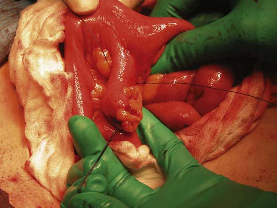
FIGURE 96–2 Close-up view of the diverticulum. Note that traction sutures have been placed to facilitate manipulation.
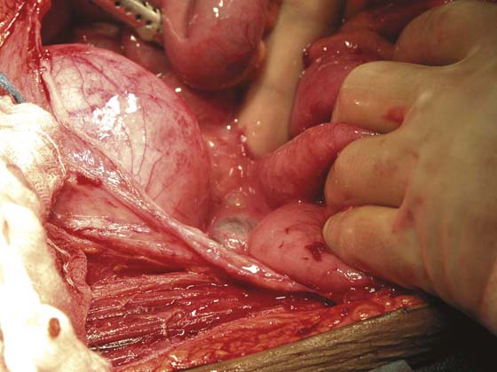
FIGURE 96–3 A still more magnified view of the ileal diverticulum. Note the cecum in the foreground.
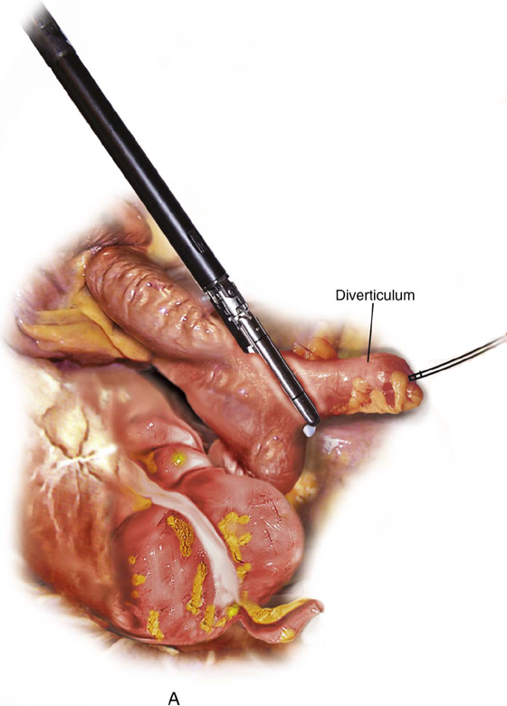
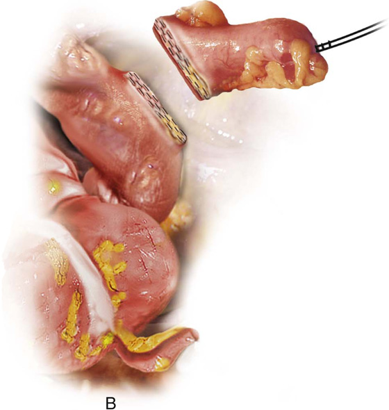
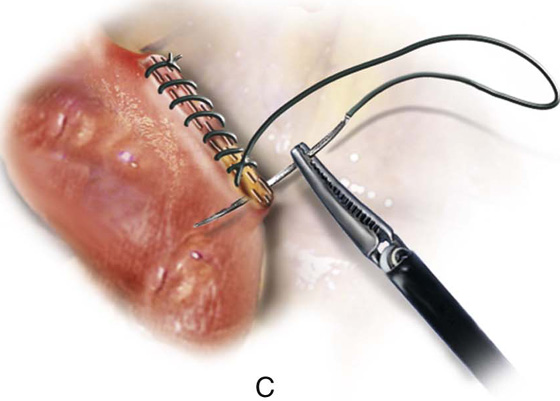
FIGURE 96–4 The base of the diverticulum is cross-clamped by a stapling device. The diverticulum is stapled and cut. The staple line may be oversewn and imbricated with 2-0 silk.


