Maternal medicine

Published on 09/03/2015 by admin

Filed under Obstetrics & Gynecology

Last modified 22/04/2025

Print this page

rate 1 star rate 2 star rate 3 star rate 4 star rate 5 star
Your rating: none, Average: 0 (0 votes)

This article have been viewed 1271 times

Maternal medicine

Suzanne V.F. Wallace, Henry G. Murray and David James

Introduction

Maternal medicine encompasses the spectrum of medical conditions a woman can present with in pregnancy. Some of these may predate pregnancy and others may develop during pregnancy. Currently in the UK, with improvements in other areas of obstetric care, most maternal deaths are now caused by medical conditions. In the most recent edition of the Confidential Enquiry into Maternal Deaths (2006–2008) venous thromboembolism was responsible for maternal death in 0.79 per 100 000 maternities, whilst cardiac disease was responsible for maternal death in 2.31 per 100 000 maternities.

There are an increasing number of women with medical conditions in pregnancy. Women with significant pre-existing medical conditions that in the past may have led to voluntary or involuntary infertility (for example, cystic fibrosis) are now becoming pregnant in increasing numbers. In addition, more women are older when embarking on pregnancy and have more acquired problems such as obesity and hypertension.

Whether a woman is known to have a medical condition prior to pregnancy or develops one within pregnancy, the key to successful management is to have a framework to ensure that all the implications of the condition are considered (Fig. 9.1). This enables robust pregnancy plans to be made whether a disease is common or rare. Multi-disciplinary and multi-professional team-working are also essential elements in caring for these women.

Minor complaints of pregnancy

Minor complaints of pregnancy by definition do not cause significant medical problems. However, minor medical complaints are often not perceived as minor by the women affected and can have considerable impact on a woman’s quality of life in pregnancy. In addition, many of the symptoms of minor conditions of pregnancy are the same as those of significant pathological disease that needs to be excluded. Symptoms frequently relate to physiological adaptations of the body to pregnancy and women should be reassured (once pathology has been excluded) that these symptoms represent normal pregnancy changes.

Abdominal pain

Abdominal pain or discomfort is common in pregnancy and is usually transient. The physiological causes include:

It is important to differentiate physiological abdominal pain from pathological causes in women with severe, atypical or recurrent pain. These include:

Social causes, particularly domestic abuse, should be considered in women who present with recurrent episodes of abdominal pain where organic pathology has been excluded.

Once a pathological cause has been excluded reassurance is often a successful management option and analgesics are rarely required.

Heartburn

Gastro-oesophageal reflux is more likely to occur in pregnancy because of delayed gastric emptying, reduced lower oesophageal sphincter pressure and raised intragastric pressure. It affects up to 80 % of pregnant women, particularly in the third trimester. The differential diagnoses are:

Conservative management includes dietary advice to avoid spicy and acidic foods and to avoid eating just prior to bed. Symptoms can be improved by changing sleeping position to a more upright posture. If conservative measures are not successful antacids are safe in pregnancy and can be used at any time. Histamine-receptor blockers, such as ranitidine, and proton pump inhibitors have a good safety profile in pregnancy and can be used if antacids alone are insufficient to improve symptoms.

Pelvic girdle dysfunction (symphyseal pelvic dysfunction, SPD)

Raised levels of relaxin in pregnancy increase joint mobility to allow expansion of the pelvic ring for birth. However, in some women this effect can be exaggerated and cause discomfort either at the symphysis, the hip or at other points around the pelvis; this usually worsens with increasing gestation. Women often describe characteristic pain on walking or standing with tenderness over the pelvic ring. Urinary tract infection should be excluded with anterior pain.

Physiotherapists will advise on exercises to improve stability, techniques for minimizing symptoms during daily activities and positions for birth. A pelvic girdle support may improve symptoms. As with back pain, simple analgesics can be used. The problem usually resolves after pregnancy

Medical problems arising in pregnancy

Anaemia

Anaemia commonly occurs in pregnancy. While in many developed countries it is mild and quickly and easily treated resulting in minimal complications, in some countries it is severe and a major contributor to maternal death.

Aetiology

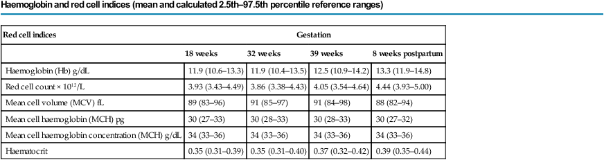
Pregnancy causes many changes in the haematological system, including an increase in both plasma volume and red cell mass; the former is greater than the latter with the result that a ‘physiological anaemia’ often occurs. There is an increased iron and folate demand to facilitate both the increase in red cell mass and fetal requirements, which is not always met by maternal diet. Iron deficiency anaemia is thus a common condition encountered in pregnancy, particularly in the third trimester. Table 9.1 shows the changes of haemoglobin and red cell parameters in normal pregnancy.

Table 9.1

Haemoglobin and red cell indices (mean and calculated 2.5th–97.5th percentile reference ranges)

Haemoglobin and red cell indices (mean and calculated 2.5th–97.5th percentile reference ranges)

Red cell indices Gestation
  18 weeks 32 weeks 39 weeks 8 weeks postpartum
Haemoglobin (Hb) g/dL 11.9 (10.6–13.3) 11.9 (10.4–13.5) 12.5 (10.9–14.2) 13.3 (11.9–14.8)
Red cell count × 1012/L 3.93 (3.43–4.49) 3.86 (3.38–4.43) 4.05 (3.54–4.64) 4.44 (3.93–5.00)
Mean cell volume (MCV) fL 89 (83–96) 91 (85–97) 91 (84–98) 88 (82–94)
Mean cell haemoglobin (MCH) pg 30 (27–33) 30 (28–33) 30 (28–33) 30 (27–32)
Mean cell haemoglobin concentration (MCH) g/dL 34 (33–36) 34 (33–36) 34 (33–36) 34 (33–36)
Haematocrit 0.35 (0.31–0.39) 0.35 (0.31–0.40) 0.37 (0.32–0.42) 0.39 (0.35–0.44)

image

(Reproduced with permission from Shepard MJ, Richards VA, Berkowitz RL, et al (1982) An evaluation of two equations for predicting fetal weight by ultrasound. Am J Obstet Gynecol 142:47–54. © 1982 Elsevier.)

Implications on pregnancy

Iron-deficiency anaemia mainly affects the mother. With mild anaemia the fetus is usually unaffected despite the reduced oxygen-carrying capacity of the mother. However the baby is more likely to have iron deficiency in the first year of life because of a lack of development of fetal iron stores in utero. With severe anaemia there is an increased risk of preterm birth and low birth weight.

The implications to the mother in all trimesters are the risk of developing symptomatic anaemia that can cause fatigue, reduced work performance and an increase in susceptibility to infections. If anaemia persists to the time of delivery, there will be a lack of reserve if significant blood loss occurs. There is a strong association between severe anaemia and maternal mortality. The risk of requiring blood transfusions peripartum is also raised.

Management

Prompt recognition and treatment of developing anaemia optimizes a woman’s haemoglobin levels in pregnancy and reduces the risk of commencing labour anaemic.

Although most anaemia in pregnancy is secondary to iron deficiency, consideration should be given as to whether there is an underlying anaemia condition or if folate deficiency could also be involved. If there is clinical suspicion that iron deficiency is not the cause of the anaemia or if a woman has failed to respond to iron supplementation then the iron status should be assessed by either ferritin or zinc protoporphyrin levels, folate measured and haemoglobin electrophoresis performed to exclude haemoglobinopathies. Oral iron supplementation is recommended as first line treatment. This is better absorbed if taken with ascorbic acid (for example, orange juice) and if tea and coffee are avoided at the time of ingestion. Dietary advice should also be given. Compliance with iron supplementation is often poor due to the side effects of constipation and gastric irritation. If iron is either not tolerated or if improvements in haemoglobin are not seen despite iron therapy then parenteral iron can be considered.

Adequate continued postnatal treatment is essential to reduce the risk of a woman entering a further pregnancy anaemic.

Gestational diabetes

Gestational diabetes is an increasingly common antenatal condition occurring in 2–9 % of all pregnancies.

Risk factors

Risk factors are the same as those for type 2 diabetes and are listed in Box 9.1. This list is taken from the NICE Clinical Guideline ‘Diabetes in pregnancy’ (2008). The Guideline only recommends that some of the risk factors should be used for screening in practice. The presence of one or more of these risk factors should lead to the offer of a75 g oral glucose tolerance test (OGTT). However, many clinicians feel that the presence of any risk factor, rather than a subset, should trigger the offer of an OGTT.

Box 9.1

Women at increased risk of glucose intolerance in pregnancy

• Previous large infant (more than 4.5 kg, or above the 95th centile for gestational age)*

• Previous gestational diabetes*

• First-degree relative with diabetes*

• Obesity (BMI more than 30 kg/m2)* or booking weight more than 100 kg

• Specific ethnic background:

• Macrosomia in current pregnancy (variably defined in different studies, e.g., fetal abdominal circumference measured with ultrasound >90th centile, or fetal weight estimated using formulae based on ultrasound measurements)

• Glycosuria ≥1+ on more than one occasion or ≥2+ on one occasion

• Previous unexpected perinatal death

• History of polycystic ovary syndrome

• Polyhydramnios

• FBG more than 6.0 mmol/L or random blood glucose more than 7.0 mmol/L

Key: * Risk factors in bold are those that the NICE Clinical Guideline recommends should be used for screening in practice during pregnancy in the form of a 75 g oral glucose tolerance test.

(National Institutes for Health and Clinical Excellence (2008) Diabetes in pregnancy: Management of diabetes and its complications from pre-conception to the postnatal period. NICE Publication Guideline 63, p 1–38.)

Clinical features and diagnosis

Gestational diabetes may be asymptomatic. As such, a screening programme needs to be in place that can either be universal or selective. Most units prefer a selective approach for practical and financial reasons. Selective screening is offered to the at-risk groups listed in Box 9.1. As described above, screening is by an 75 g OGTT at 28 weeks, or if very high risk, early in the second trimester and then repeated at 28 weeks (if normal at the first test). In the OGTT, a fasting glucose level is first measured, then a 75 g loading dose of glucose is given and a further glucose level taken at 2 hours post-sugar load. There is an ongoing debate as to the levels of glucose at which gestational diabetes should be diagnosed. Table 9.2 indicates the two most commonly used diagnostic criteria.

Table 9.2

Diagnostic criteria for gestational diabetes using a 75g oral glucose tolerance test

Diagnostic criteria Normal fasting value (venous plasma glucose) Normal 2 hour value (venous plasma glucose)
WHO 1999a One or more abnormal values required <7.0 mmol/L <7.8 mmol/L
IADPSGb One or more abnormal values required <5.1 mmol/L <8.5 mmol/L

image

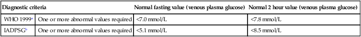
Comment: In a given population, use of the IADPSG criteria results in more diagnoses of ‘gestational diabetes’ than using the WHO criteria. WHO, World Health Organization; IADPSG, International Association of Diabetes and Pregnancy Study Groups.

aWorld Health Organization: Definition, Diagnosis and Classification of Diabetes Mellitus and its Complications: Report of a WHO Consultation. Part 1: Diagnosis and Classification of Diabetes Mellitus. Geneva, World Health Organization, 1999.

bMetzger, B.E., Gabbe, S.G., Persson, B., et al. International association of diabetes and pregnancy study groups recommendations on the diagnosis and classification of hyperglycemia in pregnancy. Diabetes Care 2010;33:676–682.

Implications on pregnancy

Gestational diabetes is predominantly a disease of the third and sometimes second trimester (Table 9.3). In the mother, the presence of gestational diabetes increases the risk of recurrent infections and of pre-eclampsia developing. For the fetus there is increased risk of polyhydramnios and macrosomia, the latter being related to the degree of glucose control. There is an increased risk of stillbirth. Considering birth, women with gestational diabetes are more likely to have an induction of labour. If vaginal birth occurs, shoulder dystocia, an instrumental birth and extended perineal tears are more common. Women with diabetes are more likely to have a caesarean section. Babies are more likely to need admission to the neonatal unit. They are at increased risk of neonatal hypoglycaemia due to the relative over-activity of the fetal pancreas in utero. This is less likely to occur if maternal blood sugars are well controlled around the time of birth. Maternal glucose readily crosses the placenta whilst insulin does not.

Table 9.3

Effect of gestational diabetes on pregnancy

  Maternal risks/complications Fetal/neonatal risks/complications
First trimester
Second trimester
Third trimester
Pre-eclampsia
Recurrent infections
Macrosomia
Polyhydramnios
Stillbirth
Labour Induction of labour
Poor progress in labour
 
Delivery Instrumental birth
Traumatic delivery
Caesarean section
Shoulder dystocia
Postnatal   Neonatal hypoglycaemia
Neonatal unit admission
Respiratory distress syndrome
Jaundice
Longer term Type 2 diabetes later in life Obesity and diabetes in childhood and later life

Whilst for the majority of women gestational diabetes will resolve post-pregnancy, in some women this diagnosis is the unmasking of type 2 diabetes and diabetic care will need to continue. Women who have had gestational diabetes remain at higher risk of developing type 2 diabetes later in life. For the babies, fetal programming effects increase the risk of obesity and diabetes in later childhood.

Management

Multi-disciplinary teams consisting of obstetricians, diabetic physicians, diabetic specialist nurses and midwives and dieticians should manage diabetes in pregnancy.

Antenatally, the aim is to reduce the risk of complications by achieving good glucose control. Initially, this is by dietary measures aiming to avoid large fluctuations in glucose levels: consuming increased amounts of low glycaemic index carbohydrate and lean protein and avoiding high glycaemic index carbohydrate foods. If this is unsuccessful then medication can be used. Metformin and glibenclamide are increasingly used in pregnancy and may reduce the need for insulin, but a number of women with gestational diabetes will need insulin to optimize control. The aim is that preprandial/fasting capillary glucose levels should be between 4.0 and 6.0 mmol/L and that 2-hour post-prandial value should be between 6.0 and 8.0 mmol/L. Randomized-controlled trial evidence has demonstrated that treatment of gestational diabetes with the aim of achieving normoglycaemia in the woman improves outcomes. Serial growth scans are advised to alert to increasing macrosomia.

Delivery at term is recommended to reduce the risk of stillbirth. This may need to be brought forward depending on the degree of diabetic control, the presence of macrosomia or if other conditions have arisen, such as pre-eclampsia. In labour, blood glucose should be regularly measured and hyperglycaemia treated to reduce the risk of neonatal hypoglycaemia. The fetus should be continuously monitored. Diabetic therapy can be discontinued with the delivery of the placenta. The baby will need blood glucose measurements to look for hypoglycaemia, and feeding should be commenced early to assist the baby in maintaining its sugar level.

Postnatally, all diabetic treatment should be discontinued and capillary glucose testing continued. In the majority of women these values will be normal, indicating that this was genuine gestational diabetes. If they remain elevated then there is a suspicion of type 2 diabetes and referral to a diabetic team is indicated. Women should be advised of the long-term implications of gestational diabetes and the need for regular screening by, for example, an annual OGTT by their general practitioner. Advice on reducing other lifestyle risks associated with diabetes may also be appropriate.

Infections acquired in pregnancy

Women will encounter infections in pregnancy just as they would outside of pregnancy. However, the relative immunosuppressive conditions of pregnancy can affect the way the body responds to the infection.

Chicken pox

Chicken pox is a highly infectious childhood illness caused by Varicella zoster virus; it has significant implications on both the mother and fetus. Pregnant women are particularly at risk of developing a varicella pneumonia that has a high maternal and fetal mortality rate. If acquired early in pregnancy, there is a 1–2 % risk to the fetus of congenital varicella syndrome (eye defects, limb hypoplasia and neurological abnormalities). If acquired near term there is a risk of neonatal varicella that has a significant mortality risk.

If a non-immune pregnant woman is exposed to chicken pox, she can be offered zoster immunoglobulin to reduce the risk of infection. If a woman becomes infected, aciclovir should be given to reduce the risk of maternal complications. Ultrasound imaging can screen for congenital varicella syndrome. With infection at term, delivery should ideally be delayed to allow time for passive transfer of antibodies to the fetus. Care should be taken to avoid contact with other non-immune pregnant women.

Parvovirus B19

Infection with parvovirus B19 is also known as erythema infectiosum, fifth disease or ‘slapped cheek syndrome’. A common childhood illness, maternal symptoms can include fever, rash and arthropathy, but often effects are minimal. In contrast, there are potentially significant fetal effects as parvovirus infects rapidly dividing cells and can cause miscarriage in early pregnancy and fetal anaemia and heart failure (‘fetal hydrops’) later in pregnancy.

Management includes the use of simple analgesics and antipyretic agents for the maternal symptoms and avoidance of contact with other pregnant women. If the infection is contracted after 20 weeks, serial Doppler ultrasound scanning of the blood flow in the fetal middle cerebral artery can detect fetal anaemia (blood flow increased) that may need to be treated with in utero blood transfusions.

Human Immunodeficiency Virus (HIV) Infection

HIV is a virus that weakens the immune system and over time AIDS (acquired immune deficiency syndrome) may develop. HIV also increases the risk of catching other infections and developing cancers. However, people with HIV infection may be asymptomatic for many years. The number of people living with HIV worldwide is increasing and a significant proportion of these are women of reproductive age. With advancing disease, highly active antiretroviral therapy (HAART) has been shown to reduce morbidity and mortality from HIV infection.

Implications of the disease on pregnancy

The main concern in pregnancy is the high risk of vertical transmission (up to 45 %) of HIV from mother to baby without medical intervention. This can occur transplacentally in the antenatal period, during vaginal birth and postnatally through breastfeeding. The risk is highest in advanced disease, at seroconversion and with high viral loads. In women who do not breastfeed, transmission rates fall to less than 25 %. With medical intervention in the form of multiple anti-retroviral therapy it is possible to reduce vertical transmission further to less than 2 %.

In addition there are increased risks of miscarriage, fetal growth restriction, prematurity and stillbirth in women with advanced HIV disease.

Some women will already be on HAART prior to pregnancy and this should be reviewed to consider the safety of individual medications in pregnancy. Many women will be treatment naïve.

Women who are taking HAART and have viral loads less than 400 copies/mL can deliver vaginally as there is a very low risk of vertical transmission. However, those who are not taking HAART and/or have viral loads of ≥400 copies/mL or more should be advised to have a caesarean section to reduce the risk of vertical transmission.

Management

Women with HIV who become pregnant should be managed jointly by a specialist obstetrician and HIV physician. Input from the paediatric team should occur antenatally to discuss neonatal screening and treatment.

Women should be regularly assessed clinically and with blood measurements of viral load and CD4 count.

The initial package of care for women with HIV in pregnancy involves anti-HIV medication, caesarean section and avoiding breastfeeding. The use of anti-HIV drugs in pregnancy has been shown to reduce the risk of vertical transmission. Some women will already be taking HAART for their own health needs and this should continue. In treatment naïve women, anti-HIV medication should commence in the second trimester and continue until birth. Regimes used include zidovudine monotherapy and HAART (nucleotide analogues and protease inhibitors appear relatively safe, non-nucleoside reverse transcriptase inhibitors should be avoided). However, HAART is the recommended treatment of choice. Whilst caesarean section is still advocated for women with non-suppressed disease, women with a viral load of <400 copies/ml who have taken HAART in pregnancy can now opt for vaginal birth without increasing transmission. Invasive procedures should be avoided in pregnancy and labour, for example amniocentesis, the use of fetal scalp electrodes and fetal scalp blood sampling.

Neonatal screening for HIV infection commences at birth and continues until 12 weeks. Babies require neonatal antiretroviral treatment as postexposure prophylaxis for several weeks. Women should be strongly advised not to breastfeed.

Confidentiality is an issue for some women with HIV whose families may not know their status. Women should be reassured that confidentiality can and will be maintained despite the increased medical intervention.

Acute viral hepatitis

Seven hepatitis viruses have been identified, the most common being hepatitis A, B and C. All can present similarly with general malaise, nausea, vomiting and pyrexia together with hepatic dysfunction; however with hepatitis B and C a significant proportion can be asymptomatic (up to 80% of women with hepatitis C). Hepatitis A is spread by the faeco-oral route while B and C are transmitted by a blood-borne route. They can be differentiated by serological tests. Hepatitis A is usually cleared after the initial infection, hepatitis B can be cleared, can persist as a carrier state or can lead to chronic infection, and hepatitis C commonly leads to chronic infection and a long-term risk of cirrhosis and liver failure.

The incidence of hepatitis in pregnancy has a wide geographical variation. In the UK, 1–4% of women will be infected with hepatitis B or C.

Pregnancy does not usually change the course of an acute hepatitis infection. A small number of chronic hepatitis B carriers may suffer a reactivation of the disease state during pregnancy. There is some evidence that pregnancy in women with hepatitis C may cause acceleration of the disease progression.

Hepatitis usually does not impact on the pregnancy itself. In women who have a severe acute infection during pregnancy, there is an increase in the incidence of spontaneous preterm labour. The main concern is the risk of transmission to the neonate. With hepatitis A this can happen if acute infection occurs in the last couple of weeks before delivery. With chronic hepatitis B and C, carriage transmission can occur perinatally. In women with chronic hepatitis C, vertical transmission will occur in 1 in 20 births.

Management in pregnancy relates to prevention, identification and reduction of the risk of vertical transmission. The risk of hepatitis A infection can be reduced by hygiene measures and consideration of immunization for women in areas of endemic hepatitis A infection. Women at risk of hepatitis B and C should be counseled regarding risk-taking behaviour (particularly, intravenous drug use). Hepatitis B immunization can be offered before pregnancy, however there is currently no effective immunization against hepatitis C.

Women can be screened for hepatitis B and C in pregnancy. This may be universal or selective screening based on a woman’s history. Identification antenatally is important to reduce vertical transmission. In women with hepatits C, co-infection with HIV should be excluded.

Vertical transmission of hepatitis B and C is not reduced by either caesarean delivery or avoidance of breast-feeding. Thus vaginal delivery is advocated (unless there are other obstetric indications for caesarean delivery), but with avoidance of interventions that may increase blood contact, such as fetal scalp electrode siting or fetal blood samples. Babies of mothers with hepatitis B can be treated with hepatitis B immunoglobulin and early hepatitis B immunization, which reduces transmission rates to 5–10%. There are limited options to reduce transmission rates with hepatitis C, but early identification of infected neonates ensures adequate follow up for the risk of chronic liver disease.

Tuberculosis

Tuberculosis remains a world health issue with at least 8 million new cases per year and up to 2 million deaths. Although the developed world has low rates of infection, higher rates are found in refugees and travelers to and from endemic areas. The two main risks to the fetus are the use of certain anti-tuberculous drugs and if the mother has severe respiratory illness with sustained hypoxia. Mycobacterium tuberculosis rarely crosses the placenta. The risks to the woman from untreated TB are the same as in non-pregnant patients. Tuberculin testing should be undertaken if the disease is suspected. Chest X-ray and sputum culture should be performed in those who test positive. If the diagnosis is confirmed then multiple therapy, as in the non-pregnant patient, is indicated. Streptomycin is the only drug that is absolutely contraindicated in pregnancy because of the risk of fetal ototoxicity.

Malaria

Malaria occurs in over 200 million people per year and results in more than 1 million deaths annually. It is a common complication of pregnancy in those countries where the disease is endemic. Women who live in endemic areas show an increased prevalence of the severe forms of the disease. The severity of disease is related to the species of parasite, the level of parasitaemia and the immune status of the individual. Plasmodium falciparum is the most virulent of the organisms, as it attacks all forms of the erythrocyte. The parasite grows in the placenta and placental malaria occurs in anywhere between 15 and 60 % of cases. Congenital malaria is rare in the infants born to mothers who have immunity as protective immunoglobulin G crosses the placenta.

The main risk of acute malaria to the woman is severe anaemia and its consequences. In the fetus, acute malaria is associated with an increased likelihood of growth restriction, miscarriage, preterm birth, congenital infection and perinatal death.

Mothers travelling to endemic areas should take prophylaxis or, preferably, not go to the area until the pregnancy is completed. They should also be advised to keep their skin covered and to use insecticides to minimize the risk of being bitten by mosquitoes.

Drug treatment of an acute attack will depend on the nature of the infection. Prophylaxis is given in the form of chloroquine phosphate at a dose of 300 mg each week, starting 1 week before travel and continuing for 4 weeks after leaving the area. Where chloroquine-resistant strains exist, a combination of chloroquine and pyrimethamine with sulfadoxine can be used, or proguanil and mefloquine. These drugs need to be taken with a folic acid supplement. Although chloroquine can cause retinal and cochleovestibular damage in high doses in both the mother and the fetus, it has never been shown to be associated with an increased incidence of birth defects where it has been taken for prophylaxis.

Acute pyelonephritis and urinary tract infections

Asymptomatic bacteriuria occurs in 2–10 % of all sexually active women. When pregnant, 12–30 % of this group of women will develop pyelonephritis from ascending infection due to structural and immune changes to the renal tract. If the bacteriuria is treated with antibiotics, the risk of later development of acute ascending urinary tract infection can be minimized. Nevertheless, approximately 1 % of all pregnancies are complicated by an episode of acute pyelonephritis. The common organism is Escherichia coli and this should be treated aggressively with antibiotics according to known sensitivity. Most community-acquired infections are usually sensitive to amoxicillin or cefuroxime. Additional treatment with fluid replacement, pain relief and bed rest may also be of benefit. Pyelonephritis in pregnancy must not be underestimated as over 15 % of women will develop a bacteraemia, with a small proportion of these progressing to septic shock and/or preterm labour.

Thromboembolic disease

Venous thromboembolism (VTE) is one of the leading causes of maternal mortality in the developed world. VTE is around 10 times more common in pregnancy than when not pregnant.

Clinical features and diagnosis

VTE presents as in non-pregnant individuals: deep vein thrombosis (DVT) with swelling and tenderness of a leg and pulmonary embolism (PE) with respiratory symptoms (shortness of breath and pleuritic chest pain) or collapse. In pregnancy, DVT is more likely to occur on the left (90 %) due to compression of the left common iliac vein by the right common iliac artery and ovarian artery (the vein is not crossed on the right). The majority of DVTs in pregnancy occur in the ileofemoral veins, so lower limb symptoms may not be as obvious. Clots in the ileofemoral veins are more likely to embolise than those in the calf.

D-Dimer measurements are of limited help in pregnancy, although the negative predictive value is high, a positive result does not help to establish a diagnosis as it can increase with the physiological changes in the coagulation system that occur in pregnancy.

Radiological investigations should be performed as in non-pregnant individuals. Doppler ultrasound of the lower limb veins or MRI of the pelvic veins should be performed to assess for DVT. Spiral artery CT or venous perfusion scanning are used to diagnose PE. Although care must be taken when undertaking radiological examinations in pregnancy because of the radiation risk to the fetus, ultimately if an investigation needs to be done to establish a diagnosis it should be done.

Management

All women should have an individual risk assessment undertaken for VTE in pregnancy. Depending on the risk score, a plan for thromboprophylaxis can be made to reduce the risk of thrombosis occurring. For women with low risk this may not require additional measures; for some women postnatal prophylaxis may be required and for those at highest risk antenatal prophylaxis may be recommended with graduated elastic compression stockings and low molecular weight heparin (LMWH). In this last scenario, thromboprophylaxis should be started as early as possible in pregnancy.

If acute VTE occurs in pregnancy, prompt management is vital and heparin therapy should be commenced empirically whilst awaiting investigations. If a DVT or PE are excluded on subsequent diagnostic testing (see above) then the heparin can be discontinued.

LMWH has been used extensively in pregnancy as it does not cross the placenta and has a good safety profile. In contrast, warfarin crosses the placenta and is associated with an embryopathy if used in the first trimester and with fetal intracranial bleeding when used in the third trimester. Most women with venous thromboembolism can be adequately managed on LMWH so warfarin is rarely needed in this scenario. On most occasions heparin therapy can be temporarily stopped around the time of birth to reduce the risk of postpartum haemorrhage and to enable regional anaesthesia to be given if required (LMWH use is associated with epidural haematoma). Simple measures such as avoiding dehydration and using graduated elastic compression stockings should also be employed.

Postnatally, women traditionally continue on heparin prophylaxis or treatment for 6 weeks. If an acute venous thromboembolic event has occurred in this pregnancy it is likely that heparin prophylaxis will be needed in future pregnancies. Women should also be advised to avoid oestrogen-containing contraceptives.

Liver disease

Obstetric cholestasis

Management

The pruritus of obstetric cholestasis can be difficult to treat. Topical emollients are safe for use in pregnancy but provide little symptomatic relief. Antihistamines are sometimes used for their sedative ability but have little impact on the itch itself. Ursodeoxycholic acid has been shown to improve both pruritus and liver function, but long-term safety data is lacking. In spite of this it is the mainstay of antenatal treatment. In view of the potential risk of clotting abnormalities, oral water-soluble vitamin K supplementation can be used, particularly for those women whose clotting tests suggest an abnormality.

The best way to monitor the fetus antenatally has not yet been established. Methods such as serial growth ultrasound scans and cardiotocographs (CTGs) that can detect problems with placental function are not predictive of at-risk fetuses in obstetric cholestasis. Consequently delivery once fetal maturation is reached is often recommended to reduce the small risk of late stillbirth.

Postnatally, women are usually advised to avoid oestrogen-containing contraceptives which can precipitate further symptoms.

Acute fatty liver of pregnancy

Pre-existing medical conditions and pregnancy

An increasing number of women are now entering pregnancy with pre-existing medical conditions. Ideally these women should be offered preconceptual counselling to allow the implications of pregnancy with their specific medical condition to be discussed and a plan put in place. This may involve deferring pregnancy until a specific target in the disease management is met. However, this opportunity is frequently missed.

Renal disease in pregnancy

Pregnancies complicated by chronic renal disease are rare (0.15%), however they are associated with a significant risk of adverse maternal and fetal outcomes. In the majority of cases, the risks and management relate to the degree of renal impairment and not to the underlying cause of the renal disease.

Implications of pregnancy on the disease

In women with chronic renal disease, pregnancy can cause a deterioration of renal function. Mostly this will recover after the end of the pregnancy, but for some women this will lead to a permanent reduction in renal functioning and a shorter time to end stage renal failure. The likelihood of renal deterioration depends on baseline creatinine as shown in Table 9.4.

Table 9.4

Maternal renal function and chronic renal disease in pregnancy

  Serum creatinine (mmol/L) Loss of >25% renal function in pregnancy (%) Deterioration of renal function post-partum (%)
Mild renal impairment <125 2 0
Moderate renal impairment 124–168 40 20
Severe renal impairment >177 70 50

image

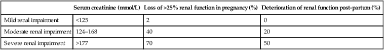
(Data from Williams D, Davidson J. Chronic kidney disease in pregnancy. Br Med J 2008;336:211–215.)

Management

Women with chronic renal disease should ideally be seen for pre-pregnancy counseling to discuss the implications of a potential pregnancy so that informed decisions can be made. For some women the risk of deterioration to end stage renal failure and a requirement for dialysis will be too great.

Pregnant women with renal disease should be offered care in multidisciplinary clinics that include an obstetrician and a renal or obstetric physician. Initial review should involve assessment of baseline renal function, blood pressure and proteinuria. Low dose aspirin (75 mg) from 12 weeks until delivery should be offered to reduce the risk of pre-eclampsia. Women already on anti-hypertensive treatment may need their medications reviewed to ensure that they are appropriate for pregnancy. Careful surveillance of blood pressure, renal function and for urinary tract infection is required throughout pregnancy. Growth scans should be arranged in the third trimester to assess fetal growth. For women with proteinuria, prophylactic heparin may be required to reduce the risk of venous thromboembolism. All women are at increased risk of urinary tract infections, women with chronic renal disease and the presence of more than one confirmed urinary tract infection may benefit from the use of prophylactic antibiotics.

Diabetes mellitus

Diabetes is one of the commonest pre-existing medical conditions women are seen with in pregnancy. The incidence of pregnant women with pre-existing diabetes is around 0.4 %. The majority of these have type 1 diabetes; however, with changes in population demographics there are an increasing number of women with type 2 diabetes. Women with type 2 diabetes tend to be older, be more obese and have more unplanned pregnancies than women with type 1 diabetes. Rates of complications in pregnancy are similar in both groups of women.

Implications of the disease on pregnancy (Table 9.5)

Many of the complications seen with gestational diabetes are also seen with women with pre-existing diabetes. However, there are additional complications related to abnormal glucose homeostasis in the periconception period and in the first trimester, as well as complications related to long-standing underlying vascular disease.

Table 9.5

Risks of pre-existing diabetes in pregnancy

  Maternal concerns Fetal/neonatal concerns
First trimester Increased insulin requirements Miscarriage
Fetal abnormality
Second trimester
Third trimester
Pre-eclampsia
Recurrent infections
Worsening retinopathy if vascular disease
Macrosomia
Polyhydramnios
Stillbirth
Growth restriction
Labour Induction of labour
Poor progress in labour
Preterm delivery
Delivery Instrumental birth
Birth trauma caesarean section
Shoulder dystocia
Postnatal Return to pre-pregnancy control within hours of birth Neonatal hypoglycaemia
Neonatal unit admission
Respiratory distress syndrome
Jaundice
Longer term Diabetes in childhood (2–3 % if mother has type 1; 10–15 % if mother has type 2)

There is an increased risk of congenital abnormality in women with pre-existing diabetes, particularly neural tube defects and congenital heart disease; the likelihood of this occurring is related to the level of glycaemic control periconceptionally and in early pregnancy. Women with an HbA1c above 10 % have up to a 25 % chance of a fetal abnormality being present. Women are at an increased risk of fetal loss throughout pregnancy; again this is related to glycaemic control.

Although fetal macrosomia is the most common fetal growth pattern in diabetes, in women with pre-existing vascular disease and those who develop early pre-eclampsia, fetal growth restriction can be a problem.

Women with hypertension and diabetic nephropathy are at high risk of developing pre-eclampsia (approximately 30 %).

Management

Preconception counselling in diabetic women enables a woman to be informed about pregnancy and diabetes and also allows women to consider the best time to try to conceive. As many of the complications of diabetes relate to the level of glycaemic control, the aim is to get the HbA1c less than 6.1 % before conception. If this is achieved the complication rate of pregnancy in women with diabetes is not much greater than the normal population. Medications can be reviewed. Insulins, both traditional and the newer agents, have been shown to be safe in pregnancy. Metformin is usually continued but other oral hypoglycaemic agents usually stopped. Consequently many women with type 2 diabetes will require insulin in pregnancy. However, some of the medications used to treat the complications of diabetes are not safe, for example, angiotensin-converting enzyme (ACE) inhibitors used in the treatment of diabetic nephropathy should be stopped in pregnancy. Women with diabetes should take a higher (5 mg rather than the normal 400 µg) periconceptual dose of folic acid in view of the increased risk of neural tube defects.

Multi-disciplinary team working is key in managing women with diabetes. Obstetric diabetes clinics will often consist of an obstetrician, endocrinologist, diabetes specialist nurse, dietician and specialist midwife. Women will be seen regularly throughout pregnancy by this team.

The metabolic goal during pregnancy is to maintain blood glucose as close to the normal range as possible, while avoiding severe hypoglycaemia. This involves increasing capillary blood glucose monitoring and tightening control more than is usual outside of pregnancy. The target levels are the same as given in gestational diabetes section (above). Because women are encouraged to keep glucose control tight they can experience unpleasant attacks of hypoglycaemia. Various measures (oral glucose preparations and/or IM glucagon) need to be in place to deal with this complication.

Fetal assessment for abnormalities involves combined testing for chromosomal problems (if the mother wishes) in the first trimester and a routine anatomy scan at 20 weeks. Additional scanning to look at cardiac anatomy is sometimes recommended in view of the increased risk of congenital cardiac disease. Regular serial growth scans can detect both macrosomia and fetal growth restriction.

For maternal wellbeing, low dose aspirin from the second trimester can reduce the risk of pre-eclampsia developing. In women with vascular disease care should be taken to keep blood pressure well controlled to reduce the risk of disease deterioration. All women with pre-existing diabetes should have an ophthalmic assessment in each trimester for evidence of development of worsening of diabetic retinopathy.

Women with diabetes should give birth in a hospital with neonatal facilities. Delivery plans will depend on the stability of diabetes in pregnancy and maternal and fetal wellbeing; however, delivery at around 38–39 weeks is usually recommended. Vaginal birth is often planned but caesarean section rates are high in this group of women. The fetus should be monitored continuously. The neonates are at risk of neonatal hypoglycaemia. This risk can be reduced by strict glycaemic control in labour and a sliding scale infusion of insulin–dextrose is often required to achieve this control in women with pre-existing diabetes.

Postnatally, women return to pre-pregnancy treatment regimens as soon as they are delivered and eating and drinking.

Thyroid disease in pregnancy

Thyroid disorders of various types complicate approximately 1–1.5 % of pregnancies. Increased oestrogen in normal pregnancy leads to an increase in thyroid-binding globulin that necessitates an increased production of thyroid hormone to maintain free T4 and T3 levels. These changes, along with a fall in iodine levels in the maternal plasma due to increased renal loss, result in an enlargement of the thyroid gland of 10–20%. A fall in the thyroid-stimulating hormone (TSH) levels is also a feature of the first half of pregnancy, which may be explained by thyroid stimulatory effects of human chorionic gonadotrophin.

Hypothyroidism

Hypothyroidism is the commonest thyroid problem to occur in pregnancy and complicates around 1 % of pregnancies. Most cases have a basis in autoimmune diseases, where autoantibodies like thyroid peroxidase, and those associated with Hashimoto’s disease cause gland destruction and fibrosis. Hypothyroidism may also be iatrogenic as the consequence of thyroidectomy, radio-iodine ablation or excessive doses of antithyroid drugs.

Management

Thyroid function testing in these women should be performed every trimester, and replacement should be adjusted as T4 levels fall due to the increase in maternal extracellular fluid levels. The diagnosis of inadequate treatment/hypothyroidism in the mother is made by a raised level of TSH.

Where hypothroidism is secondary to treatment for hyperthyroidism, neonatal surveillance for neonatal thyroid dysfunction secondary to transplacental transmission of thyroid antibodies.

Iodine deficiency in many countries is endemic. Untreated it is associated with poor fetal outcomes from miscarriage, stillbirth, neonatal death and congenital abnormalities including cretinism. All pregnant women should be encouraged to ensure an adequate iodine intake in pregnancy; if necessary through supplementation. Where the mother is receiving adequate replacement therapy, the outcome for the infant is normal.

Hyperthyroidism

Hyperthyroidism occurs in approximately 0.2 % of pregnancies, 95 % of which are due to Graves’ disease. Diagnosis is made by the finding of elevated T4 and T3 levels associated with a lowered TSH level.

Obesity

The impact of obesity in pregnancy is increasing. Prevalence is dependent on the population served, though a national survey in the UK in 2009 found that in 9.3 per 100 000 maternities women had a BMI of more than 50 (UK Obesity Surveillance System). Women with obesity often feel stigmatized, however obesity is associated with significant medical problems in pregnancy that must be addressed. Obese women are more likely to suffer with co-morbidities such as hypertension, sleep apnoea, diabetes and cardiovascular disease all of which can increase further the risks of pregnancy.

Implications of obesity on pregnancy (Table 9.6)

Antenatally, obesity is associated with a number of maternal and fetal complications. In the first trimester the risk of miscarriage and congenital abnormality (especially neural tube defects) is increased, the aetiology of this has not been established. Throughout pregnancy there is an increased risk of venous thromboembolism. Later in pregnancy obese women are more likely to develop pre-eclampsia and gestational diabetes. In addition, many minor complications of pregnancy such as gastro-oesophageal reflux and pelvic girdle dysfunction are more likely to occur in women with a raised body mass index.

Table 9.6

Risks of obesity in pregnancy

  Maternal risks/complications Fetal/neonatal risks/complications
First trimester Venous thromboembolism Miscarriage
Fetal abnormality
Second trimester
Third trimester
Pre-eclampsia
Gestational diabetes
Venous thromboembolism
Macrosomia
Stillbirth
Difficulty with fetal assessment
Labour Induction of labour
Poor progress in labour
Delivery Instrumental birth
Traumatic birth Caesarean section
Anaesthetic complications (difficulties with intubation or epidural insertion)
Shoulder dystocia
Postnatal Postpartum haemorrhage
Venous thromboembolism
Neonatal unit admission
Neonatal death
Longer term Childhood obesity
Juvenile diabetes

Obesity is associated with fetal macrosomia; however, maternal adiposity can have significant impact on the ability to accurately determine fetal size both clinically and using ultrasound. There is an increased risk of stillbirth and neonatal death. In the long term, children of obese mothers are more likely to have childhood obesity and juvenile diabetes.

Obese women are more likely to have an induction of labour, to have poor progress in labour and a caesarean section. This higher rate of caesarean births in obese women is thought to be secondary to a combination of fetal macrosomia, co-morbid conditions and the hormonal effect of adipose tissue on labour.

The risks of caesarean section, both anaesthetic and obstetric, are higher in women with a higher body mass index. If vaginal birth is achieved, shoulder dystocia and extended perineal tears are more frequent. There is a higher risk of postpartum haemorrhage.

Management

Ideally, preconceptual counselling would allow women to defer pregnancy until a nearer normal body mass index is achieved, but this rarely occurs.

Women with obesity should have hospital-based care because of the associated risks in pregnancy and at birth. Support from a dietician should be offered with the aim of achieving a more healthy diet rather than weight reduction. Folic acid should be taken until 12 weeks. Some authorities recommend a higher dose (5 mg) in view of the increased risk of neural tube defects but evidence for this is lacking. A thorough assessment for other risk factors for pre-eclampsia and venous thromboembolism should be performed. Based on this aspirin to reduce the risk of pre-eclampsia or thromboprophylaxis to reduce the risk of venous thromboembolism may be appropriate. A glucose tolerance test to screen for gestational diabetes should be offered in the late second trimester.

The efficacy of routine ultrasound screening for anomalies is reduced in obese women because of poor visualization. Furthermore, although clinical assessments of fetal growth are limited by maternal habitus there is little evidence that ultrasound provides a more accurate assessment again because of poor visualization.

In view of the potential complications of labour and birth, obese women should deliver in a hospital unit. If there are no other contra-indications to vaginal birth this should be planned.

Thrombophilia

Thrombophilia can be heritable or acquired. Inherited thrombophilias are found in approximately 15 % of the Caucasian population, the most common being factor V Leiden. The most common acquired thrombophilia is antiphospholipid syndrome that is associated with a number of adverse outcomes in pregnancy. Thromophilias are responsible for 20–50 % of venous thromboembolic events in pregnancy.

Management

Women with thrombophilia should be ideally seen in a combined obstetric–haematology clinic. Input from a haematologist is key when planning care for the large spectrum of disorders with their varying risks of thrombosis and pregnancy-related morbidity. A full assessment of all risk factors for venous thromboembolism in addition to the thrombophilia will allow decisions about the appropriate prophylaxis to be made: whether this be antenatal and/or postnatal LMWH or avoiding dehydration and the use of graduated compression stockings.

As previously discussed, LMWH is safe for use in pregnancy but care should be taken to make a plan for its use around the time of birth to minimize the risks of bleeding and ensure that a woman has the full range of analgesic options available to her. In terms of the other obstetric risks of thrombophilias, with the exception of antiphospholipid syndrome, it is unclear if treatment with aspirin or heparin improves pregnancy outcome. However, for women with antiphospholipid syndrome there is some evidence that the use of aspirin (and possibly heparin) does reduce the incidence of pregnancy complications.

Epilepsy

Epilepsy affects approximately 1 % of the obstetric population.

Medications

Different anti-epileptic medications have different effects on the fetus. Sodium valproate appears to have the greatest risk and should be avoided if possible in women of reproductive age. The risks to the fetus of anti-epileptic medication include congenital abnormalities (mainly neural tube defects and congenital heart abnormalities), in utero growth restriction and more subtle long-term neurodevelopmental effects. The effects are increased when more than one anti-epileptic drug is being taken. Carbamazepine and lamotrigine are considered to be the safest anti-epileptic drugs for use in pregnancy.

Often anti-epileptic drug levels fall in pregnancy and rise in puerperium, so drug doses may need to be altered to maintain seizure control.

Some anticonvulsant medications induce vitamin K deficiency that can lead to an increased risk of haemorrhagic disease of the newborn.

The overall aim is to be on the fewest medications at the lowest dose commensurate with the epilepsy remaining controlled.

Management

Ideally women with epilepsy should be seen preconceptually for counselling about their risks in pregnancy and to review their medications. Medications may need to be altered and sometimes it is appropriate to advise delaying pregnancy until a ‘safer’ drug regimen is established. Women should be advised not to abruptly stop their medication because of fears about the fetus and it should be emphasized that the risk of uncontrolled epilepsy is likely to be greater than the risks of the medications being taken. A higher 5 mg dose of folic acid is recommended periconceptually and in the first trimester due to the increased risk of neural tube defects.

Women should be managed by a multi-disciplinary team with the aim of avoiding seizures in pregnancy. Combined screening for chromosomal disorders and anatomy scanning can be performed as normal. Serial growth scans may be required, particularly if a woman is on more than one medication. If anti-epileptic medication that induces vitamin K deficiency is being taken, vitamin K can be given to the mother in the last few weeks of pregnancy and the baby can receive intramuscular vitamin K just after birth to reduce the risk of hemorrhagic disease of the newborn.

Although women worry about seizures occurring in labour, given the tiredness and stress of this time, this is uncommon, but delivery in a hospital unit is advisable.

Women with epilepsy should be given advice antenatally and after delivery regarding safe practices when looking after their newborn, such as not bathing the baby on their own and changing the baby on the floor rather than a high changing table.

Breastfeeding is safe for women on most anti-epileptic medications.

Migraine

Headaches are common in pregnancy. The most common are migraine and those due to tension. New onset headaches, especially those associated with focal or abnormal neurological signs, impaired intellect and pain that impairs sleep, need specialist assessment.

In women who suffer migraine before pregnancy, the frequency of attacks drops by 50–80 % during pregnancy, but increases again in the puerperium. If an attack does occur the initial treatment comprises simple analgesia, avoidance of light, bed rest and various coping mechanisms. If these simple measures do not work and the migraine is persistent then more potent analgesics, β-blockers and/or tricyclic antidepressants have all been used with success. The ergot derivatives often used as prophylaxis/treatment outwith pregancy are contraindicated due to their vasoconstrictive effects.

Cardiac disease

There has been a large increase in cardiac disease in pregnancy in recent years. Although some of this is explained by women who themselves have had congenital heart disease now having children, the majority is acquired. Cardiac disease is now the main cause of indirect maternal death in the UK. A multitude of cardiac conditions can be encountered in pregnancy, including valvular lesions, congenital heart disease, cardiomyopathies, arrhythmias and ischaemic heart disease.

Implications of pregnancy on the disease

Pregnancy puts a great strain on the maternal cardiovascular system. The necessary rise in cardiac output can result in deterioration of some conditions, such as aortic stenosis, as these women have a fixed cardiac output. In other conditions, such as regurgitant lesions, pregnancy can be well tolerated.

Many symptoms of cardiac disease are also symptoms of pregnancy, such as breathlessness, palpitations and syncope; cardiovascular signs are also mimicked by pregnancy (bounding pulse, systolic murmur) and as a result it can be difficult to diagnose a new cardiac condition or deterioration in a known cardiac condition.

Depending on the underlying heart condition, women can be at risk in pregnancy of the following conditions:

Management

Multi-disciplinary management between obstetricians, cardiologists and obstetric anaesthetists should ideally start at the preconception phase. For some women with poor cardiac functional status pregnancy may not be advisable. The risk of maternal death can be extremely high in some conditions, for example, in women with Eisenmenger’s syndrome maternal death rates of 40–50 % are described.

Although the New York Heart Association classification provides some information about possible prognosis (Box 9.2) care plans should be individualized. Antenatally, stressors such as anaemia and infection should be minimized. Medication may need to be altered in some women and anticoagulation may also be required. Fetal surveillance may include serial growth scans and Doppler measurements as well as screening for cardiac defects. Maternal surveillance may involve regular echocardiograms.

Labour is a problematic time and attempts should be made to minimize pain and ensure fluid balance is diligently maintained. The haemodynamic changes that occur in the immediate postpartum period mean that this is often the most risky time for women with cardiac disease and careful surveillance and joint management by the obstetric, cardiac and anaesthetic teams is vital.

Respiratory disorders

Respiratory disease, predominantly asthma, is common in pregnancy. As with the assessment of cardiac disease, differentiating physiological changes from pathological ones can be difficult as women experience a sense of breathlessness (dyspnoea) that increases from early pregnancy to peak at 30 weeks.

Asthma

Asthma is an increasingly common disorder and can be expected to affect 5–10 % of pregnant women.

Cystic fibrosis

Although cystic fibrosis (CF) is ultimately a fatal disease, the life expectancy of a sufferer has markedly increased in the last 30 years due to early diagnosis and improvements in treatment. The incidence of CF is 0.05–0.1 % of births in Caucasian populations in which 5 % of adults carry the recessive gene. The increased life expectancy has provided opportunities for women with CF to consider pregnancy. As a result, women with CF account for 0.4–0.8 % of pregnancies in US and UK, with up to 80 % achieving a live birth.

Management

Ideally pregnancies should be planned and pre-pregnancy advice sought. This involves genetic counselling, the optimization of treatment of lung and gastrointestinal function, and an assessment for pulmonary hypertension. Significantly raised pulmonary pressures are associated with a high maternal mortality and pregnancy should be avoided. High-dose folate (5 mg/day) should be taken periconceptually to reduce the risk of fetal anomaly.

Multidisciplinary care is essential, including obstetricians, respiratory physicians and obstetric anaesthetists. Pulmonary function tests should be performed at the start of pregnancy and repeated according to symptoms. Nutritional support is mandatory and specialist dieticians should be able to advise on the necessary supplements. Chest infections should be treated promptly.

Women should be screened for gestational diabetes with a glucose tolerance test.

Autoimmune disease

Autoimmune disease is five times more common in women than men. Systemic lupus erythematosis (SLE), scleroderma, antiphospholipid syndrome and thyroid disorders (discussed above) all can have an effect on placental function and result in miscarriage, fetal growth restriction, early onset severe pre-eclampsia, thrombosis, and fetal death. Some autoimmune conditions, such as rheumatoid arthritis and Crohn’s disease, improve in the altered steroid environment of pregnancy, but there is a serious increased risk of relapse during the puerperium.

Systemic lupus erythematosis

SLE is a multisystem disorder characterized by periods of relapse and remission. The diagnosis of SLE is dependent on the serological finding of the antinuclear antibody (ANA) in the serum and at least 4 of 11 other clinical or laboratory criteria published by the American Rheumatology Association, including rash, renal impairment, arthritis, and thrombocytopenia.

Haemoglobinopathies

Sickle cell syndromes

These genetic disorders involve abnormalities in haemoglobin synthesis resulting in abnormal S haemoglobin being produced. The disease spectrum can range from the relatively asymptomatic sickle cell trait where women are heterozygous for the sickle gene, through to homozygous sickle cell disease where women can have regular sickle cell crises. Although there is a strong link with certain ethnicities, especially those from sub-Saharan Africa and the Middle East, sickle cell syndromes are now seen throughout the world.

Management

Women whose partners also carry the sickle gene can be offered prenatal diagnosis if desired. All women with sickle cell syndromes should be advised to take a higher dose (5 mg) of folic acid to reduce the risk of neural tube defects as their haemolytic anaemia increases their risk of folate deficiency. In women with sickle cell disease, low-dose aspirin should be considered to reduce the risk of pre-eclampsia and prophylactic antibiotics to reduce the risks of infection. Serial growth scans should be performed to look for evidence of growth problems. Anaemia can worsen during pregnancy and blood transfusions may be required to maintain an adequate haemoglobin level. If crises occur they should be treated promptly to reduce the risk to the fetus.

During pregnancy and labour, dehydration should be avoided and the need for venous thromboembolism prophylaxis should be regularly assessed depending on other risk factors.

The thalassaemias

These disorders are associated with a reduction in the rate of production of the alpha- and beta-globin chains of haemoglobin. In alpha-thalassaemia, the degree of impairment depends on the number of alpha-globin genes absent with one absent causing minimal symptoms and four being incompatible with life. Most of the women with alpha-thalassaemia who become pregnant will have one or two alpha genes missing and will have mild anaemia. In beta-thalassaemia individuals can be homozygous or heterozygous resulting again in a spectrum of symptomatology. Women with homozygous beta-thalassaemia rarely become pregnant; however, women with heterozygous beta-thalassaemia have minimal symptoms and no impairment to pregnancy.

Conclusions

It is essential to have a framework for considering the implications of medical conditions in pregnancy. These now are responsible for an increasing number of maternal deaths and adequate understanding is essential if this trend is to be reversed.

image Essential information

Infections acquired in pregnancy

• Some infections in pregnancy can adversely affect the mother and the fetus though not alwas equally seriouslyVertical transmission of HIV can be reduced to a minimum by anti-retroviral therapy during pregnancy. If virus is detectable at the end of pregnancy elective delivery by caesarean section is recommended

• The main management strategy in women with either hepatitis, A, B or C is to implement a variety of measures to prevent vertical transmission, though an elective caesarean section does not appear to help this

• The main risk of tuberculosis in pregnancy is on the health of the woman. Placental transfer is rare. Steptomycin is the only anti-tuberculous drug that is contraindicated

• Asymptomatic and symptomatic bacteriuria are common infections in pregnancy and prompt recognition and treatment is necessary to prevent progression to peyelonephritis

• Some infections can be prevented by prior immunization and some can be treated effectively during pregnancy