28
Mastocytosis
(urticaria pigmentosa)
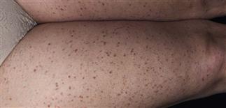
Very small 1–3-mm pigmented macules over the bilateral thighs of this adult woman characteristic of urticaria pigmentosa. Usually asymptomatic. May be misdiagnosed as vasculitis.
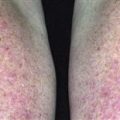
There are many brownish-red macules and patches. The infant was irritable and had difficulty sleeping. Scratching lesions caused itching hives and flushing.
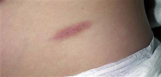
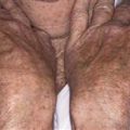
A typical solitary lesion. This brown oval plaque had been present for months. There were no symptoms. There was no family history of mastocytosis.
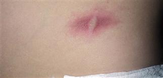
Stroking the lesion produced a wheal and intense flushing around the hive (Darier sign). Stroking releases histamine from mast cell granules, confirming mastocytosis. No biopsy was performed.
DESCRIPTION
Mast cell proliferation. Increased numbers of mast cells in various organs. Most often seen in young children and usually confined to skin. Often involves skin and internal organs in adults. Mast cells store histamine in granules. Histamine released by scratching lesions or ingesting certain agents. Cause unknown.
HISTORY
• Onset occurs between birth and 2 years in 55% of cases. • Incidence: 1 in 1000 to 1 in 8000 live births. • Typically improves gradually; usually clears spontaneously by puberty. • Mast cell disease that begins after age 10 usually persists for life. • When disease is systemic, gastrointestinal tract and skeletal system most commonly involved, and there can be associated mast cell leukemia. • Pruritus is most common complaint. Scratching lesions or emotional upset stimulates histamine release and episodes of itching. When large numbers of mast cell lesions are present, pruritus can be severe and difficult to treat.
PHYSICAL FINDINGS
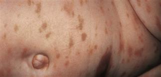
• Most common presentation is pediatric-onset, localized cutaneous disease. • Brown, slightly elevated, non-blanchable macules and patches averaging 0.5–1.5 cm in diameter. • Lesions vary in number. Most infants have one or a few. May resemble café-au-lait spots. Other children have several or many lesions that may be concentrated on the trunk. • Erythema or whealing occurs when lesions are scratched (Darier sign). • Blisters seen in infants. • Biopsy shows mast cell infiltrates in the dermis. Special stains performed on skin biopsy specimens, such as toluidine blue or Giemsa, useful for identifying histamine granules.
TREATMENT
• No cure; therapy is to relieve symptoms. • Skin lesions may be treated with a strong topical corticosteroid for 1–6 weeks. Occlusion enhances penetration and is useful for localized symptomatic plaques. • Intralesional injections with triamcinolone (Kenalog) 10 mg/cc are very effective. Solution may be diluted with saline to avoid inducing atrophy at injection site. • Corticosteroids may decrease or eliminate the mast cells. • H1 (fexofenadine, loratadine, cetirizine, hydroxyzine, diphenhydramine) and H2 antihistamines (cimetidine, ranitidine, famotidine) decrease pruritus, flushing, gastrointestinal symptoms. • Doxepin has potent H1 activity and can be used if antihistamines fail. • Oral disodium cromoglycate (400–800 mg q.d.) may alleviate pruritus, whealing, flushing, diarrhea, abdominal and bone pain. • Oral psoralen plus ultraviolet A therapy given four times each week controls itching and whealing. Reserved for adults not responsive to other treatment. • Avoid non-immunologic agents that stimulate histamine release, such as aspirin, non-steroidal anti-inflammatory drugs, codeine, morphine, polymyxin B, vancomycin, succinylcholine, estrogens, radiocontrast media.