Vaginal Masses

Published on 10/03/2015 by admin

Filed under Obstetrics & Gynecology

Last modified 22/04/2025

Print this page

rate 1 star rate 2 star rate 3 star rate 4 star rate 5 star
Your rating: none, Average: 0 (0 votes)

This article have been viewed 3298 times

Vaginal Masses

Synonyms/Description

None

Etiology

The most common vaginal masses are benign.

Vaginal Cysts

Vaginal wall cysts tend to be embryologic in nature and often asymptomatic. These cysts include Gartner’s duct cysts, Müllerian cysts, epithelial inclusion cysts (ectopic epithelium), urethral diverticula, and cysts resulting from a blocked gland Bartholin duct cyst) or obstructed Müllerian duct anomaly. A complete vaginal septum will typically be diagnosed during menarche when a hematometra develops from the obstructed menstrual flow (see Hematometra and Hematocolpos). Gartner’s duct cysts are remnants of the mesonephric ducts and are most often discovered incidentally. They are located along the anterolateral aspect of the vagina and are typically clear unilocular cysts. Bartholin’s glands are mucus-secreting glands in the posterolateral aspect of the vaginal opening, near the rectum. Bartholin’s duct cysts result from blockage of the duct and swelling from accumulated secretions.
Kondi-Pafiti and colleagues studied 40 cases of benign vaginal cysts. Of these, 12 cases were Müllerian cysts (30.0%), 11 were Bartholin’s duct cysts (27.5%), 10 were epidermal inclusion cysts (25.0%), 5 were Gartner’s duct cysts (12.5%), 1 was an endometrioid cyst (2.5%), and 1 was an unclassified cyst (2.5%). Mean patient age was 35 years (range 20 to 75). Most of the patients (31 cases, 77.5%) were asymptomatic, and the Bartholin’s duct cyst was the more frequently symptomatic.

Vaginal Solid Masses

Fibroids may occur in the vagina, originating from the smooth muscle cells of the anterior vagina or vesicovaginal septum. These are usually solid rounded masses that are well encapsulated and not particularly vascular. Implants of endometriosis are commonly found in the rectovaginal septum or along the posterior wall of the bladder, and may indent or involve the vaginal wall. Malignant masses in the vagina are very rare. Metastatic spread (such as lymphoma or melanoma) accounts for the most common malignant masses in the vagina, followed by primary squamous cell carcinoma. Primary vaginal cancers represent only 1% to 2% of all gynecologic malignancies, and 85% of these primary vaginal malignancies are squamous cell carcinoma. Other rare primary vaginal cancers include adenocarcinoma, melanoma, lymphoma, and sarcomas.
Postoperative or radiation changes may result in vaginal lesions secondary to inflammation and fibrosis. Fistulas between the vagina and rectum can present with vaginal symptoms.

Ultrasound Findings

The best way to evaluate the vagina sonographically is by placing a high-frequency transducer (such as the transvaginal probe) on the perineum and by looking down the vagina, rectum, and urethra simultaneously. Once the vaginal probe is actually inserted into the vagina, it will bypass any vaginal pathology, and the vaginal findings will be obscured and undetectable.
A 3-D acquisition taken from the perineum is important to generate the coronal view of the floor of the pelvis. Using that reconstructed view, the vagina, urethra, and rectum can be seen in cross-section and their relationship with one another evaluated. This view can demonstrate the location of the mass within the floor of the pelvis, specifically showing the relationship of the mass to the vagina, urethra, and rectum. This reconstructed view of the pelvic floor is also ideal to demonstrate defects in the vaginal wall such as fistulas (see Figure V1-9), and is increasingly being used in urogynecology. Similar to any other pelvic mass, the appearance of the vaginal mass, such as gray-scale texture, contour, and degree of vascularity using color flow Doppler, provides clues as to the diagnosis. The location of the mass is also important, keeping in mind that masses in the anterior compartment may be urologic and those in the posterior compartment may be gastrointestinal. The vagina, urethra, and rectum are in close proximity to one another, sharing walls that may be affected by the mass. Refer to the differential diagnosis section that follows for a description and comparison of these masses and their sonographic appearance.

Differential Diagnosis

A clear and asymptomatic cyst in the lateral wall of the vagina is likely to be a Gartner’s duct cyst. Fluid (or blood) in a hemi-hematocolpos secondary to a vaginal septum typically has low-level echoes indicating unclotted blood, much like an ovarian endometrioma. A complex cyst with solid elements seen anterior to the vagina suggests a urethral diverticulum. When the lesion is symptomatic, peripheral color flow may be seen, owing to inflammation. A complex cyst in the posterior-lateral wall of the vagina is likely to be a Bartholin’s duct cyst and has only peripheral color flow. The most common rounded and focal solid mass (with limited color flow) is a fibroid in the vaginal wall.
Although vaginal cysts are usually benign, most solid vaginal masses with abundant color flow tend to be malignant. The appearance of the solid mass is not useful to determine the specific tissue diagnosis, as sarcomas, lymphomas, and other lesions look similar to one another. Malignant vaginal masses are typically completely solid, abundantly vascular, and irregular in contour. They may extend into the surrounding tissues such as the pelvic side walls.

Clinical Aspects and Recommendations

The treatment of vaginal masses depends on the diagnosis. Benign cysts are treated depending on the patient’s symptoms. Asymptomatic Gartner’s duct cysts are typically monitored with follow-up ultrasounds, whereas a symptomatic Bartholin’s duct cyst requires drainage or marsupialization. Urethral diverticula may be found incidentally or when they become symptomatic, and are usually managed by a urogynecologist or urologist. Treatment of malignant vaginal masses depends on the specific type of malignancy and usually involves a multidisciplinary team including gynecologic and medical oncology.

Figures

image

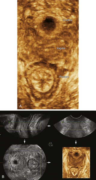
Figure V1-1 A and B, 3-D reconstructed view of the floor of the normal pelvis, showing the urethra, vagina, and rectum en face. B demonstrates the multiplanar view of the pelvic floor, showing the acquisition planes. The 3-D volume was acquired from the perineum and sweeping side to side. The A plane in the upper left-hand corner shows the acquisition view looking straight down the vagina. The B plane shows the same view at right angles from the A plane. The C plane is the reconstructed view of the floor of the pelvis, which is crucial to evaluating the perineal structures, including the length of the vagina and its relationship to neighboring structures.


image

Figure V1-2 A and B, Gartner’s duct cyst: Long axis view looking down the vagina from the perineum, showing a clear unilocular cyst just posterior to the urethra. B is a 3-D reconstructed image of the pelvic floor, showing that the cyst is located along the left side of the vagina.


image

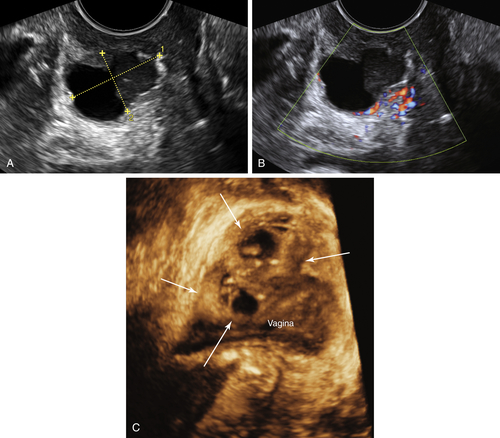
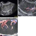
Figure V1-3 Urethral diverticulum. A and B, 2-D view of a complex cystic mass with irregular borders and internal debris. Note that the vascularity is only in the peripheral aspect of the mass. The mass was located anterior to the vagina and lateral to the urethra. The patient was quite symptomatic, particularly upon voiding. C, 3-D reconstructed view of a different case of a urethral diverticulum. Note the complex, multicystic mass anterior to the vagina where the urethra should be. The exact location of the urethra is obscured and likely encapsulated by this cystic mass, which was quite symptomatic.


image

Figure V1-4 Bartholin’s duct cyst. (A) 2-D and (B) 3-D views of a heterogeneous solid mass located anterolateral to the rectum. Note that the interior of the mass has no discernible blood flow.

image

Figure V1-5 Vaginal fibroid. Magnified view of a small, rounded, solid mass in the wall of the vagina.


image

Figure V1-6 A to C, Leiomyosarcoma. Multiple views of a solid and very vascular mass located along the right side of the vagina and extending posteriorly toward the right buttock.


image

Figure V1-7 A and B, Myxoid spindle cell sarcoma. Very vascular, lobulated and irregular solid mass along the lateral and posterior aspect of the vagina. Note that the mass has a fingerlike extension into the surrounding side wall, indicating the aggressive behavior of the tumor.

image

Figure V1-8 Vaginal lymphoma. Long axis view from the perineum, looking down the vagina, showing a lobulated irregular vascular mass extending the entire length of the vagina.


image

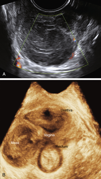
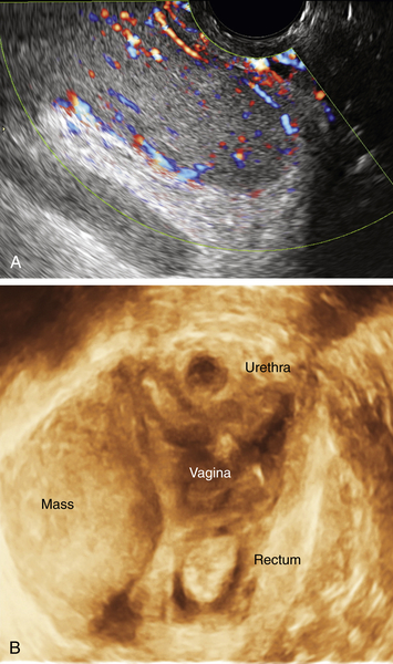
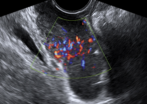
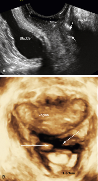
Figure V1-9 A and B, Recto-vaginal fistula. 2-D and 3-D views of the perineum looking down the vagina in a patient with Crohn’s disease who had clinical evidence of a fistula. Arrows demonstrate the location of the fistula, which was identified first on the 3-D reconstruction (B) and then recognized on standard 2-D imaging (A). An MRI done the same day had been read as negative.


Suggested Reading

Dai Y., Wang J., Shen H., Zhao R.N., Li Y.Z. Diagnosis of female urethral diverticulum using transvaginal contrast-enhanced sonourethrography. J Int Urogynecol. January 31, 2013  [Epub ahead of print].

Elsayes K.M., Narra V.R., Dillman J.R., Velcheti V., Hameed O., Tongdee R., Menias C.O. Vaginal masses: magnetic resonance imaging features with pathologic correlation. Acta Radiol. 2007;48:921–933.

Fletcher S.G., Lemack G.E. Benign masses of the female periurethral tissues and anterior vaginal wall. Curr Urol Rep. 2008;9:389–396.

Hwang J.H., Oh J.M., Lee N.W., Hur J.Y., Lee K.W., Lee K.J. Multiple vaginal Müllerian cysts: a case report and review of literature. Arch Gynecol Obstet. 2009;280:137–139.

Kondi-Pafiti A., Grapsa D., Papakonstantinou K., Kairi-Vassilatou E., Xasiakos D. Vaginal cysts: a common pathologic entity revisited. Clin Exp Obstet Gynecol. 2008;35:41–44.