Bladder Masses

Published on 10/03/2015 by admin

Filed under Obstetrics & Gynecology

Last modified 22/04/2025

Print this page

rate 1 star rate 2 star rate 3 star rate 4 star rate 5 star
Your rating: none, Average: 0 (0 votes)

This article have been viewed 1558 times

Bladder Masses

Synonyms/Description

Bladder tumor
Focal bladder lesion

Etiology

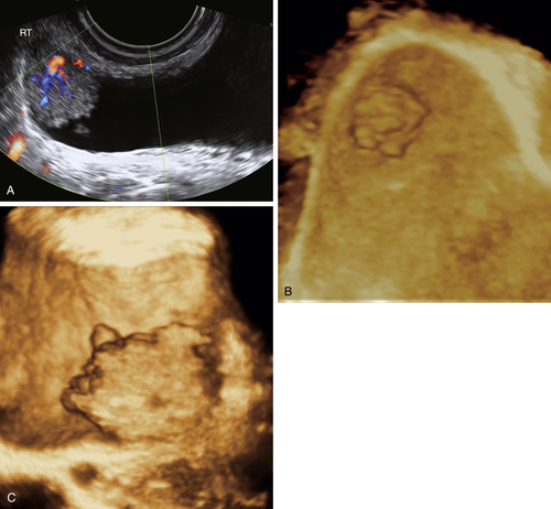
Transitional Cell Cancer

In the United States, bladder cancer is reportedly the fourth most common malignancy. The vast majority of bladder neoplasms arise from the epithelium, with urothelial (transitional cell) carcinoma accounting for 90% of cases. Squamous cell carcinoma is rare and accounts for 2% to 15% of bladder cancers. The least common is adenocarcinoma, which may be primary or metastatic to the bladder.

Fibroma

Fibroma (leiomyoma) is the most common benign tumor of the bladder, although it represents only 0.4% of all bladder neoplasms. It is most prevalent among women in the third to fifth decades of life.

Endometriosis

Endometriosis occurs as a fusiform mass in the bladder wall and is covered in the section on endometriosis.

Diffuse Bladder Wall Thickening

Diffuse bladder wall thickening is seen in cases of severe chronic cystitis or in chronic bladder outlet obstruction where the bladder becomes trabeculated (more common in males with large prostates).

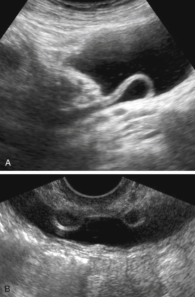
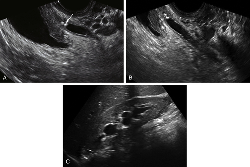
Findings Specifically Related to the Ureteral Orifice

 Ureterocele
 Ureteral reimplantation site
 Stone at the ureteropelvic junction (UPJ) with edema of ureteral orifice

Urethral Diverticula

Urethral diverticula occur just under the bladder along the urethra. They can be quite painful, especially when the patient voids.

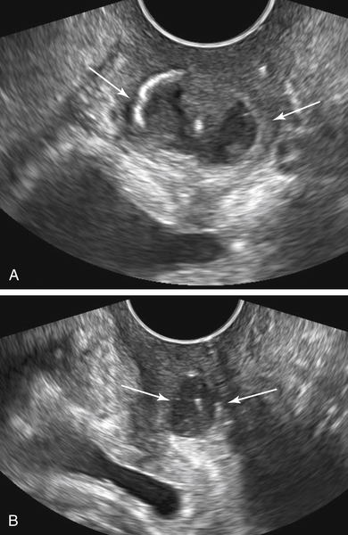
Other Bladder Masses
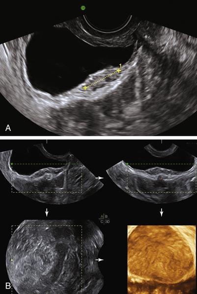
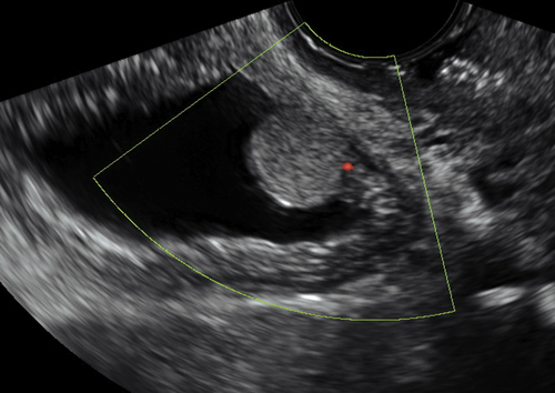
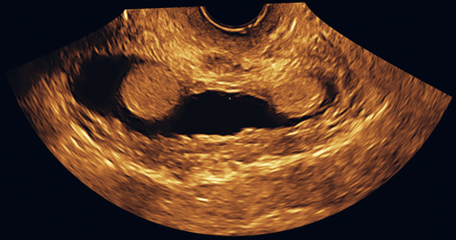
There are many benign tumors such as paraganglioma, plasmacytoma, hemangioma, neurofibroma, and lipoma that can occasionally (rarely) occur in the bladder. Malignant neoplasms reported in the bladder include rhabdomyosarcoma, leiomyosarcoma, lymphoma, osteosarcoma, and metastatic tumors such as melanoma.

Ultrasound Findings

Bladder masses are typically located in the bladder wall. Because these arise from the submucosal portion of the bladder wall, they typically appear as smooth intramural lesions. Transitional cell carcinoma is a focal mucosal lesion, which is fungating and extends into the lumen of the bladder with an irregular surface. Color Doppler usually reveals abundant blood flow, as with other pelvic malignancies. Bladder wall lesions are typically fusiform with an intact mucosal surface and focally widen the wall of the bladder. Endometriosis of the bladder wall (likely the most common diagnosis in gynecologic patients) has spotty blood flow by Doppler and a smooth inner and outer wall. Lesions involving the ureteral orifice can be cystic, such as ureteroceles (which are usually asymptomatic); in the case of reimplantations, there will be a surgical history.
Urethral diverticula are complex masses along the urethra, just under the bladder (see Vaginal Masses). These may indent the floor of the bladder and be quite painful during the ultrasound. They are best seen by placing the transducer on the perineum and looking cephalad toward the bladder. They are usually cystic with varying amounts of solid area and calcification, depending on chronicity of the lesion.

Differential Diagnosis

Mucosal lesions with an irregular surface and protruding into the bladder are usually transitional cell carcinomas. Lesions that are solid and completely contained within the bladder wall may be endometriosis (fusiform with little detectable blood flow) or fibroma (rounded and ball-like) versus other rare solid rounded tumors.
If the bladder wall is diffusely abnormal and thickened, etiologies may include long-standing bladder dysfunction or obstruction or chronic cystitis. It is normal for the bladder wall to appear thickened and trabeculated if the bladder is underfilled.
If the lesion is along the urethra, it is likely to be a urethral diverticulum.

Clinical Aspects and Recommendations

Bladder lesions can cause dysuria, frequency, and hematuria, but often may be asymptomatic. Bladder malignancies typically present with hematuria because of their location in the mucosal layer of the bladder. Pedunculated intraluminal masses may lead to obstruction of urine flow or inability to completely empty the bladder. If a bladder mass arises in proximity to one of the ureters, it can obstruct the ureter, thus presenting with flank pain and hydronephrosis. Lesions that develop outside the wall of the bladder not impinging on the lumen may remain asymptomatic for a long time. There are no general management recommendations because this depends on the type of lesion diagnosed.

Figures

image

Figure B1-1 Transitional carcinoma of the bladder. A, A 2-D view with color Doppler showing abundant vascularity. B and C, The same tumor using 3-D surface ultrasound. Note the fungating mucosal surface lesion typical of bladder carcinoma.


image

Figure B1-2 A, Fusiform bladder wall mass (calipers) in a patient with endometriosis. B, The 3-D volume view of the bladder wall mass, showing a smooth mucosal surface.

image

Figure B1-3 Ureterocele. A, A longitudinal view of the ureterocele implant into the bladder. B, The patient has bilateral ureteroceles.


image
image
image

Figure B1-4 Patient with surgically reimplanted ureters. Note the homogeneous rounded structures at the site of reimplantation, 2-D and 3-D.


image

Figure B1-5 Urethral diverticulum (arrows). A, Note the complex mass, partly calcified, indenting the floor of the bladder. B, The long axis view of the mass alongside the urethra.


image

Figure B1-6 A and B, Two small stones stuck in the distal end of the ureter. C, The associated hydronephrosis.


Suggested Reading

Fasih N., Prasad Shanbhogue A.K., Macdonald D.B., Fraser-Hill M.A., Papadatos D., Kielar A.Z., Doherty G.P., Walsh C., McInnes M., Atri M. Leiomyomas beyond the uterus: unusual locations, rare manifestations. Radiographics. 2008;28:1931–1948  Review.

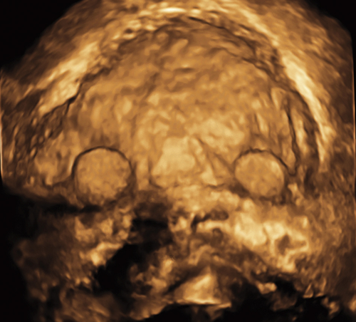
Kocakoc E., Kiris A., Orhan I., Poyraz A.K., Artas H., Firdolas F. Detection of bladder tumors with 3-dimensional sonography and virtual sonographic cystoscopy. J Ultrasound Med. 2008;27:45–53.

Wong-You-Cheong J.J., Woodward P.J., Manning M.A., Davis C.J. From the archives of the AFIP: inflammatory and nonneoplastic bladder masses: radiologic-pathologic correlation. Radiographics. 2006;26:1847–1868  Review.

Wong-You-Cheong J.J., Woodward P.J., Manning M.A., Sesterhenn I.A. From the archives of the AFIP: neoplasms of the urinary bladder: radiologic-pathologic correlation. Radiographics. 2006;26:553–580  Review.