Chapter 41 Manipulation
 Therapeutic Keys
• Manipulation is a passive manual maneuver that introduces movement beyond the passive range of motion (ROM) through the elastic barrier, but does not exceed the anatomic barrier.1 Mobilizations are passive stretches with or without oscillations over which the patient can exert control.2 Chiropractic adjustments are techniques that range in force from a near imperceptible force to high-velocity thrusts causing joint cavitation (popping noise).
• As a prudent starting point, initial evaluation should determine if manipulation of the patient is appropriate, seeking out “red flags” that contraindicate manipulation, such as fracture, infection, neoplasm, progressive neurologic deficit, cord pressure, or cauda equina syndrome.3,4
• A correct differential diagnosis is key to selection of patients, and the functional assessment is key to the selection of appropriate manual medicine techniques.5
• If gross signs of inflammation are present in a joint (heat, swelling, redness, and pain), and repeated movement to end range (ER) worsens the signs, manipulation in that direction would most likely aggravate the condition.
• When bringing the joint complex to tension, if the pain peripheralizes, it is a relative contraindication to manipulation.
• The practitioner should be alerted that an adverse outcome is likely from manipulation when repetitive motion with progressive force in the direction of the manipulation peripheralizes pain and reduces function.
• Relaxation techniques (heat, muscle work, and calming environment) are helpful to patients complaining of anxiety, muscle tension, stiffness, and aching before manipulation.6
• If pain is the chief complaint, icing the area for 5 minutes causes surface anesthesia and 20 minutes causes sedation of the actions of the muscle spindle cells. Sedation of a muscle reflex arc before manipulation of a fixated painful joint may facilitate treatment.2
• If the patient reports an increase in pain or stiffness after manipulation, ice the area of treatment for 10 to 30 minutes to reduce spasm and pain.7,8 The patient should be placed into a position that centralizes pain when icing.
• “The goal of manipulation is to restore maximal pain-free movement of the musculoskeletal system and postural balance.”9
• Do not treat muscle spasm as a primary condition. Muscle spasm is almost always a response of the body to a noxious stimulus. Find the cause and treat it.10
• Trigger points are myofascial irritations that are frequently caused by underlying joint fixations. Manipulation can irritate a trigger point and can precipitate a muscle spasm later that day.11
 Historical Perspective
Manipulations are depicted in prehistoric cave drawings and Chinese statues, circa 2700 BC,12 but Hippocrates is credited with the earliest recorded written physician’s prescription of manipulative treatment methods, which changed little until the sixth century. He advocated key principles of judicious use of force, direction of thrust, and proper levering of joints.13
During the Dark Ages, priests provided medical treatment at their monasteries. Kessler stated: “Friar Moultan, of the order of St. Augustine, wrote The Complete Bonesetter. The text, which was revised by John Turner in 1656, suggests that manipulation was practiced in medical settings throughout the Middle Ages and Renaissance.”14
Three main concepts developed during the 1700s still have a major influence on manipulation today. The first held that “vertebral luxation” (bone out of place) was responsible for spinal deformity. The second, the mainstream medical opinion, maintained that “caries of the spine” caused spinal deformity, which was treated with bloodletting and rest, while condemning extension and manipulation as both useless and dangerous and citing concerns about the potentially disastrous effects of manipulating tuberculous, neoplastic, rheumatic, or fractured joints. The third held that muscles were the main cause of problems, and treatment should be complete rest or active exercise, as the case warranted.13
It is interesting to note the following:
• Hippocrates railed against the abuse of manipulative therapy by physicians and others of his time.
• Physicians of the late 1700s assailed one another’s methods of treatment (e.g., in The Lancet, December 16, 1826, the banner on page 347 appropriately read “THE YELLOW JOURNAL”).
• Surgeons held “bonesetters” in great contempt “when they condescend[ed] to speak at all of bonesetters and their works.”12
• Bonesetters held their secrets and passed them from father to son.
• Financial competition was noted early in the literature. “It is known to most practitioners of surgery, and has been made known to many to their great cost and loss, that a large portion of the cases of impaired mobility or usefulness of limbs after injury fall into the hands of a class of men called ‘bonesetters’.”15
• Although there has been a great deal of animosity, and claims of superiority made by the various practitioners of manipulative treatment even to this day, “specific conclusions cannot be derived from the scientific literature for or against either the efficacy of spinal manipulative therapy or the pathophysiologic foundations from which it is derived.”16
 Schools of Thought in Manipulation
Bonesetters of England
Bonesetters of England generally held that a bone was out of place and had a “feel” for what was wrong. Hutton described the information gained from a bonesetter as “bring[ing] some spoils out of the camp of the Philistines.”15
Chiropractic
D.D. performed the first chiropractic manipulation on a deaf janitor whose hearing had been lost when he stooped over and felt something give in his back. D.D. reasoned that if the deafness occurred from something slipping out, restoring the vertebra to its correct position should cure the condition: “With this new objective in view, a half-hour’s talk persuaded Mr. Lillard to allow me to replace it and his hearing was restored.’’17
Although there are multiple schools of thought in chiropractic, each with its own strengths and weaknesses, the literature supports the need to combine therapeutic exercise with manipulation. Rehabilitation of the Spine, A Practitioner’s Manual, 2nd ed. edited by Craig Liebenson,4 is considered a landmark publication by many in the field and addresses this topic at length.
Naturopathic
Several naturopathic schools in the past were associated with chiropractic and eclectic schools of medicine. The genesis of the naturopathic profession is well documented in Chapter 4, History of Naturopathic Medicine.
Osteopathic
Andrew Still left the practice of allopathic medicine and started a school of osteopathy in Kirksville, Missouri. It is highly probable that the first chiropractor, D.D. Palmer, went to this school and learned some of the techniques, but it is not well documented. The famed “equal but separate” movement of the osteopaths led to a 1921 resolution, submitted at the American Osteopathic Association convention, that allowed entrance of chiropractors with advanced standing into their schools. Still, before his death, saw the defection of his osteopathic profession into the ranks of medical orthodoxy.18–20 Interestingly, manipulation is now only an elective segment in some American osteopathic schools, whereas in England, where osteopathy is not part of the medical establishment, manipulation is still the mainstay of osteopathic practice.
Allopathic
James Cyriax, James Mennell, and John Mennell were brilliant physicians who worked to reintegrate manipulation into medical practice. They wrote valuable texts on manipulative therapy, although they did not totally agree on the effects they achieved with manipulation. Mennell held to the correction of lost joint play and denied the effect on the intervertebral disc,21 whereas Cyriax claimed reduction of a protruding disc.10
“Controversy and contention” best describe the higher levels of the respective schools of medical thought. The impression one gets in reading through the literature is intolerance of others’ ideas expressed in ad hominem attacks. The mistake “lay” manipulators and “nonphysicians” make is not ineffectiveness, but in their willingness to seek training outside the fraternal order of the “medical” brotherhood; to address the public directly rather than communicating exclusively within the order; and, worst of all, to openly compete, economically and politically, against the fraternal order.13,15
Donald B. Tower, in the chairman’s summary at the National Institute of Neurological and Communicative Diseases and Stroke conference in 1975,22 noted a physician who received little credit for his early contribution to the field: J. Evans Riadore, a London physician who wrote a treatise on the irritation of spinal nerves in 1843. He attributed many diseases to this condition, stating: “If any organ is deficiently supplied with nervous energy of blood, its functions immediately, and sooner or later its structure, become deranged.” This was a viewpoint subsequently echoed by osteopaths and chiropractors.22
The fifth edition of Spinal Manipulation by Bourdillion et al,23 the first authors who were medical manipulators, has been largely reworked from previous editions and heavily influenced by osteopathic methods.
Physical Therapy
James Cryiax influenced Robin McKenzie, a New Zealand physical therapist who subsequently developed a systematic approach called mechanical diagnosis and treatment (MDT) of musculoskeletal conditions. It is a process of evaluation and treatment of musculoskeletal disorders based on a mechanical history and the patient’s symptomatic and mechanical response to movement, positions, and loading.4 McKenzie’s approach is often prejudicially dismissed as “extension exercises for the low back.” He proposed a new tack in the approach to back pain that was found to be highly effective in populations with acute and chronic back pain as well as low in cost.24 MDT includes McKenzie’s observations that most conditions are self-resolving and that focus should be on prevention and recovery of function by teaching patients proper posture and self management. After years of studying, performing, and researching manipulation, he felt that only 20% of patients needed manipulative therapy, and 80% could self-treat using ER loading strategies learned from books or practitioners.25
 Is Manual Medicine the Right Treatment?
Patients with somatic pain caused by psychosocial factors often seek a physical cause and treatment for their pain. If the presenting complaints do not seem to follow a mechanical pattern and the pain diagram is nonanatomic, consider the possibility of psychosocial factors. Psychosocial workplace factors associated with risk of spinal injury include job dissatisfaction, stressful working conditions as perceived by the employee, employer practices reported as being unfair, poor coping skills, lack of recognition at work, low supervisor support, a high frequency of job problems, and negative beliefs of or attitudes toward the consequences of having “low back trouble.”26,27 A major factor in identifying the incidence of future pain is the patient’s perception of being disabled.28 If the patient’s history and records include previous evaluation by multiple providers with conflicting and confusing reports from the patient, caution is advised in approaching the management of the patient. If in doubt, seek a consultation with an astute colleague, physiatrist, or appropriate specialist.
Is It Safe to Move?
A clinician must answer the question, “Is it safe to ‘move the patient’ using conservative therapies, exercise, mobilizations, and manipulations?” Red flags have been shown to have many false positives, resulting in unnecessary additional diagnostic testing. Recent studies showed less than 1% of patients presenting to general practitioners with conditions that warranted further diagnostic evaluation. Red flags are a source of unnecessary medical interventions. When the medical history and examination indicate a serious disease, neurologic compromise, or progressive neurologic deficit, further evaluation or consultation is warranted. Although the question of safety is usually answered by the provider’s clinical training, a general outline of triage is presented as a starting point to help determine if further evaluation is indicated before initiating or continuing care.29
Signs of Neoplasm
The combination of age over 50 years, a history of cancer, unexplained weight loss of more than 10 kg within 6 months, failure to improve after 1 month of conservative care, and an elevated erythrocyte sedimentation rate (ESR) have a sensitivity of 100% in identifying patients with a neoplasm. Night pain, especially pain that prevents the patient from getting sleep or “drives them from the bed” also generates a high degree of suspicion.30,31
Signs of Infection/Inflammation/Illness
Heat, swelling, pain, redness, and loss of function are the cardinal signs of inflammation. Acute inflammation lasts from 1 to 3 days and chronic inflammation usually is considered from 7 days to 7 weeks, but may last up to 12 months. The stages between acute and chronic inflammation overlap, and a continuum exists.32 The clinical picture may be confused by chronic pain.
Laboratory tests may show signs such as an elevated C-reactive protein, ESR, or other tests for inflammatory arthritides. Signs of infection may be as simple as a low grade fever, chills, a shift to the left in the white blood cell count, signs of a worsening chronic illness, or frank presentation of an acute illness.33 Clinical signs are pain that is constant 24 hours a day, described as aching, throbbing, or burning, and aggravated by movement with no position that gives relief.
Signs of Neurologic Disorder or Progressive Neurologic Deficit
A loss of sensation, decreased or absent deep tendon reflex, loss of muscle strength, and positive nerve root tension are signs of nerve root involvement. If only these neurologic findings are present and do not become progressively worse, appropriate conservative care may be effective.34 Dizziness, drop attacks, diplopia, dysarthria, dysphagia, numbness, nystagmus, and nausea are more concerning signs of central nervous system involvement. Loss of bladder or bowel control or saddle anesthesia requires urgent evaluation with magnetic resonance imaging of the lumbar spine. If progressive neurologic deficit of weakness, loss of sensation, and loss of function, or the previously mentioned signs and symptoms are present, a consultation with a competent colleague, physiatrist, or neurosurgeon is indicated.
Signs of Fracture
A fracture typically has an obvious traumatic cause when the patient is young or middle-aged. In older patients, pathologic fractures are a concern, especially if there is a history of prolonged or repeated steroid use. Be wary of occult fracture after motor vehicle accidents or athletic injuries and nontraumatic fractures in the elderly. The red flags suggesting vertebral fracture are age more than 50 years, female sex, major trauma, pain and tenderness, and a distracting painful injury. In particular, look above and below the site of injury, as the force of trauma may be transmitted and cause a fracture some distance from the site of impact.35 In a pelvic fracture, there is always a fracture in two places in the ring.36
Is the Best Treatment Movement or Rest?
Pain can be confusing and lead one to conclude that inflammation is present. If pain is constant aching, throbbing, or burning, it suggests that the pain has a chemical cause, and it is inflammation. When the pain goes away, even for 30 minutes in a day, it suggests that the pain is due to mechanical pressure on tissue. To illustrate this point, bend your finger backward until you feel strain and hold it. At first you may only feel discomfort, but after an extended period of time, it will become painful. Next, try bending the finger backward beyond strain until it hurts. The pain you feel is due to abnormal stresses on a normal tissue; there is nothing wrong with your finger! You do not need drugs, modalities, or manual therapy. Simply stop overstraining the soft tissues. Robin McKenzie used this seemingly simple example to teach the consequences of sustained ER loading as a common cause of pain—mechanical pain rather than pain from inflammation.25,37
Which Direction to Move?
Orthopedic Tests
• The examiner can make the patient worse by forcing tests or performing them incorrectly.
• The least stressful tests should be done first. If the first test causes severe pain, most of the other motions will be painful afterward and confuse the findings.
• The test by definition has a positive response, which correlates to a specific condition that must be produced when the test is performed for the test to be positive (e.g., Lindner’s test and Soto Hall’s test are performed in the same way, but the findings to report a positive test are different).
• The test must reproduce the pain of the primary complaint and be positive by the test’s definition.
• Tests are centered in allopathic medicine to diagnose pathology, fracture, moderate to severe sprain/strain, and dislocation. Although the patient feels pain from the movements of these tests, these are not true positive tests just because the patient reports the movement hurts.
When multiple orthopedic tests are indiscriminately performed on one visit, without an understanding of the mechanism of action of the stress these maneuvers put on the tissues, the unwitting examiner merely subjects the patient to the trauma of a series of painful maneuvers that only serve to confuse the practitioner and aggravate the patient’s condition. Detailed texts that cover this area include those by Magee38 or Evans39; such texts are helpful and should be referred to for additional information. It is even more helpful to attend programs in which experienced practitioners teach skills and knowledge to inexperienced practitioners.
Selective Tissue Tension Tests
This is an introduction to the differential diagnosis of soft tissue injuries by means of active and passive movements using the selective tissue tension tests of James Cryiax10; this topic is covered in depth in Kessler14 and Magee.38
Passive Movements
• Normal—the amount of motion and feel of the tissues are appropriate for age
• Abnormal—normal end-feels that are not normal for the joint and ROM that are being tested (e.g., the feeling of bone on bone is the normal end-feel at the elbow when extended but is abnormal when it occurs at the ER of knee extension)
• Pathologic—these end-feels are only present in joints that have undergone pathologic changes
Normal End-Feels
Capsular end-feel. This is a firm, “leathery” feeling, felt, for example, when the normal shoulder is at full external rotation. When felt in conjunction with a capsular pattern of restriction, and in the absence of significant inflammation or effusion, it indicates capsular fibrosis.
Bony end-feel. This feels abrupt, as when moving the normal elbow into full extension. When accompanying a restriction of movement, it may suggest hypertrophic bony changes, such as those that occur with degenerative joint disease, or possible malunion of bony segments after healing of a fracture.
Soft tissue approximation end-feel. This is a soft end-feel, as when fully flexing the normal elbow or knee. It may accompany joint restriction in the presence of significant muscular hypertrophy.
Muscular end-feel. This more rubbery feel resembles what is felt at the extremes of straight-leg raising (SLR) from tension on the hamstrings. It is less abrupt than a capsular end-feel.
Abnormal End-Feels
The tissue involved is determined by the response to the passive ROMs and by moving the joint to ER and into closed pack position.
Muscle or tendon. Active and passive movements are limited or painful, or both. Pain is caused by active contraction or passive stretching of the inflamed or torn muscle-tendon unit.
Ligaments. Both active and passive movements are limited or painful, or both, in the same direction due to stretch of the inflamed or torn ligament.
Capsular pattern. Only joints that are controlled by muscles have a capsular pattern. Pain is caused with both active and passive motions, and the limitation of motion is in a specific proportion that is listed in tables in Kessler’s text.14
McKenzie’s Mechanical Diagnosis and Therapy (MDT)
MDT is an invaluable system for practitioners who perform manual therapy. Rather than a technique, it is a method of diagnosis determined by response to repeated movement and pressure to joints and soft tissue that, once mastered, allows rapid assessment and selection of patients for treatment. It also identifies those not likely to respond to manual therapy so the provider’s treatment program can be altered, or the patient can quickly receive an appropriate referral. Resources available are classes from the McKenzie Institute on MDT, McKenzie’s textbooks,40,41 and a chapter in Liebenson’s text.4 Only a few of the many helpful concepts from the McKenzie method are discussed in the following. Keep in mind that McKenzie asserted that only 20% of patients needed manipulative therapy, and prevention is key to success in his model.
Centralization and Peripheralization
A cervical spine problem can refer pain to the shoulder blade, arm, forearm, and hand. A low back problem can refer pain to the sacroiliac joint (SIJ), buttock, thigh, leg, and foot. When the referred pain from the spine is brought closer to the midline in response to movements, position, or load, and remains reduced, it is called “centralization.”24 When the pain moves out from the spine in response to movements, position, or load, it is called “peripheralization.” When a movement produces centralization, it is a motion to pursue for treatment. When a movement causes peripheralization, it is a motion to avoid, as it will likely worsen the condition. If no movement or position centralizes the pain, it is a poorer prognosis for response to the treatment method being used. Although this phenomenon is typically associated with vertebral disc problems, it can be observed in many musculoskeletal problems.
 Pretreatment Assessment
Functional Assessment
Often the focus is on injury, pathology, and disease, but the majority of musculoskeletal pain is from pathomechanics due to postural and repetitive strain, disuse, deconditioning, fatigue, and nutritional factors that must be addressed if treatment is to be successful. Once the portion of the patient’s condition that is a musculoskeletal problem has been established, the mechanical fault should be determined. Manipulative treatment without correction of the mechanical fault results in prolonged treatment and recurrence. Detecting this is sometimes easy, whereas at other times it is a mystery that takes careful investigation, as discussed in McKenzie,37a Greenman,1 Lewit,5 and Liebenson.4
Establish the baseline of the location of the complaint and its response to motion and then repeat motions to ER. In the cervical spine, test flexion, extension, rotation, and lateral bending. In the lumbar spine, test flexion, extension, and lateral translation. The testing procedures are described in detail with photos in Liebenson’s text.4
Cervical Spine
There is considerable discussion of the topic of vertebrobasilar stroke (VBS) after manipulation of the cervical spine. Tests done in the office have not proved effective, but the history should alert the practitioner to the possibility of VBS insult, and manipulation is contraindicated. The presence of the signs listed in the section on “Signs of Neurologic Disorder” are important signs that indicate not to use mobilization or manipulation. The most important risk factors identified by Terrett42 were dizziness, unsteadiness, giddiness, vertigo, and sudden severe pain in the side of the head or neck, which is different from any pain the patient has had before. If the practitioner elects to follow the pattern of the progression of force, the occurrence of these symptoms in the early mobilizations contraindicate progressing the force and manipulation. However, VBS has occurred during an examination simply by having the patient turning his or her head. The incidence is small, but missing this clinical presentation is devastating, though not clearly caused by the evaluation or manipulative treatment.43
Lumbar Spine and Sacroiliac Joint
Assessing Radiating Leg Symptoms
Some additional ideas to finding the area of lesion in the lumbar spine are as follows:
• Support Adams (the belt test)—With the patient standing, the doctor has him or her bend forward and notes the level of pain. The doctor then secures the patient’s pelvis by hugging the anterior superior iliac spines with the arms, pressing the patient’s sacrum into the doctor’s hip. If the patient’s pain is decreased, it indicates the lesion is in the pelvis (probably the SIJ or hip); if pain increases, it is in the lumbar spine.
• Patrick’s test—Flexion, abduction, and external rotation of the hip are blocked; this causes pain over the inguinal fossa and into the thigh when a hip lesion is present.
Assessing the Sacroiliac Joint
The SIJ is the “dumping ground” for pain from the lumbar spine, just as the shoulder blade is the “dumping ground” for pain from the cervical spine. The key to determining if the problem is in the SIJ is to rule out all lumbar problems first. Second, Laslett45 determined that when two or more of the following SIJ tests produce concordant pain, it is highly likely that the SIJ is the pain generator.
1. Gapping test—patient is supine. The examiner crosses his or her arms, placing heels of the hands on the anterior superior iliac spine, forcefully pressing laterally and down into the table to “gap” the SIJ, attempting to stretch the anterior SIJ ligaments.
2. Compression test—The patient is lying on his or her side with the painful SIJ up. The pressure is directed to the opposite iliac crest, attempting to compress the anterior SIJ and stretch the posterior SIJ ligaments.
3. Posterior shear or “thigh thrust”—The patient is supine. The practitioner’s hand, a small block, or sand bag is placed under the patient’s sacrum. The hip is flexed to 90 degrees (perpendicular to the floor), the knee is maximally flexed, and a thrust is applied down the shaft of the femur. One should avoid adduction of the hip, as this will cause pain in normal patients.
4. Pelvic torsion (Gaenslen’s test)—The patient is supine at the edge of the table with one thigh extended over the edge of the table. On the other side, the hip and knee are flexed to the chest. Overpressure is applied to the extended thigh to accentuate the posterior rotation of the opposite side. This should be performed on both sides.
5. Sacral thrust—With the patient lying prone, the examiner thrusts down on the sacrum.
6. Cranial shear—With the patient prone, cranial pressure is applied to the apex of the sacrum with the examiner’s hands, whereas the painful side ankle is placed between the examiner’s knees and is tractioned caudally.
Techniques for correction of these fixations are described in Maigne,46 Bourdillion,23 and Maitland.2
 Treatment Concepts
Notes on the Art of Manipulation
When a movement causes pain, it should not be performed unless the diagnosis of the patient’s condition is sure and the motion will cause no harm.46
Barrier Concepts
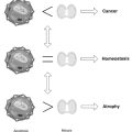
Joint motion is described from neutral to an ER that is ultimately limited by the anatomic barrier, which, if exceeded, causes tissue trauma. The extent of the active ROM can be increased by passive motion to a point at which all of the tissues around the joint have been brought to tension, called the elastic barrier.1 Beyond the elastic barrier is a small ROM referred to as the paraphysiologic space.47 It is within the paraphysiologic space that joint cavitation, the “popping” sound, occurs. When joints and soft tissues are dysfunctional, alterations of ROM may occur both within the ROM and at the end of it. If one focuses only on working in the paraphysiologic space, one severely limits the effects that can be made on dysfunction.
• Edematous and boggy end-feel—indicates joint effusion and possibly inflammation. Muscle spasms should not be treated as a primary condition, since they are almost always the body’s response to a noxious stimulus or pathophysiologic fault. Cryotherapy, positive galvanism, pulsed ultrasound, and gentle, frequently repeated movements within the active ROM are indicated.
• Springy and taut end-feel—ligamentous fixations (as felt in the normal knee in lateral bending stress tests and drawer tests). Manipulation works well on these types of fixations. Surging sinusoidal and interferential current, ultrasound, and moist hot packs facilitate the treatment. One can follow with cryotherapy in the subacute stages.
• Bone-on-bone end-feel—usually indicative of degenerative joint disease (as felt in the normal elbow in extension). If there is fine crepitus, mild degenerative joint disease (DJD) may be present; coarse crepitus indicates moderate DJD, and joint creaking may be advanced DJD. Diathermy, contrast baths, and other naturopathic therapies indicated in osteoarthritis are helpful.
Performing Manipulation
1. Test the direction of manipulative thrust with repeat mobilization to ER to ascertain that the pain does not peripheralize, the movement does not increase PDM, and the movement does not result in increased obstruction to motion from mobilization, using a progression of force to determine the effect of the direction and force applied.
2. Stabilize the area to be manipulated: A point of stabilization is created by one hand and the physician’s body weight, while the other hand performs the manipulation. Minor corrections of the position of the stabilized part or hand, or both, are frequently interpreted by an observer as twisting or wrenching motions. Twisting and wrenching are difficult to control and may injure the patient.
3. Bring the joint to tension, removing the periarticular tissue slack. Mobilization with tissue slack not taken up is safe, but manipulation when tissue slack is present invites injury.
4. Thrust only when the patient is relaxed and the fixation is felt.
The manipulative thrust can be described according to the following:
Troubleshooting Technique
Common problems blocking successful manipulation can be overcome by the following:
• Learning joint play analysis on the peripheral joints and practicing manipulations of those joints before attempting to manipulate the spinal joints.
• Practicing the manipulative procedure with repeated light oscillatory movements while modifying the direction to feel the changes in joint tension, thus gaining experience, confidence, and knowledge without inflicting injury.
• Being sure of the procedure; otherwise, the patient may sense the physician’s hesitation and guard, preventing the manipulation and increasing the risk of a poor outcome.
• Feeling the fixation of the joint as it is brought to tension; otherwise, the manipulation traumatizes the tissues unnecessarily or does not gain the desired result.
• Learning the arthrology of the joint to be manipulated so as not to put the joint in a “locked” or jammed position that will cause the force of the manipulation to be dispersed to the surrounding tissues and joints.
1. Greenman P. Principles of manual medicine. Baltimore: Williams & Wilkins; 1996. 39-44
2. Maitland G.D. Peripheral manipulation, 3rd ed., London: Butterworth-Heinemann, 1991.
3. Bigos S., Bowyer O., Braen G., et al. Acute low back problems in adults. Rockville, MD: US Department of Health and Human Services, Public Health Service, Agency for Health Care Policy and Research; 1994.
4. Liebenson C., ed. Rehabilitation of the spine: a practitioner’s manual, 2nd ed., Baltimore: Williams & Wilkins, 2007.
5. Lewit K. Manipulative therapy in rehabilitation of the locomotor system. Boston: Butterworth-Heinemann; 1985.
6. Arnell P., Beattie S. Heat and cold in the treatment of hypertonicity. J Can Phys Assoc. 1972;24:61–67.
7. Stamford B. Giving injuries the cold treatment. http://www.physsportsmed.com/issues/1996/03_96/cold.htm#avoid. Accessed 12/29/2003
8. Rizzo T.D. Using RICE for injury relief. http://www.physsportsmed.com/issues/1996/10_96/rizzo.htm. Accessed 12/29/2003
9. Dvorak J., Dvorak V., Schneider W. Manual medicine. Heidelberg, Germany: Springer Verlag, 1985.
10. Cryiax J. Textbook of orthopaedic medicine, vol. 1, 8th ed., London: Bailliére Tindall, 1982.
11. Travell J.G., Simons D.G. Myofascial pain and dysfunction: the trigger point manual. Baltimore: Williams & Wilkins; 1983.
12. Schafer R. Chiropractic health care: a conservative approach to health restoration, maintenance, and disease resistance. Des Moines: Foundation of Chiropractic Education and Research; 1976.
13. Lomax E. Manipulative therapy. In: Buerger A.A., Tobis J.S. Approaches to the validation of manipulation therapy. Springfield, IL: CC Thomas, 1977.
14. Kessler R.M. Management of common musculoskeletal disorders: physical therapy principles and methods. Philadelphia: Harper & Row; 1983.
15. Hood W.P. On the so-called “bone-setting,” its nature and results. Lancet. 1871;1:304–310. 344-349
16. Goldstein M., ed. The research status of spinal manipulative therapy: a workshop held at the National Institutes of Health, February 2-4, 1975. Bethesda, MD: US Department of Health, Education, Welfare, Public Health Service, National Institutes of Health, National Institute of Neurological and Communicative Disorders and Stroke, 1975.
17. Palmer D.D. The science, art and philosophy of chiropractic. Portland, OR: Portland Printing House; 1910.
18. Gibbons R. The evolution of chiropractic: medical and social protest in America. Notes on the survival years and after. In: Haldeman S., ed. Modern developments in the principles and practice of chiropractic. New York: Appleton-Century-Crofts; 1980:16–19.
19. Northup G. History and development of osteopathic concepts: osteopathic terminology. In: Goldstein M., ed. The research status of spinal manipulative therapy: a workshop held at the National Institutes of Health, February 2-4, 1975. Bethesda, MD: US Department of Health, Education, Welfare, Public Health Service, National Institutes of Health, National Institute of Neurological and Communicative Disorders and Stroke; 1975:43–51.
20. Wardwell W.I. Discussion: the impact of spinal manipulative therapy on the health care system. In: Goldstein M., ed. The research status of spinal manipulative therapy: a workshop held at the National Institutes of Health, National Institute of Neurological and Communicative Disorders and Stroke, February 2-4, 1975. Bethesda, MD: US Department of Health, Education, Welfare, Public Health Service, National Institutes of Health, National Institute of Neurological and Communicative Disorders and Stroke; 1975:20–21.
21. Zhon D.A., Mennell J.M. Musculoskeletal pain: diagnosis and physical treatment. Boston: Little, Brown and Company; 1976.
22. Tower D.B. Chairman summary: evolution and development of the concepts of manipulative therapy. In: Goldstein M., ed. The research status of spinal manipulative therapy: a workshop held at the National Institutes of Health, National Institute of Neurological and Communicative Disorders and Stroke, February 2-4, 1975. Bethesda, MD: US Department of Health, Education, Welfare, Public Health Service, National Institutes of Health, National Institute of Neurological and Communicative Disorders and Stroke; 1975:59.
23. Bourdillion J.F., Day E.A., Bookhout M.R. Spinal manipulation, 5th ed., Boston: Butterworth-Heinemann, 1992.
24. McKenzie R. The lumbar spine. Waikanae, New Zealand: Spinal Publications; 1981.
25. McKenzie R., Kubey C. Seven steps to a pain-free life: how to rapidly relieve back and neck pain using the McKenzie method. New York: Plume; 2000.
26. Krause N. Psychosocial job factors, physical workload, and incidence of work-related spinal injury: a 5-year prospective study of urban transit operators. Spine. 1998;23:2507–2516.
27. Feuerstein M., Berkowitz S.M., Huang G.D. Predictors of occupational low back disability: implications for secondary prevention. J Occup Environ Med. 1999;41:1024–1031.
28. Estlander A.M., Takala E.P., Viikari-Juntura E. Do psychological factors predict changes in musculoskeletal pain? A prospective, two-year follow-up study of a working population. J Occup Environ Med. 1998;40:445–453.
29. Williams C.M., Maher C.G., Hancock M.J., et al. Low back pain and best practice care: a survey of general practice physicians. Arch Intern Med. 2010;170:271–277.
30. Deyo R.A. Diagnostic evaluation of LBP: reaching a specific diagnosis is often impossible. Arch Intern Med. 2002;162:1444–1447.
31. Henschke N., Maher G.C., Refshauge K.M. Screening for malignancy in low back pain patients: a systematic review. Eur Spine J. 2007;16:1673–1679.
32. Robbins S.L., Cotran R.S. Pathologic basis of disease, 2nd ed., Philadelphia: WB Saunders, 1979.
33. Wasson J., et al. The common symptom guide: a guide to the evaluation of 100 common adult and pediatric symptoms, 2nd ed., New York: McGraw-Hill, 1984.
34. Beneliyahu D.J. Chiropractic management and manipulative therapy for MRI documented cervical disk herniation. J Manipulative Physiol Ther. 1994;17:177–185.
35. Waddell G. An approach to backache. Br J Hosp Med. 1982;28(187):190–191. 193-194
36. Henschke N., Maher G.C., Refshauge K.M. A systematic review identifies five “red flags” to screen for vertebral fracture in patients with low back pain. J Clin Epidemiol. 2008;61:110–118.
37. McKenzie R., Kubey K. Seven steps to a pain-free life: how to rapidly relieve back and neck pain using the McKenzie method. New York: Plume; 2000.
37a. McKenzie R. Treat your own back, 7th ed., Waikanae, New Zealand: Spinal Publications, 1997.
38. Magee D.J. Orthopedic physical assessment, 2nd ed., Philadelphia: WB Saunders, 1992.
39. Evans R.C. Illustrated essentials in orthopedic physical assessment. St. Louis: Mosby; 1994.
40. McKenzie R. The lumbar spine. Waikanae, New Zealand: Spinal Publications; 1997.
41. McKenzie R. The cervical and thoracic spine. Waikanae, New Zealand: Spinal Publications; 1990.
42. Terrett A.G.J. Vertebrobasilar stroke following manipulation. West Des Moines, IA: National Chiropractic Mutual Insurance Company; 1996.
43. Cassidy J.D., Boyle E., Côté P., et al. Risk of vertebrobasilar stroke and chiropractic care. Spine. 2008;33:S176–S183.
44. Butler D. Mobilization of the nervous system. New York: Churchill Livingstone; 1991.
45. Laslett M., Williams M. The reliability of selected pain provocation tests for sacroiliac joint pathology. Spine. 1994;19:1243–1249.
46. Maigne R. Orthopedic medicine, a new approach to vertebral manipulation. Springfield, IL: CC Thomas; 1972.
47. Sandoz R. Some physical mechanisms and effects of spinal adjustments. Ann Swiss Chirop Assoc. 1976;6:91–141.
Bourdillion J.F., Day E.A., Bookhout M.R. Spinal manipulation, 5th ed., Boston: Butterworth-Heinemann, 1992.
A good text that covers the basics. It is exceeded in many areas by Greenman’s text.
Greenman P. Principles of manual medicine, 2nd ed., Baltimore: Williams & Wilkins, 1996.
A great text that covers the material in a clear, understandable, and usable format.
Haldeman S., ed. Principles and practice of chiropractic, 2nd ed., New York: Appleton-Century-Crofts, 1992.
The second edition has major changes and new contributors, and is an excellent source of information on history, research, diagnosis, and treatment of all phases of manipulation, with special attention to the spine.
Liebenson C., ed. Rehabilitation of the spine: a practitioner’s manual, 2nd ed., Baltimore: Lippincott Williams & Wilkins, 2007.
Liebenson brought together many fine contributors who address the cutting edge methods of manual medicine. It covers much of what is needed in clinical practice. Very useful and readable. The second edition is a major update and a gold mine of information.
Maitland G.D. Vertebral manipulation, 4th ed., London; Boston: Butterworth-Heinemann, 1977.
A physical therapist’s approach, with emphasis on patient selection, pretreatment assessment, assessment during treatment, assessment after treatment, and therapeutic approach for each area of the spine.
Maitland G.D. Peripheral manipulation, 3rd ed., London; Boston: Butterworth-Heinemann, 1991.
The same approach as the previous book, but for the extremities. The author suggests learning the extremities before attempting to learn spinal manipulation. After all, a joint is a joint.
Dagenais S., Haldeman S. Evidence-Based Management of Low Back Pain. St. Louis: Elsevier Mosby, 2007.
The North American Spine Society’s up-to-date, clinically oriented evidence-based medicine text. It details and references the description, theory, efficacy and harms of various treatment interventions for low back pain that are readily transferable to many musculoskeletal conditions.
Henschke N., Maher C.G., Refshauge K.M., et al. Prevalence of and screening for serious spinal pathology in patients presenting to primary care settings with acute low back pain arthritis. Rheumatism. 2009;60:3072–3080.
An in depth look at the prevalence of red flags in primary care practice and how some red flags are positive for patients without serious pathology.