Management of acute coronary syndrome

Published on 10/04/2015 by admin

Filed under Surgery

Last modified 22/04/2025

Print this page

rate 1 star rate 2 star rate 3 star rate 4 star rate 5 star
Your rating: none, Average: 0 (0 votes)

This article have been viewed 959 times

Problem 27 Management of acute coronary syndrome

The pain has been present for 2 hours, is a retrosternal ‘heaviness’ graded at 6 out of 10 and radiates to both arms. It began while the patient was sitting watching television. He has not made specific attempts to relieve the pain and does not think changes in posture or inspiration alter the intensity. The only accompanying symptom is mild dyspnoea but this is much less prominent than the pain. On questioning, the man says he experienced a similar discomfort while walking quickly uphill a week ago, but it had resolved after a few minutes of rest.

The patient has never been diagnosed with, or been investigated for, coronary artery disease.

He has a 20 pack-year smoking history and quit about 20 years ago. He also has hypertension. He has been tested for hypercholesterolaemia and diabetes and not been diagnosed with either of these conditions.

Current medication is perindopril 10 mg daily for hypertension.

The patient is mildly hypertensive at 150/90 mmHg in both arms, has a regular heart rate of 90/min, oxygen saturation of 96% on room air, two heart sounds with no murmur, gallop or rub, non-elevated JVP and a clear chest to auscultation.

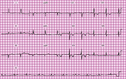
You interpret the ECG (Figure 27.1), and your consultant asks you your opinion.

You institute urgent therapy, as per guidelines. Five minutes after administration of the GTN the chest pain has improved but not resolved, and he is given 10 mg IV morphine which results in complete resolution. He is admitted to the coronary care unit for continuous ECG monitoring.

Thirty minutes later the troponin-I comes back at 1.8 µg/L (normal range <0.04 µg/L).

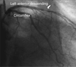
The day after presentation the patient is taken to the cardiac catheterization laboratory where a coronary angiogram is performed (Figure 27.2). It shows a severe stenosis of the left anterior descending coronary artery and this is treated with percutaneous coronary intervention (PCI) with the use of a bare metal stent. A left ventriculogram is also performed and shows normal left ventricular systolic function. No heparin is required after a PCI, so the heparin infusion is stopped.

The day after the PCI (48 hours after admission), the patient is asymptomatic, has no abnormalities on cardiovascular examination and ECG monitoring has shown no arrhythmias.

He is discharged the day after the PCI, with no complications. You see him in clinic after 6 weeks, and he is actively participating in the cardiac rehabilitation programme at your hospital. You advise him that he is doing well, and to continue his current medications.

Answers

A.1

A.2

A.3

A.4 An acute coronary syndrome (ACS).

This patient has symptoms which have been shown to be predictive of coronary artery disease (CAD), namely retrosternal location of pain, radiation to the arms, recent discomfort on physical exertion with relief from rest. He also has two well-established risk factors for CAD. Unlike some other causes of chest pain, CAD often has relapsing symptoms, so that patients presenting with chest pain will often have a history of investigation or treatment for CAD and this will often have included revascularization either surgically or percutaneously.

A.5 Current guidelines recommend that a patient presenting with symptoms that potentially represent an acute coronary syndrome should have an ECG within 10 minutes of presentation. The most important task on examining the ECG is to find or exclude ST segment elevation in a pattern consistent with an ST elevation myocardial infarction (STEMI) or find a new left bundle branch block (LBBB), because these patients are given a diagnosis of STEMI and require emergency reperfusion therapy (either thrombolysis or percutaneous coronary intervention). Crucially, no other investigation is required to make this diagnosis and institute reperfusion therapy.

Other investigations performed at presentation include a chest X-ray, cardiac biomarkers (troponin), electrolytes and creatinine, full blood count and blood glucose level.

A.6 Sinus rhythm, normal axis and anterior T wave inversion.

A.7 The patient is having a non-ST elevation acute coronary syndrome (NSTEACS) (a syndrome being a collection of signs and symptoms).

All acute coronary syndromes can be put into one of two categories: (1) STEMI or (2) non-ST elevation ACS (NSTEACS). We have diagnosed this patient with an ACS and he does not have ST segment elevation, so we must diagnose a NSTEACS at this point.

A.8 It is certainly possible, but we cannot make that diagnosis until the cardiac biomarker result is available. A NSTEMI is diagnosed when a patient has symptoms consistent with an acute coronary syndrome accompanied by cardiac biomarkers above the upper limit of normal. No matter what abnormalities are present on the ECG, a NSTEMI cannot be diagnosed unless the cardiac biomarkers are elevated, and we do not know that yet.

A.9 The patient should be placed on electrocardiographic monitoring and given guideline-based treatment for a NSTEACS:

A.10 The biomarker elevation enables us to now diagnose a NSTEMI – i.e. a myocardial infarction.

Patients with a NSTEACS have a variable short-term risk of death or myocardial infarction depending on how many high-risk features are present. The best known of these features are cardiac biomarker elevation (troponin) and ischaemic ECG changes. But other variables have also been shown to be important for risk stratification including age, congestive cardiac failure, haemodynamic compromise, ventricular arrhythmias, previous known coronary disease and diabetes. Summation of high-risk features can be performed to produce a risk score, the TIMI and GRACE scores being the best known examples.

The elevated troponin level (NSTEMI) in our patient immediately puts him in a high-risk category. However, it is important to note from the risk variables mentioned in the previous paragraph that it is possible to be high risk without biomarker elevation.

A.11 Probably not at this point. Although trials have shown that these agents reduce myocardial infarction in high-risk NSTEACS patients, recent trials have shown no coronary benefit and an increased risk of bleeding from their routine use in these patients prior to coronary angiography. Their use can be left to the discretion of the interventional cardiologist at the time of angiography.

A.12 Yes. Patients with a high-risk NSTEACS benefit from early coronary angiography to guide coronary revascularization during their initial hospitalization. Guidelines recommend angiography be performed within 48–72 hours in high-risk patients if they remain pain free and haemodynamically stable. The revascularization can be performed by percutaneous coronary intervention (PCI) or by coronary artery bypass grafting (CABG), depending on the distribution of coronary disease. Generally, the more widespread the coronary disease, the more suitable the patient is for CABG rather than PCI.

A.13 The day after the PCI in the absence of a complication. Patients with a NSTEACS treated with PCI are at low risk of developing recurrent ischaemia or life-threatening arrhythmias more than 24 hours after the onset of chest pain. They are generally monitored electrocardiographically for a total of 48 hours and are able to be discharged 24 hours after the PCI provided they are stable and the PCI was performed without complication.

A.14 Discharge medications include the following combination:

A.15 Treatment of the hypertension should continue with a target blood pressure of <130/80 mmHg, as is recommended for patients with established coronary disease.

The patient should be advised to call an ambulance if he has chest pain that has not resolved 10 minutes after GTN.

He should attend a cardiac rehabilitation service for advice on physical activity, nutrition, exercise and alcohol consumption. Trial evidence supports this recommendation.

He should consume about 1000 mg of omega-3 polyunsaturated fatty acids per day, through a combination of oily fish and fish oil capsules or liquid. Trial evidence supports this recommendation.

You should advise him about the legality of driving, and consult your local guidelines. This is important, and often overlooked. Providing him with a copy of the driving guidelines may be helpful.