137
Malignant melanoma, lentigo maligna
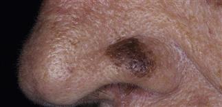
Melanoma represents 4% of all cancers in men and 3% of all cancers in women. This innocuous pigmented lesion on the nose was shown to be lentigo maligna on biopsy.
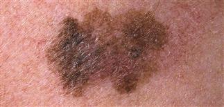
It cannot be overemphasized that melanomas vary considerably—no single color or change is diagnostic. This melanoma has areas of inflammation (red), regression (white), and deeper pigment (blue).
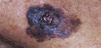
Lesions tend to be greater than 6 mm in diameter, flat, and asymmetric, with varying coloration. This advanced lesion shows ulceration.
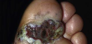
Lesions appear and tend to spread laterally within the skin over a few years, before nodules develop within the lesion and necrosis develops, as shown here in this acral lentiginous melanoma.
DESCRIPTION
An increasingly common malignancy of melanocytes.
HISTORY
• Lifetime risk of melanoma is 1 in 75. Risk factors include skin types 1 or 2, atypical nevi, personal or family history of melanoma, history of blistering sunburn, large congenital nevi. • Thirty percent of melanomas develop within a pre-existing nevus. • The thinner the melanoma, the better the prognosis.
PHYSICAL FINDINGS
Melanomas vary in appearance. No single color or change is diagnostic. When melanoma develops in a pre-existing lesion, there is usually a focal color change. Four clinical subtypes of melanoma are recognized. • Superficial spreading melanoma. Most common subtype. Occurs most often on trunk and extremities. Lesions usually flat, asymmetric, with varying colors. Tends to spread laterally. • Nodular melanoma (10–15%). Tends to occur on extremities as raised, brown to black, rapidly growing papules. • Lentigo maligna and lentigo maligna melanoma (5–10%). Represent in situ melanoma and progression to invasive melanoma. Develop over years on sun-exposed white skin, most often on face. Lesions flat, brown, mottled. • Acral lentiginous melanoma (7%). Occurs on hands and feet, including nails of people with darker skin types. Lesion similar in appearance to lentigo maligna.
Amelanotic melanoma (2%). Describes a non-pigmented melanoma of any subtype. The lesion is an innocent-appearing, enlarging, pink-red papule.
TREATMENT
• Biopsy report should state diagnosis, anatomic site, Breslow level (vertical thickness), whether margins are involved. Breslow level is single most important prognostic factor. Ulceration is the second most important prognostic factor. • Palpate regional nodes. Suspicious nodes should be evaluated by nodal biopsy. Best biopsy technique is complete excision of entire lesion. Shave biopsy not recommended. May consider incisional or punch biopsy for large lesions or lesions in cosmetically important areas. The most suspicious area should be included in biopsy. • Once melanoma confirmed by biopsy, reexcision with appropriate surgical margins is determined by Breslow. Melanoma in situ requires margin of 0.5 cm. A 1.0-cm margin recommended for tumors up to 2.0 mm thick; a margin of 2.0 cm recommended for tumors up to 4.0 mm thick. Sentinel node biopsy is performed for thicker tumors. Sentinel lymph node status predicts risk of recurrence and mortality. • Adjuvant treatment for advanced disease often recommended with interferon. • Follow-up examination is performed at regular intervals. Includes visual examination of entire skin surface, palpation of regional and distant nodes, and palpation of liver.


