194 Malignant melanoma
Salient features
History
• Age (<50 and location on trunk has better prognosis)
• Family history (a patient with a dysplastic naevus with two relatives having malignant melanoma has a 300 time increased chance of having a malignant melanoma)
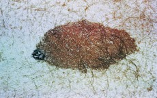
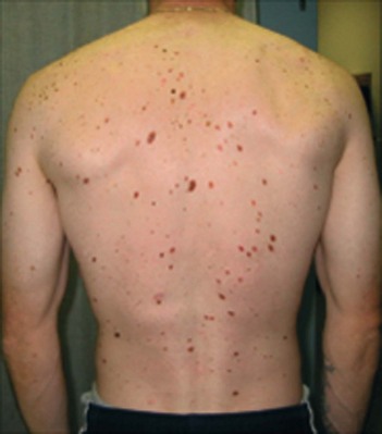
• Remember MacKie’s four independent risk factors for melanoma: freckles, moles (Fig. 194.1), atypical naevi (Fig. 194.2) and a history of severe sunburn.
Examination
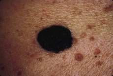
• Irregular and asymmetrical, with a fuzzy border where the pigment appears to be leaking into the surrounding skin (Fig. 194.3). (The American Cancer Society mnemonic to suspect malignancy in a mole is ABCDE: asymmetry, border irregular, colour variegation and diameter >6 mm; elevated lesions are more suspicious.)
Questions
What are the different types of malignant melanoma?
Depending on the clinical picture and histological findings these include:
• superficially spreading malignant melanoma (commonest type)
• acral lentiginous melanomas (arising on the palms, soles and nail beds)
• malignant melanoma on mucous membranes
• lentigo maligna melanoma (arising in lentigines in older individuals)
• amelanotic (non-pigmented) melanoma
• miscellaneous: melanoma arising from blue naevi, congenital and giant nevocytic naevi.
Advanced-level questions
What are the patterns of growth of malignant melanoma?
• Radial growth: where the growth is horizontal within the epidermis and superficial dermal layers (e.g. lentigo maligna, acral/mucosal lentiginous, superficial spreading types). During this stage of growth, the tumour does not have the capacity to metastasize.
• Vertical growth: occurs with time, and the melanoma grows downward into the deeper dermal layers as an expansile mass lacking cellular maturation; this correlates with the emergence of a clone of cells with metastatic potential. The nature and extent of this vertical growth phase determines the biological behaviour of malignant melanoma.
What is the treatment of such lesions?
• Excision: following histological examination the area is usually re-excised with margins dictated by the thickness of the tumour
• Elective lymph node dissection is controversial; in general, it is indicated only in melanomas of intermediate thickness with one draining chain of lymph nodes
• Intralesional injection of avirulent herpes simplex virus (but capable of replication) may be of therapeutic benefit in malignant melanoma; this is an experimental therapy (Lancet 2001;357:525)
• Interferon-alfa and vaccine therapy may reduce recurrences.
What are the prognostic factors in these patients?
• Tumour thickness: the deeper the melanoma the more likely is metastasis. Thin melanomas (<0.76 mm) approach 100% cure rate whereas only 20–30% survive for 5 years if the lesion has a depth >1.6 mm
• Anatomical location: melanomas in the TANS areas (thorax, upper arms, neck and scalp) show a higher relative risk.


