55. Malignant Atrophic Papulosis
Definition
Malignant atrophic papulosis (MAP) was originally described as a variant of thromboangiitis obliterans, but was quickly recognized to be a distinct disease entity. MAP is characterized by narrowing and eventual occlusion of the lumen of small-caliber blood vessels by intimal proliferation and thrombosis. Ultimately, ischemia and infarction of the affected organ or organ system can occur. There are two distinct forms: (1) malignant, which can affect the heart, lungs, eyes, kidneys, pericardium, central nervous system, peripheral nervous system, and gastrointestinal system; and (2) benign, which is limited to the skin. Malignant atrophic papulosis is also known as Degos syndrome.
Incidence
The incidence of MAP is rare. Only about 200 cases have been reported worldwide since its initial description in 1941. Malignant atrophic papulosis is found among both sexes, predominantly in young adults and in Caucasians of Europe and North America, although cases have been documented in Japan, India, and continental Africa.
Etiology
The etiology of MAP remains unknown. There have been suggestions of autoimmune, hypersensitivity, viral, and genetic bases for this disease, but to date none have been confirmed. It is also theorized that MAP is not a single entity, but the product of several disease processes.
Signs and Symptoms
• Abdominal distention
• Abdominal pain
• Aphasia
• Bowel infarction
• Cerebral edema
• Constipation
• Constrictive pericarditis with calcification
• Decreased platelet aggregation
• Diarrhea
• Dizziness
• Facial or extremity paresthesias
• Fatigue
• Fistula(e)
• Gaze palsy
• Headache
• Hemiplegia
• Impaired fibrinolytic activity
• Increased intracranial pressure
• Malabsorption symptoms
• Nausea
• Paraplegia
• Peritonitis
• Pleural effusion
• Pleuritis
• Seizures
• Severe restrictive pulmonary disease
• Spastic paralysis
• Uncal herniation
• Viscus perforation
• Vomiting
• Weakness
• Weight loss
 |
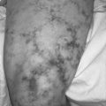
| Malignant Atrophic Papulosis. Malignant atrophic papulosis, also called Degos’ disease, in a patient with systemic lupus erythematosus. |
Medical Management
Since the initial description of MAP in 1941, various medications have been used to treat it, including corticosteroids, immunosuppressants, sulfonamides, tetracyclines, aspirin, dipyridamole, anticoagulants, penicillin, and interferon alfa-2a. As yet, none of those medications has demonstrated clearly any superior effectiveness in alteration of the disease process. The prognosis is that this disease is typically fatal within 2 to 3 years after the diagnosis of systemic involvement. Frequently surgical intervention is required to correct or treat gastrointestinal bleeding, perforated viscus, infarcted bowel, or possible intracranial bleeding.
Complications
• Bowel ischemia
• Cerebral infarction
• Epidural hemorrhage
• Gastrointestinal bleeding
• Intestinal perforation
• Intracerebral hemorrhage
• Neuropathy
• Pericarditis
• Peritonitis
• Pleuritis
• Spinal cord infarction
• Subdural hemorrhage
Anesthesia Implications
Because of the frequency of gastrointestinal symptoms and involvement, the patient with MAP may be either acutely or chronically dehydrated. Concomitantly the patient’s electrolyte concentrations may be significantly altered from normal levels. If the situation allows, it would be prudent to correct the fluid and electrolyte imbalances before administering anesthetia.
Both heart tones and breath sounds should be auscultated preoperatively. The patient with significant pericardial involvement may demonstrate a friction rub. Electrocardiograph and chest x-ray are also necessary to fully evaluate the patient’s pulmonary and cardiac status. The anesthetist may find any manner of alterations in the electrocardiograph as a result of small infarcts over time. The presence of restrictive lung disease will require the anesthetist to reduce the peak inspiratory pressures and prolong the inspiratory time. Usually this is best accomplished by selecting the pressure-controlled ventilation option available on most late-model anesthesia machines and reducing the peak inspiratory pressure for each ventilation.
The anesthetist should also have bleeding times determined preoperatively for the patient with MAP. Both fibrinolytic activity and platelet aggregation are altered by MAP. As a result, the anesthetist should anticipate greater blood volume losses than would usually be anticipated with a procedure.
Both the peripheral and central nervous systems may be significantly altered by MAP. The anesthetist should thoroughly document any functional deficits of the peripheral nervous system preoperatively. More significant is the focus on the effects MAP may produce in the central nervous system. These effects may be quite extensive. The lesions may ultimately produce cerebral edema, which can culminate in increased intracranial pressure. The anesthetist must be astute and thorough in documenting signs and/or symptoms of increased intracranial pressure. Anesthesia-related medications should be selected with the patient’s increased intracranial pressure in mind. Appropriate medications include thiopental, propofol, and narcotic analgesics. The patient should be mildly hyperventilated throughout the procedure to reduce the degree of cerebral edema. The severity of the increased intracranial pressure will be a determining factor in deciding whether to extubate the patient at the end of the procedure or to maintain mechanical ventilatory support for a period of time—an eventuality that must be thoroughly discussed with the patient and/or family preoperatively.