Lymphadenopathy
Lymphadenopathy is a common presenting condition. It may be localised or generalised. The causes are multiple but a careful history and clinical examination will often simplify the diagnosis. Lymphadenopathy, especially cervical, is extremely common in children who are otherwise healthy. Painful tender nodes are usually associated with infection. Firm or hard, painless nodes are commonly the seat of malignancy. Only the commoner causes, which the student would be expected to know, are described in this section.
History
Infection
Local infections will be usually obvious, e.g. dental abscess with cervical lymphadenopathy. With generalised lymphadenopathy, there may be a history of malaise, lethargy and fever. Check for exposure to TB, which may be a cause of lymphadenopathy in the immunocompromised patient. Check for visits abroad. Check if there are animals in the household, e.g. cats (cat scratch fever) or dogs (toxoplasmosis). In the case of cat scratch fever, the scratches are often healed before the patient presents. Note any history of local trauma.
Primary malignancy
There will normally be a history of malaise, fever or night sweats. The patient may have noticed lumps at several sites. Spontaneous bruising and bleeding associated with thrombocytopenia may also be present.
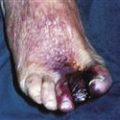
Secondary malignancy
The primary may be obvious or may be very small and have not been noticed by the patient (e.g. a malignant melanoma in an inaccessible site). There may be a history of malignancy that has been treated several years previously with metastases presenting late (e.g. axillary lymphadenopathy or cervical lymphadenopathy occurring several years after an apparently curative operation for carcinoma of the breast). Occasionally the lymph node swelling is distant from the primary, e.g. cervical lymphadenopathy (left supraclavicular fossa) from testicular cancer; Virchow’s node in gastric cancer.
Other conditions
Sarcoidosis causes bilateral hilar lymphadenopathy but may present with lymphadenopathy at other sites. Check for a history of SLE or rheumatoid arthritis.
Examination
Examine the enlarged lymph nodes. Are they painful and tender, suggesting infection, or are they firm and painless, suggesting malignancy? Check the sites draining to these nodes for a site of infection or primary malignancy. Check for linear scratches suggestive of cat scratch disease. Examine for bruising. Examine all other sites of potential lymphadenopathy: cervical, axillary, inguinal, popliteal and epitrochlear nodes. Check for splenomegaly and hepatomegaly. Carry out a full general examination.
General Investigations
■ FBC, ESR
Hb ↓ blood dyscrasias. Platelets ↓ dyscrasias. WCC ↑ infection, leukaemia. ESR ↑ tumour, infection. Blood film. Leukaemia.
■ LFTs
Malignant infiltration of the liver.
■ Clotting screen
Blood dyscrasias.
■ Viral titres
Viral infections, e.g. EBV, HIV.
Specific Investigations
■ Antibody screen
SLE, rheumatoid arthritis.
■ Serum angiotensin-converting enzyme (ACE)
Sarcoidosis. Positive in 50–80% of patients with active sarcoidosis.
■ Toxoplasma screen
Toxoplasmosis.
■ OGD
Virchow’s node with gastrointestinal malignancy.
■ CT
Nodal distribution. Staging of Hodgkin’s disease.
■ FNAC of lymph node
Malignancy versus other conditions.
■ Biopsy
Lymph node, e.g. Hodgkin’s disease, secondary deposit. Biopsy of local lesion, e.g. infective versus malignant.