222 Lymphadenopathy
Salient features
History and examination
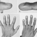
Use the mnemonic ALL AGES to approach a patient with lymphadenopathy:
• As soon as you feel a group of lymph nodes, examine drainage areas for obvious pathology. For example:
• Examine other lymph node areas in a systemic manner: submental, submandibular, deep cervical (upper and lower), occipital, posterior triangle, supraclavicular, axillary, epitrochlear and inguinal.
• Examine the mouth for the following signs:
• Examine the abdomen for liver and spleen.
• Examine the chest and do a chest radiography: for bronchogenic carcinoma, TB.
Questions
What are the causes of regional lymphadenopathy?
Cervical lymphadenopathy. Causes include infections and malignancies. Infectious causes include bacterial pharyngitis, dental abscess, otitis media, infectious mononucleosis, cytomegalovirus, gonococcal pharyngitis, toxoplasmosis, hepatitis and adenovirus. Malignancies include non-Hodgkin disease, Hodgkin disease and squamous cell carcinoma of head and neck.
Virchow node (anterior left supraclavicular lymph node; also known as Troiser’s ganglion). Enlargement suggests the presence of a thoracic or abdominal neoplasm (Am J Surg 1979;138:703). Common causes include carcinoma of breast, bronchus, lymphomas and GI neoplasms.
Delphian node (a midline prelaryngeal lymph node). Heralds thyroid disease, laryngeal malignancy or lymphoma.
Axillary lymphadenopathy. Causes include infections and malignancies. Infectious causes include staphylococcal, streptococcal infections of the arm, cat-scratch fever, tularaemia and sporotrichosis. Malignant causes, including Hodgkin’s disease, non-Hodgkin lymphoma, carcinoma of breast and melanoma, are common.
Epitrochlear lymphadenopathy. The most common causes are lymphoma/chronic lymphatic leukaemia and infectious mononucleosis. Other diagnoses include HIV, sarcoidosis and connective tissue disorders. In developing countries, secondary syphilis, lepromatous leprosy, leishmaniasis and rubella are important causes.
Inguinal lymphadenopathy. In adults, some degree of lymph node enlargement is not uncommon. In those who walk outdoors with footwear, benign reactive lymphadenopathy is common. Malignant causes include lymphoma, malignant melanoma, carcinoma of external genitalia. Benign causes include cellulitis, syphilis, chancroid, genital herpes and lymphogranuloma venereum.
Node of Cloquet (also known as Rosenmüller node). A deep inguinal lymph node located near the femoral canal. When palpable it may be mistaken for an inguinal hernia.
What are the causes of generalized lymphadenopathy?
Advanced-level questions
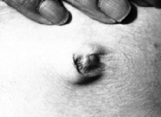
What do you know of Sister Joseph’s nodule?
It is a classic sign of gastric adenocarcinoma i n the umbilical area and may represent a metastatic deposit or an enlarged anterior abdominal wall lymph node (Fig. 222.1).
How is Hodgkin’s disease staged?
Stage I: one lymph node region involved
Stage II: more than two lymph node regions involved on one side of the diaphragm
Stage III: lymph nodes involved on both sides of the diaphragm
Stage IV: involvement of extranodal sites(s) beyond that designated as ‘E’ below.
How is a patient with Hodgkin’s disease treated?
• Localized disease (i.e. stages IA, IIA) is treated with radiotherapy. Five-year cure rates are in excess of 80% and relapses are treated with chemotherapy.
• Disseminated disease (stages IIIB, IV) is treated with combination chemotherapy with the ABVD regimen (adriamycin, bleomycin, vincristine, dacarbazine) alternating with the MOPP regimen (mechlorethamine, oncovin, procarbazine and prednisolone). Five-year survival rates are about 50%. Patients who relapse after initial chemotherapy may be cured with autologous bone marrow transplantation.
• In stages IIB and IIIA, optimal management is controversial but current evidence suggests an advantage to combination chemotherapy (N Engl J Med 1993;328:560).
How are non-Hodgkin’s lymphomas classified?
A working classification is as follows.
Low grade or favourable. There is a slow, indolent course with good response to minimal therapy and long survival. These include small lymphocytic, plasmocytoid, follicular small cleaved cell and follicular mixed cell. Patients are usually treated with alkylating agents such as chlorambucil or a CVP regimen (cyclophosphamide, vincristine and prednisolone).
High grade or unfavourable. This includes immunoblastic, small non-cleaved (Burkitt’s and non-Burkitt’s), lymphoblastic and true histiocytic lymphoma. The mainstay of therapy is combination chemotherapy; traditionally the CHOP regimen (cyclophosphamide, doxorubicin, vincristine and prednisolone) is used. In highly aggressive lymphomas, autologous bone marrow transplantation may increase the chances of cure.
Intermediate grade. Follicular large cell, diffuse small cleaved cell, diffuse mixed cell, diffuse large cell.
Others. Cutaneous T cell (mycosis fungoides, adult T cell leukaemia/lymphoma).
What symptoms of Non-Hodgkins’s lymphoma merit urgent referral to an oncologist?
Symptoms of non-Hodgkin lymphoma needing urgent referral include (Lancet 2003; 362:139–46):
What are the risk factors in localized Hodgkins’ lymphoma?
What are the poor prognostic factors of advanced Hodgkin’s lymphoma?
Hasenclever index is an international prognostic score including the following seven factors:
What are the late effects of Hodgkin’s lymphoma and its treatment?
What are the investigations done before initiation of treatment on non-Hodgkins’s lymphoma?
Essential procedures (Lancet 2003; 362:139–46):
• A full history, recording growth rate, symptoms present, performance status
• A detailed physical examination, with special attention to all node-bearing areas including Waldeyer’s ring
• Adequate surgical biopsy specimen, allowing immunophenotyping and examined by a skilled pathologist
• Bone marrow aspirate and trephine, to include molecular genetic analysis if available.
Optional procedures, depending on clinical picture:
• Endoscopy, e.g. for gastric MALT lymphoma
• Plain radiographs, bone scan or MRI
• Positron electron tomography (PET)