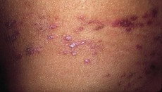
169 Lupus pernio
Salient features

Advanced-level questions
How may sarcoid present?
• Acute sarcoidosis usually presents in the third decade, characterized by erythema nodosum, parotid enlargement and hilar lymphadenopathy.
• Chronic sarcoidosis usually presents in the fifth decade with an insidious onset characterized by fatigue, dyspnoea, arthralgia and lupus pernio. Bone cysts are another feature.
How would you investigate such a patient?
• FBC and ESR: leukopenia, eosinophilia and raised ESR
• Chest radiography. Three stages of sarcoidosis are defined depending on radiographic findings, although these stages do not indicate chronology of disease:
• Slit-lamp examination of the eyes
• Kviem’s test: the antigen is cultured in human spleen and thus, increasingly, it is recommended that this test should not be performed because of the risk of transmitting viral infections
• Mantoux test: skin anergy is present in 70%
• Serum ACE levels: raised in 40–80% with active disease, although this finding is neither sensitive nor specific enough to have diagnostic significance
• Lung function tests: usually restrictive changes with decreased lung volumes and diffusing capacity; occasionally airflow obstruction
• Serum calcium levels: hypercalcaemia in 10% of patients
• Bronchoalveolar lavage: usually characterized by an increase in lymphocytes and a high CD4:CD8 T cell ratio
• Histological confirmation is by the presence of non-caseating granulomas with typical manifestations (fibrotic response develops over time) in biopsy of lung (transbronchial), lymph node, skin, liver, gums or minor salivary glands.
What are the specific indications for systemic steroids in sarcoidosis?
• Progressive deterioration in lung function, particularly transfer factor and vital capacity. Serial evaluation should be performed every 2 months initially, gradually increasing the intervals between follow-up to about 4–5 months; patients should be followed for at least 2 years after steroids are stopped
What is Löfgren syndrome?
Erythema nodosum, hilar adenopathy and polyarthralgias in a patient with sarcoidosis.
What are the diagnostic features of cardiac sarcoidosis?
• Histologic diagnosis group: cardiac sarcoidosis is confirmed when histologic analysis of operative or endomyocardial biopsy specimens demonstrates epithelioid granuloma without caseating granuloma (Dis Mon 2009;55:675–92).
• Clinical diagnosis group: in patients with a histologic diagnosis of extracardiac sarcoidosis, cardiac sarcoidosis is suspected when item (a) and one or more of items (b) to (e) are present:


