Lung Cancer: Epidemiology, Surgical Pathology, and Molecular Biology

Published on 29/05/2015 by admin

Filed under Pulmolory and Respiratory

Last modified 22/04/2025

Print this page

rate 1 star rate 2 star rate 3 star rate 4 star rate 5 star
Your rating: none, Average: 0 (0 votes)

This article have been viewed 1348 times

Chapter 65 Lung Cancer

Epidemiology, Surgical Pathology, and Molecular Biology

Epidemiology

Incidence and Survival

Lung cancer currently is the highest cause of cancer mortality in the United States and even surpasses the sum of the next four cancer types in both men and women. In terms of incidence, (the number of new cases of cancer in a given year), lung cancer is second only to breast cancer in women and prostate cancer in men. In the United States, estimates of new cases of lung cancer for 2010 were approximately 116,000 in men and 105,000 in women, with approximately 160,000 deaths. Lung cancer deaths accounted for 31% and 27% of overall cancer deaths in men and women, respectively, and are more numerous than deaths due to breast, prostate, and colon cancers combined. In terms of overall causes of death in men, lung cancer is the second most common cause of death, after vascular disease, with deaths due to heart attacks and stroke. The overall 5-year lung cancer survival rate is between 15% and 20% as a consequence of its late stage at onset of symptoms and relatively aggressive clinical behavior. Like other malignancies, lung cancer is a disease of aging, with increasing rates in persons older than 50 years of age, and rarely is diagnosed in those younger than 40 years. Graphing lung cancer incidence according to chronologic age of diagnosis on a logarithmic scale demonstrates a straight line for both men and women, indicating that the carcinogenic pathways probably are similar for both genders and that steroid hormones (estrogen and androgens) do not play a major role in carcinogenesis.

Lung cancer more typically is present in men than in women, and in African Americans than in white Americans; these disparities in incidence probably are due to smoking patterns. The disparity extends to younger persons as well; young African American men and women have significantly higher lung cancer rates than their white counterparts. The incidence of lung cancer in adult African American and in white American male adults in 2008 was 101 and 69 per 100,000, respectively. Both African American and white American women had similar cancer incidences of approximately 55 cases per 100,000. Other ethnic and racial groups, such as Hispanic persons, Native Americans, and Asian or South Pacific Americans have a lower incidence than that for white Americans.

Lung cancer incidence is highest in the developed nations of North America (United States and Canada), the European Union, and countries of the former Soviet Union, where cigarette availability and smoking rates are highest. In 2008, lung cancer was diagnosed worldwide in approximately 1.6 million people and led to cancer deaths in 1.4 million, and a majority of new cases are reported from China and less developed countries. Since the 1970s, lung cancer incidence has been decreasing in the United States, Canada, United Kingdom, and Australia but has been increasing in Japan and India. Unfortunately, during this same time, lung cancer incidence has been increasing among women in the United States, Canada, Denmark, Norway, and Sweden. Worldwide, lung cancer incidence is more than double for men relative to women, and the relative risk is even higher in Western Asia, Central and Southern America, and Southern Africa.

Although lung cancer is increasing in large developing nations, such as China and India, the socioeconomic demographic features are important in appreciating lung cancer incidence. The risk of lung cancer is inversely associated with education and income and appears to be more closely linked to smoking than risk of disease from occupational or environmental exposures. Data from developing nations are not as robust and accurate as those from Western countries, and analysis of the socioeconomic setting of smoking also involves confounding variables, such as local diet and environmental carcinogens and inhalant exposures.

When the mortality rates (deaths due to disease within a calendar year) for lung cancer in women and men are plotted against the calendar years for the last 50 years, one notes a marked difference in the pattern of the curves (Figure 65-1). The incidence rates for men show a marked rise from the early 1950s to the beginning of the 1990s, whereas rates for women lag by about a quarter of a century and then show an identical rise, essentially parallel to those for men, and continuing into the 21st century. By the 1950s, death due to lung cancer far exceeded prostate and colon cancer deaths among men, and by 1990, lung cancer deaths among women exceeded breast and gynecologic cancer deaths. Usually cancer mortality follows cancer incidence; however, in the case of lung cancer, owing to the very high cancer fatality rate (mortality from cancer for persons given a diagnosis of cancer), lung cancer mortality exceeds the most common cancers in both men and women. As a result of the Surgeon General’s 1964 report on smoking, men began to stop smoking in the succeeding 20 to 30 years; the incidence and mortality of lung cancer began to decline from a peak in the early 1990s to a level that approximates the mortality rate of the 1970s. The phenomenon of social smoking among women lagged behind that for men; subsequently, both the incidence of lung cancer in women and its associated mortality continue to rise into the present day.

image

Figure 65-1 A, SEER incidence and U.S. death rates for cancer of the lung and bronchus (men and women). Joinpoint Regression analyses for whites and blacks from 1975 to 2007 and for Asian/Pacific Islanders, American Indians/Alaska Natives, and Hispanics from 1992 to 2007. API, Asian Pacific Islander; AI/AN, American Indian/Alaska Native. B, SEER-delay adjusted rates for cancer of the lung and bronchus mortality differences among men and women of all races in the United States (1975-2007). SEER, Surveillance, Epidemiology, and End Results [Program].

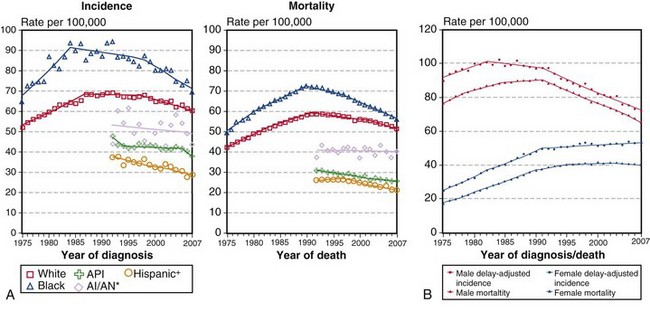
(A, Incidence data for whites and blacks are from the SEER nine areas—San Francisco, Connecticut, Detroit, Hawaii, Iowa, New Mexico, Seattle, Utah, Atlanta. Incidence data for Asian/Pacific Islanders, American Indians/Alaska Natives, and Hispanics are from the SEER thirteen areas—the SEER nine areas plus San Jose–Monterey, Los Angeles, Alaska/Native Registry, and Rural Georgia. Mortality data are from U.S. mortality files, National Center for Health Statistics, Centers for Disease Control and Prevention [CDC]. Rates are age-adjusted to the 2000 U.S. standard population [19 age groups, Census P25-1103]. Regression lines are calculated using the Joinpoint Regression program Version 3.4.3, April 2010, National Cancer Institute. Joinpoint analyses for whites and blacks during the 1975-2007 period allow a maximum of 4 joinpoints. Analyses for other ethnic groups during the period 1992 to 2007 allow a maximum of 2 joinpoints. *Rates for American Indians/Alaska Natives are based on the contract health service delivery area [CHSDA] counties. †Hispanic is not mutually exclusive of whites, blacks, Asians/Pacific Islanders, and American Indians/Alaska Natives. Incidence data for Hispanics are based on the NHIA [North American Association of Central Cancer Registries/NAACCR Hispanic Identification Algorithm] and exclude cases from the Alaska Native Registry. Mortality data for Hispanics exclude cases from Connecticut, the District of Columbia, Maine, Maryland, Minnesota, New Hampshire, New York, North Dakota, Oklahoma, and Vermont. B, Data from SEER nine areas and U.S. mortality files [National Center for Health Statistics, CDC]. Rates are age-adjusted to the 2000 U.S. standard population [19 age groups, Census P25-1103]. Regression lines are calculated using the Joinpoint Regression Program Version 3.4.3, April 2010, National Cancer Institute [http://seer.cancer.gov/statfacts/html/lungb.html].

Although lung cancer typically refers to the malignant bronchogenic epithelial tumors of the lung, namely, squamous cell carcinoma, adenocarcinoma, large cell carcinoma, and small cell (neuroendocrine) carcinoma, the relative incidence, distribution, and frequency of these tumors over time are characterized by marked differences. In the 1970s, squamous cell carcinoma predominated in incidence over the other bronchogenic carcinomas. Of interest, the age-adjusted incidence rate of adenocarcinoma, among both men and women, continued to rise, so that beginning in the late 1990s and continuing into the early 2000s, adenocarcinomas have become the most frequently encountered lung cancers. The two histologic subgroups of lung cancer most strongly associated with smoking, squamous cell carcinoma and small cell carcinoma, have shown a decrease in their overall incidence, reflecting the trend of decreased cigarette smoking among men. The relative percentages of the major lung cancer histologic types are adenocarcinoma 38%, squamous cell carcinoma 20%, small cell (neuroendocrine) carcinoma 13%, and large cell carcinoma 5%. The remainder are composed of variant carcinomas, sarcomatoid (spindle cell) carcinomas, salivary-type carcinomas, neuroendocrine-carcinoid carcinomas, and others.

The histologic sequence eventuating in squamous cell carcinoma begins with smoking-induced replacement of respiratory epithelium with bronchial squamous metaplasia. Additional cellular and molecular events lead to squamous dysplasia, in situ carcinoma, and ultimately, invasive squamous cell carcinoma. Small cell carcinoma does not have a clearly defined precursor lesion, although it is presumed to be the Kulchitsky cell; however, small cell carcinomas often arise in a setting of squamous metaplasia and dysplasia, indicative of the effects of the smoking environment. Adenocarcinoma, a histologic subtype characterized by formation of malignant-appearing glands, papillae, and intracytoplasmic mucin, also arises in the context of cigarette smoking, yet approximately 25% of lung cancer cases are not attributable to direct smoking. These tumors typically are located peripherally and may be associated with scars that in some cases may be preexisting but also may represent a host desmoplastic response to the tumor. As a histologic subtype, adenocarcinomas of the lung continue to increase in incidence among both men and women.

Etiology and Risk Factors

The major risk factor associated with lung cancer in a majority of cases is cigarette smoking. The probability that a diagnosis of lung cancer will be made in a nonsmoker is less than 15%. The case for cigarette smoking as a major cause of lung cancer is made by strong epidemiologic evidence and by significant results of animal studies. Both case-control and cohort studies have demonstrated the strong association of smoking with lung cancer and have shown that the association is dose-dependent and linked to current versus past smoking behavior. Initial case-control studies investigating retrospective reviews of smoking behavior in groups with and without lung cancer calculated odds ratios of approximately 10 : 1 for smoking associated lung cancers. Later, longitudinal cohort studies demonstrated mortality ratios averaging 10 : 1 for lung cancer deaths in the group of smokers relative to nonsmokers. Moreover, the annual incidence of lung cancer showed a logarithmic relationship with age and duration of smoking. Mortality ratios also demonstrated a dose-response relationship with the number of cigarettes smoked per day. In general, smokers have a 70% increase in mortality relative to nonsmokers.

Cigarette smoking is associated with all the histologic types of bronchogenic carcinoma, however, as discussed above, adenocarcinoma, a smoking related cancer, is also the most common type among nonsmokers. Although a history of many pack-years of cigarette smoking is strongly linked to lung cancer, only a minority of smokers develop lung cancer, suggesting that host biologic and genetic factors may modulate carcinogenic pathways in the development of lung cancer. The clinical history of current or past cigarette smoking also affects the probability of developing lung cancer, with the incidence of lung cancer in heavy smokers more than twice that in light smokers, and more than five-fold that in ex-smokers. Not only is cigarette smoking a risk factor for the development of lung cancer; it is also a predictor of disease recurrence after lung cancer resection in early stage disease. The 5-year overall survival rate for smokers relative to nonsmokers with stage I lung cancer is, respectively, 76% and 92%. There is a modest but real effect of lung cancer attributable to passive or environmental tobacco smoke, based on an update of the Surgeon General’s report. Nonsmokers married to smokers incur an approximate 25% relative increase over nonexposed nonsmokers for the induction of lung cancer but of course significantly less than that for active smokers.

In addition to the dominant effect of cigarette smoking, other occupational and environmental conditions are associated with an increased risk of lung cancer. The most well-accepted industrial agents associated with lung cancer are asbestos, hexavalent chromium(VI), arsenic, nickel, and polycyclic aromatic hydrocarbons. Ionizing radiation exposure, as experienced after the atom bomb explosion in Japan (1945), in uranium mining, and with radon encountered in industrial and home settings, also shows hazard rates for lung cancer similar to those for environmental or second-hand cigarette smoke. In persons exposed to industrial chemicals and who also smoke, a synergistic effect may be operative in the development of lung cancer.

Asbestos manufacturing, more so than asbestos mining, is associated with pulmonary fibrosis and lung cancer and also the development of malignant pleural mesothelioma. Asbestos workers who also smoke have a synergistic or multiplicative risk of lung cancer over those who do not smoke. Lung cancer in asbestos-exposed persons usually manifests in a setting of pulmonary asbestosis (interstitial fibrosis). It is believed by some investigators that the fibroinflammatory scarring process of pulmonary asbestosis creates a microenvironment in which cancers develop. On the other hand, other researchers propose that the fibrogenic sequence of interstitial fibrosis due to high asbestos fiber burden may not be the same as the carcinogenic pathway of asbestos-induced malignancies. Both pulmonary fibrosis and asbestos-related bronchogenic carcinomas are dose-dependent on the lung fiber burden. Although pulmonary asbestosis tends to predominate in the lower lobes, the asbestos-induced cancers are found in all lobes of the lung. In the case of pleural diffuse malignant mesothelioma, the tumor may be attributable to asbestos exposure even in the absence of pulmonary fibrosis or smoking.

Surgical Pathology and Cytopathology

The malignant epithelial tumors of the lung consist of the most common bronchogenic carcinomas: adenocarcinoma, squamous cell carcinoma, small cell carcinoma, and large cell carcinoma, and their subgroups. Less common malignant epithelial tumors include the sarcomatoid carcinomas, neuroendocrine-carcinoid tumors, and salivary gland tumors. Most tumors of the lung are advanced at presentation and tend to be higher-stage cancers. Cancers of the central airways are commonly diagnosed and may occur as an endobronchial mass or as a parenchymal mass with endobronchial extension or extrinsic bronchial compression and a possible resultant obstructive pneumonia. Clinically, affected patients present with cough, hemoptysis, and/or shortness of breath. The central tumors may be approached by interventional bronchoscopy for diagnosis and management. Those tumors that manifest as peripheral scars or solitary pulmonary nodules may be histopathologically assessed by needle core or aspiration biopsy.

Cytologic evaluation of sputum, bronchial washings or brushings, and fine needle aspiration (FNA) biopsy specimens (transbronchial, transesophageal, or percutaneous) has long been utilized in the diagnostic workup of lung masses and as a staging procedure by sampling hilar or mediastinal nodes. The diagnostic accuracy is highly dependent on the sampling method. Cytologic approaches in the right clinical setting can provide valuable information without the need for an invasive procedure such as mediastinoscopy. More recently, it has been shown that the addition of ultrasound guidance to the bronchoscopy or endoscopy procedure improves diagnostic accuracy, with sensitivity of more than 85% and specificity of 100%.

Traditionally, the role of cytologic analysis was to first establish the diagnosis of malignancy and then to further stratify the carcinoma into two main categories of small cell carcinoma and non–small cell carcinoma, for appropriate management. With the advent of targeted therapy, however, this approach no longer provides the necessary information, because major changes have evolved in the treatment of squamous cell carcinoma and adenocarcinoma that may involve approaches dependent on the presence or absence of genetic mutations. Several studies have addressed the feasibility and accuracy of cytologic studies in this respect, especially when immunohistochemical methods are used in conjunction with cytomorphologic examination. Furthermore, it has been shown that molecular studies for EGFR/KRAS mutation analysis can be successfully carried out on cytologic preparations.

Adenocarcinoma

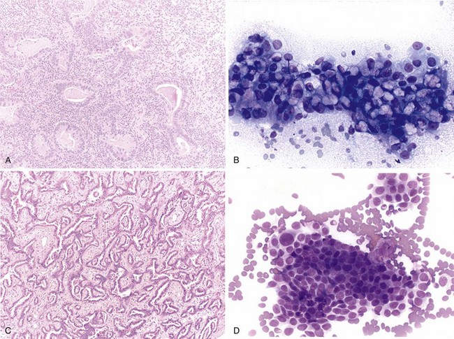
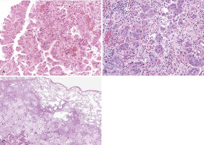
Adenocarcinomas are tumors that produce malignant-appearing glands with tubular, acinar, or papillary differentiation and whose cells may demonstrate mucin production and secretion (Box 65-1). On gross examination, the tumors may manifest peripherally with pleural retraction (puckering), as central or endobronchial masses, as diffuse pleural involvement resembling a malignant mesothelioma, arising or associated with a scar, or as a diffuse parenchymal pneumonic-like tumor. The histologic pattern and organization may show a well-differentiated acinar pattern with intracytoplasmic vacuoles or more poorly differentiated features with solid malignant growth pattern and minimal mucin expression, as demonstrated by histochemical and/or immunohistochemical staining assays. In some cases, the intracytoplasmic vacuole distends the cytoplasm and compressively deforms the nucleus to the cell margin, forming a “signet-ring” cell. The tumors may arise at various levels of the airway with malignant changes in respiratory, bronchiolar, and alveolar epithelium. Malignant transformation of these various epithelia may be associated with protean histologic and cytologic characteristics and growth patterns of invasion.

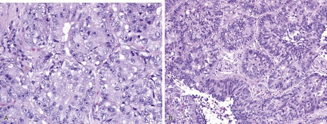
The nature of tumor growth and progression depends on whether the cell of origin is a bronchial gland, ciliated columnar cell, goblet cell, nonciliated bronchiolar cell, or type II pneumocyte. The histologic growth pattern may show a mixture of various types including acinar, papillary, solid, and lepidic (growth along alveolar interstitium), and the cytologic features may include mucinous and nonmucinous differentiation (Figures 65-2 and 65-3). The grading of adenocarcinomas is based on the histopathologic assessment of the extent of glandular differentiation and architectural pattern, cytologic expression and pleomorphism, nuclear characteristics and stratification, and mitotic activity and tumor necrosis. Most adenocarcinomas tend to be of mixed pattern, are intermediate in differentiation or moderately differentiated, and show a predominant acinar-type glandular arrangement. As expected, predominantly solid adenocarcinomas tend to demonstrate high-grade, poorly differentiated cytologic features with prominent mitotic activity and tumor necrosis. Adenocarcinomas may have clear cell features resulting from the accumulation of intracellular glycogen or mucins, and such tumors must be differentiated from metastatic clear cell renal carcinomas; the presence of intracytoplasmic lipid and glycogen is diagnostic of renal tumors of the clear cell type.

Histopathologic characteristics that correlate with a relatively poor prognosis in adenocarcinomas of the lung include a predominant solid pattern and high-grade cytologic features with mitotic figures and tumor necrosis, a micropapillary relative to a papillary histologic pattern, extension to and involvement of the visceral pleura, extensive lymphovascular tumor embolic spread, involvement of bronchial and hilar lymph nodes, and tumor-associated secondary obstructive lobar pneumonia. In addition, resection specimens that show intralobar satellite (metastatic) nodules and involvement of the surgical resection margins are associated with disease recurrence.

The histologic subtypes of adenocarcinoma appear to correlate with a history of cigarette smoking. Relative to nonsmokers, smokers more often have adenocarcinoma of the solid histologic type, and the extent of the solid component within the adenocarcinoma shows a dose-response relationship with pack-years of smoking. The solid subtype of adenocarcinoma also demonstrates a higher frequency of lymphovascular invasion and pleural involvement, and these histopathologic features are predictive for recurrence in stage I adenocarcinoma.

Historically, the most common cases of pulmonary adenocarcinoma were of the mixed type, with several histologic subtypes, and often the tumor had a so-called bronchioloalveolar carcinoma (BAC) growth margin. As originally defined, BAC is characterized by a lepidic growth pattern with atypical tumor cells (nonmucinous serous cells or mucinous cells) proliferating along the interstitial framework and not demonstrating stromal invasion or lymphovascular invasion. Tumors that manifest a BAC pattern often exhibit “ground glass” features on radiographs, and mixed-type invasive adenocarcinomas have central solid density with parenchymal distortion.

As a result of improper utilization and ambiguity of these pathologic terms, a group of international societies proposed a new classification that incorporated the histopathologic pattern, the radiographic features, and the clinical presentation and outcome. On the basis of the accumulated clinical experience with cases in which tumors 3 cm or less in diameter, with a pure lepidic growth pattern, were completely excised, followed by excellent survival, the term adenocarcinoma in situ (AIS) was adopted. These preinvasive lesions were judged to be extensions of a precursor lesion, termed atypical adenomatous hyperplasia (AAH), which also showed a lepidic growth pattern but were smaller in size (up to 5 mm in diameter). Those tumors that demonstrated an overall AIS pattern but also contained central minimal stromal invasion (less or equal to 5 mm) were designated minimally invasive adenocarcinoma (MIA). These latter cancers usually are nonmucinous and are associated with an excellent clinical outcome. In order to render a diagnosis of AIS or MIA, the entire lesion must be excised and processed for microscopic evaluation. These tumor types are excluded when histologic features of lymphovascular invasion, pleural involvement, and tumor necrosis are identified. It is recommended that the designation BAC be abandoned and that in its place, the proper histologic classification be applied, which may be AIS, MIA, adenocarcinoma with a predominant or mixed lepidic pattern, or mucinous adenocarcinoma. Tumors that were historically diagnosed as “nonmucinous BAC” may now occur as AIS, MIA, or lepidic-dominant adenocarcinomas. Many of these cases tend to be found in women, with immunohistochemical profiles of CK7 and thyroid transcription factor-1 (TTF-1) immunoreactivity and frequent epidermal growth factor receptor (EGFR) mutational status. By contrast, the historical designation of “mucinous BAC” is recommended to be changed to invasive mucinous adenocarcinoma, with immunohistochemical profiles of CK20 immunoreactivity and nonreactivity for TTF-1. These tumors frequently are associated with KRAS mutation and no EGFR mutation.

Invasive adenocarcinomas, representing a considerable majority (greater than 75% of cases), are those that exhibit significant stromal invasion and possible lymphovascular invasion and infiltration of the visceral pleura. These cancers need to be measured, because the size of the invasive component governs the tumor-node-metastasis (TNM) staging and the potential for tumor dissemination. The classification also recommends that the predominant pattern of the adenocarcinoma be designated and that the term “mixed pattern” be abandoned. The assessment of the components of the adenocarcinoma not only is important for the proper classification but also is useful for the correct identification of multiple pulmonary nodules as either multiple primary tumors or T3 or T4 tumors. Obviously, tumors that differ in histologic pattern and those that have an in situ component represent multiple primary tumors. It also is possible to differentiate whether the separate nodules are multiple T1 tumors by determining the relative distribution of the adenocarcinoma subtypes. For example, two nodules of adenocarcinoma may be regarded as separate T1 tumors if one is predominantly of the acinar subtype while the other nodule lacks the acinar pattern and is composed of predominant solid variant.

The consensus classification also addressed the recommended approach with small biopsy specimens (a common problem with fiberoptic biopsy samples) and cytologic preparations. The pathologist must make every attempt to avoid the diagnosis of “lung cancer not otherwise specified,” because the identification of a characteristic histologic pattern is important for tumor management. In a majority of cases, the distinction between adenocarcinoma and squamous cell carcinomas may be made using routine hematoxylin-eosin–stained (H&E) sections. In those cases of poorly differentiated carcinomas, mucin histochemical methods and a limited panel of immunohistochemical stains may achieve a more definitive result. Adenocarcinomas will show luminal or intracytoplasmic mucin positivity and TTF-1 immunoreactivity. Squamous cell carcinomas will lack mucin staining and will demonstrate reactivity for p63 or CK5/6 but not TTF-1.

Squamous Cell Carcinoma

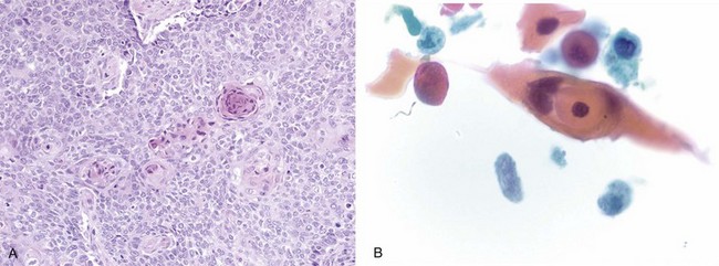
Squamous cell carcinoma is a malignant tumor arising from bronchial epithelium with a strong etiologic association with cigarette smoking, in contrast with adenocarcinoma. It most commonly is central in location and arises from the main, lobar, or segmental bronchi. Squamous cell carcinomas usually appear as white or gray masses with a variable degree of fibrosis, frequently associated with central necrosis and cavitations. Histologic hallmarks of squamous origin, namely keratinization and intercellular bridges, are seen in relation to the degree of the differentiation; although readily appreciated in well-differentiated tumors, they may be only focally present or even absent in poorly differentiated ones (Figure 65-4). In selected cases, when squamous nature cannot be ascertained by routine light microscopy, immunohistochemistry studies are invaluable. Squamous cell carcinomas usually are immunoreactive for high-molecular-weight keratin, CK5/6, and p63 and generally are nonreactive for TTF-1, CK7, and neuroendocrine markers (CD56, synaptophysin, chromogranin). Although not widely used, electron microscopy also can highlight features of squamous differentiation, such as prominent desmosomes and cytoplasmic aggregates of intermediate keratin filaments.

Recognizing histologic variants of squamous cell carcinoma, including papillary, clear cell, small cell, and basaloid types, is useful mainly in excluding the various differential diagnostic considerations. Their behavior is primarily a reflection of the degree of differentiation, with basaloid and small cell variants portending a worse outcome.

Pattern of spread is somewhat different depending on the anatomic location. Central tumors either grow along the epithelium, with or without invasion into subepithelial tissue, or expand as an endobronchial polypoid growth. In advanced cases, these tumors may directly involve hilar mediastinal tissue, including lymph nodes. Peripheral tumors, on the other hand, form a solid nodule and may directly break through the pleura into the chest wall or diaphragm.

Staging is governed by the TNM system. Regardless of the location, squamous cell carcinomas are more likely to be locally aggressive and invade adjacent structures. Distant metastasis, albeit far less common than with other subtypes of lung cancer, has a predilection for brain, liver, adrenals, lower gastrointestinal tract, and distant lymph nodes. In general, squamous cell carcinomas are associated with better prognosis and survival rate, as compared with stage-matched adenocarcinomas.

Small Cell Carcinoma and Neuroendocrine (Carcinoid) Tumors

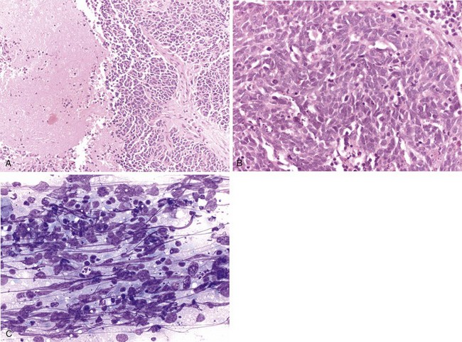
Neuroendocrine tumors, those that contain dense core neurosecretory granules composed of serotonin, histamine, and other bioactive amines, include small cell and large cell neuroendocrine carcinomas and typical and atypical carcinoid tumors. As a group, these tumors represent about one fifth of primary lung neoplasms, with small cell carcinoma being the most common in this category. These tumor types share a histologic pattern of uniform to pleomorphic cells in an organoid arrangement of tumor nests and trabecula (Figure 65-5). Carcinoid tumors may be considered as well-differentiated neuroendocrine carcinomas with little to no mitotic activity (less than 2 mitotic figures per 2-mm2 microscopic field) or tumor necrosis. Carcinoid tumors may occur in nonsmokers, in whom they manifest as a vascular rich endobronchial mass, although some carcinoid tumors may have a peripheral localization and have spindle cellular features. The tumors are low-grade malignancies with approximately 15% nodal metastases at diagnosis; nevertheless, affected patients have a greater than 90% 5-year survival. The atypical carcinoid tumors are neuroendocrine carcinomas of intermediate grade that have an organoid arrangement with more irregular and atypical cytologic pattern, identifiable tumor necrosis, and countable mitotic activity (2 to 10 mitoses per 2-mm2 microscopic field). In contrast with typical carcinoid tumors, atypical carcinoid may have nearly 50% lymph node metastases on presentation and a lower 5-year survival rate.

The high-grade poorly differentiated neuroendocrine tumors consist of the small cell (neuroendocrine) carcinoma and the large cell neuroendocrine carcinoma. The latter two have, respectively, smaller and larger cells with considerable cytologic atypia, prominent mitotic activity (more than 10 mitotic figures per 2-mm2 microscopic field), and abundant tumor necrosis. All of the neuroendocrine tumors will be immunohistochemically reactive with broad-spectrum cytokeratins, CD56, and, specifically, immunoreactive for neuroendocrine markers, such as chromogranin and synaptophysin. The high-grade neuroendocrine carcinomas are immunoreactive for TTF-1, more so than are the intermediate-grade and well-differentiated types.

Close attention to the morphologic details in conjunction with ancillary studies is important to differentiate small cell carcinoma not only from non–small cell carcinoma but also from other neuroendocrine tumors of lung, because they differ substantially in their demographics, clinical presentation, and behavior. Small cell carcinoma of lung generally is a disease of elderly men (mean age, 65 years) who are almost always current or former heavy smokers. These demographic findings are in sharp contrast with those in patients with typical carcinoid, who present at a younger age (mean, 45 to 50 years), with no difference in regard to gender or smoking history. The disparity also extends to associated prognosis and clinical behavior. Whereas typical carcinoid tumors are known for their indolent behavior and excellent survival rates, small cell carcinomas, at the other end of the spectrum, are fatal, with many patients presenting with distant metastasis at the time of diagnosis. Small cell carcinomas typically are central in location and frequently appear radiographically as hilar or perihilar masses with mediastinal lymphadenopathy and lobar collapse. Extensive necrosis is almost always present.

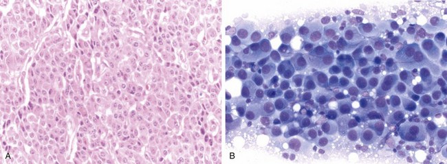
The key histopathologic features in the diagnosis of small cell carcinoma are nuclear morphologic features, such as finely granular chromatin, absence of prominent nucleoli, fragility, molding, and extensive crush artifact, as well as indistinct cytoplasmic borders and high mitotic figure count (more than 10 per 10 high-power fields) (Figure 65-6). Large zones of geographic necrosis also are common. Classically, a great deal of emphasis was placed on the small cell size or nuclear size, as suggested by the nomenclature. The usefulness of this criterion is questionable, however, in view of the wide variability in size, with frequent overlap with large cell neuroendocrine carcinomas. Large cell neuroendocrine carcinomas show a low-power neuroendocrine architecture, as seen in carcinoid tumors; however, the cell morphology closely resembles that of non–small cell carcinomas, with vesicular chromatin, prominent nucleoli, and abundant cytoplasm—features completely different from those of small cell carcinoma (Figure 65-7). Additionally, these tumors are highly mitotic (more than 10 mitotic figures per 10 high-power fields) and often show extensive necrosis, similar to that in small cell carcinomas, and in keeping with their high-grade nature.

Positive immunoreactivity with at least one neuroendocrine marker (synaptophysin, chromogranin) is required for the diagnosis of small cell carcinoma. By contrast, the main role of immunohistochemistry studies in small cell carcinoma is to exclude a wide range of other high-grade round blue cell tumors in the differential diagnosis. Cytokeratin immunoreactivity, especially the characteristic dotlike positivity, aids in excluding a lymphoid process, reactive or neoplastic, in instances of considerable morphologic overlap, particularly in small and poorly preserved samples. However, strong membranous staining with cytokeratins and diffuse staining with 34βE12 are characteristic of non–small cell carcinomas and generally are not seen in small cell carcinoma. The neuroendocrine markers synaptophysin and chromogranin, although strongly expressed in carcinoid, may be very focal in location or entirely absent in small cell carcinoma, and in such cases, CD56 is a more sensitive indicator of neuroendocrine differentiation. TTF-1, a very useful marker in establishing lung origin in adenocarcinoma, is seen in an overwhelming majority of small cell lung carcinomas, but it also may be expressed by extrapulmonary small cell carcinomas such as bladder and prostate cancers and therefore cannot be used to establish the site of origin. Although Ki-67 (MIB1) proliferation index is not an integral part of the current WHO classification of lung neuroendocrine tumors, it is of value, especially in the evaluation of small biopsy samples and cytologic specimens, when tissue is limited and poor preservation or extensive crush artifact during procurement may be inevitable. It has been suggested that Ki-67 (MIB1) percent positivity may be used as a diagnostic adjunct to morphologic examination: for typical carcinoid, less than 2%; for atypical carcinoid, less than 20%; and significantly, greater than 20% for small cell and large cell neuroendocrine carcinomas.

Traditionally, small cell carcinomas have been divided into limited-stage disease (treatable within a single radiation portal) and extensive-stage disease (contralateral or distant metastasis) with regard to prognosis. Recent large-scale analyses have shown the value of TNM staging for all neuroendocrine tumors of the lung, including carcinoid tumors.

Large Cell Carcinoma

By definition, large cell carcinoma of lung is an undifferentiated carcinoma lacking morphologic evidence of small cell carcinoma, squamous cell carcinoma, or adenocarcinoma. In other words, it represents a diagnosis of exclusion. These tumors typically are composed of sheets and nests of large polygonal cells with moderate amount of cytoplasm, vesicular chromatin and prominent nucleoli (Figure 65-7). Although the classification is purely based on morphologic grounds, judicious use of ancillary studies (as discussed previously), especially with small biopsy specimens, is of significant value in separating the poorly differentiated squamous cell carcinomas and adenocarcinomas from the truly undifferentiated large cell carcinoma. These tumors share similar demographic and clinical features with other non–small cell carcinomas of the lung, because they commonly are seen in elderly men with smoking history.

Large cell carcinomas are preferentially seen at the periphery of the lung. Several histologic subtypes are recognized: basaloid, clear cell, and lymphoepithelioma-like carcinomas. Although grouped under large cell carcinomas in the 2004 WHO classification, large cell neuroendocrine carcinoma, as strictly defined in the previous section, is best categorized as a neuroendocrine tumor, closely resembling small cell carcinoma. Nevertheless, the mere focal expression of neuroendocrine markers as detected on immunohistochemistry studies, seen in some large cell carcinomas, should not be interpreted as diagnostic for large cell neuroendocrine carcinoma. In general, large cell carcinomas are high-grade tumors that are similar in prognosis and pattern of spread to stage-matched non–small cell carcinomas, with performance status at the time of diagnosis and disease extension considered to be the most important clinical prognostic parameters. Metastases arise most commonly in the hilar and mediastinal nodes, followed by pleura, liver, bone, brain, abdominal nodes, and pericardium. The basaloid subtype carries a worse prognosis, even in the lower stages, with more common metastasis to the brain. On the other hand, lymphoepithelioma-like carcinomas generally are diagnosed at a later stage of disease but are associated with a better prognosis.

Pleural Malignancies

Diffuse malignant mesothelioma is a pleura-based cancer that may be histopathologically composed of a biphasic growth pattern of combined epithelial and sarcomatoid components. The tumor also may manifest in a monophasic form of either epithelioid cancer resembling a pleural adenocarcinoma or a spindle sarcomatoid form resembling a mesenchymal sarcoma. The diagnosis is usually but not exclusively rendered with a history of asbestos exposure, similar to that for pleural asbestosis or asbestos-related diseases and malignancies, but with a lower threshold of lung asbestos burden. Other proposed causes of diffuse malignant mesothelioma are chest radiation therapy and exposure to other mineral silicates, such as vermiculite and erionite. Erionite originally was identified in Turkey but is now appreciated to have widespread deposits in the western United States (i.e., Dakotas, Nevada, Arizona, and California). The diagnosis is based on appropriate clinical, radiographic, and histopathologic findings of a painful pleura-based tumor with scattered multiple nodules or a coalescent tumor encasing the lung. Malignant mesothelioma may obliterate the pleural cavity or be associated with an exudative pleural effusion. The tumor may invade the underlying lung and typically infiltrates through the diaphragm.

Malignant mesothelioma exhibits characteristic surgical pathology features that must be distinguished from reactive mesothelial proliferation and chronic fibrosing pleuritis. Reactive mesothelial hyperplasia frequently is present in the setting of collagen vascular diseases, drug or inflammatory reactions, and trauma or surgery. Of importance, reactive mesothelial hyperplasia lacks invasion of the stromal connective tissue and fat, does not undergo necrosis, and typically is not immunoreactive for EMA (epithelial membrane antigen) and p53 (tumor suppressor gene product). In the setting of exposure to asbestos, collagenous pleural plaques may be identified and associated with either reactive mesothelial hyperplasia or an incipient diffuse malignant mesothelioma. Separation of chronic pleuritis from malignant mesothelioma also is based on the lack of mesothelial invasion in pleuritis and the presence of an organizing zonal layered fibroinflammatory process. Desmoplastic malignant mesothelioma will, on the other hand, show stromal invasion, haphazard growth pattern, and metastatic spread. Reactive mesothelial cells and those of malignant mesothelioma are immunoreactive for cytokeratin. The histopathologic appearance of reactive mesothelial conditions is organized and layered, in contrast to the haphazard cellularity and invasion in malignant mesothelioma.

The histologic pattern of the epithelioid type of diffuse malignant mesothelioma appears as an invasive tubular and papillary malignancy that mimics a pleura-based adenocarcinoma or a metastatic adenocarcinoma. The monophasic sarcomatoid mesotheliomas resemble spindle cell sarcomas, which may manifest as primary or metastatic cancers. The most common form of malignant mesothelioma is the epithelioid variant and may show a variety of histologic features, such as tubulopapillary, glandular, or solid patterns. Cytologic examination of malignant pleural effusions shows irregular clusters of atypical mesothelial-like cells in pools of hyaluronic acid. Malignant mesotheliomas also may present in the peritoneal cavity and mimic ovarian cancers in women. Biphasic mesotheliomas are less common in the abdomen than in the pleura, and abdominal sarcomatoid mesotheliomas are rare. The distinction between malignant mesothelioma and adenocarcinoma in the pleura and peritoneal cavity is based on histologic features and supported by histochemical and immunohistochemical staining and ultrastructural findings. Mesotheliomas will be positive for Alcian blue, a stain that disappears with pretreatment with hyaluronidase. Most adenocarcinomas are histochemically positive for mucicarmine stains, although a few mesotheliomas also may stain positive. On immunohistochemical assays, malignant mesotheliomas will be immunoreactive for calretinin, keratin 5/6, WT-1 protein, and podoplanin (D2-40), in contrast with adenocarcinomas, which are immunoreactive for CEA (carcinoembryonic antigen), MOC-31, B72.3, Ber-EP4, and TTF-1. Both mesotheliomas and adenocarcinomas will be positive on assay for pancytokeratins, which will demonstrate the viability of the tissue for immunohistochemical assessments. Historically, the best diagnostic discriminator for distinguishing malignant mesothelioma from adenocarcinoma has been electron microscopic examination of the tumor. The ultrastructural appearance of malignant mesothelioma on transmission electron microscopy shows long slender microvillous processes with frequent branching and lacking a glycocalyx, in contrast with short blunt straight microvilli of adenocarcinoma, containing a glycocalyx.

Molecular Biology

The study of cellular and molecular mechanisms of lung cancer has provided insight into lung cancer biology in terms of epidemiology and predisposition to lung cancer, lung cancer carcinogenesis, and cancer progression and resistance to chemotherapy and radiation therapy. Epidemiologic investigations have demonstrated higher levels of lung cancer in patients whose family history included a diagnosis of lung cancer in a first-degree relative. In a case-control study of lung cancer in women, never-smokers with positive family history had an odds ratio of 5.7 relative to nonsmokers without a family history. Smokers without a family history of lung cancer and smokers with a family history had odds ratios of approximately 15 and 30, respectively, for the development of lung cancer relative to non-smokers without a family history of lung cancer. Among persons who are carriers of a mutated retinoblastoma (RB) gene, the mortality rate for lung cancer is 15-fold that in the general population.

The cellular predisposition to lung cancer among cigarette smokers has included protean metabolic phenotypic pathways among smokers at risk for lung cancer. It appears clear that individual metabolism must play a role in carcinogenesis: The overwhelming majority of cancers are found in smokers, but among smokers, only a minority develop lung cancer. The host factors acting on environmental smoke metabolites include the pleiotropic expressions of the cytochrome p450 oxidative enzymes. Polycyclic aromatic hydrocarbons and nitrosamines, components of cigarette smoke and carcinogenic agents, induce the expression of P450 enzymes. In turn, cytochrome P450 metabolism produces reactive oxygenated intermediates that have the capacity for DNA binding and alterations, leading to mutations and cellular dysplasia. Aryl hydrocarbon hydroxylases, as members of the mixed-function oxidases, are induced by and active on cigarette smoke chemicals, and their metabolites are capable of causing DNA mutations and malignant transformation of cells.

One of the fundamental precepts of cancer development is multistep carcinogenesis involving basic steps of initiation (transmissible DNA damage leading to cellular dysplasia with altered regulatory controls), promotion (proliferation of abnormal cellular clones to form a mass), and progression (invasion and metastases). Cellular and molecular events that drive the conversion of preneoplastic conditions to malignant tumors are activations of cellular oncogenes, deletion or inactivation of tumor suppressor genes, enhancement of survival relative to apoptotic pathways, and alterations of DNA repair mechanisms. Early studies found chromosomal 3p deletions in lung cancers, more commonly in small cell carcinomas. KRAS activation and the expression of p21, involved in activation of cellular signal transduction pathways, have been identified in lung cancers. Mutations in KRAS were identified in adenocarcinomas, especially pronounced in smokers, and were associated with relatively poor survival. Genetic amplification and mutation of the EGFR gene, associated with increased or altered protein expression, have been identified predominantly in non–small cell carcinomas. Examples of inactivation of tumor suppressor genes are loss of Rb expression in some small cell carcinomas and inactivation of p53 in non–small cell carcinomas.

Molecular biology studies have provided insight into the initiation and progression of lung cancer. Additional investigations have revealed cellular and molecular changes that modulate the tumor’s response to chemotherapy and provide acquired resistance to therapeutic management. Chemotherapeutic agents that act in the folate pathway of pyrimidine and DNA synthesis, such as pemetrexed, may demonstrate reduced efficacy when the functional level of thymidylate synthesis is overexpressed. Similarly, the overexpression of ERCC-1 (excision repair cross-complementation group 1), a repair enzymatic pathway for DNA strand breaks, modifies the therapeutic outcome with platinum drugs that intercalate in the DNA.

Tumors that appear to be markedly governed by a single or small set of pathway gene products are said to show “oncogene addiction.” Despite the complexity of genetic and epigenetic tumor cell abnormalities, these “oncogene addiction” pathway genes dominate the progressive and invasive nature of the malignancy, and interruption of their function will lead to tumor regression, thereby promoting a prolonged disease-free interval or overall survival. This molecular condition provides the pharmacologic rationale for targeted therapy. The three genes discussed next appear to be in the “driver’s seat” for tumor progression, and blocking their altered function will provide for tumor regression.

Epidermal Growth Factor Receptor Gene

EGFR is a transmembrane signaling protein that transduces extracellular signals, due to binding of EGFR ligands, into intracellular changes mediated by activating tyrosine kinase phosphorylations. EGFR, also called HER1 (human epidermal growth factor receptor 1), is in the same family as HER2, another membrane receptor that is amplified in approximately 20% of breast cancers. In breast cancer, HER2 is biologically associated with a relatively aggressive type of breast cancer, but it also is a therapeutic target for directed antibodies or small molecule tyrosine kinase inhibitors (TKIs). When EGFR is mutated, predominantly in nonsmokers, it adopts an activated state, as if constantly bound to activating ligands or growth factors. EGFR activation induces downstream signals that inhibit apoptosis and stimulate cell proliferation and migration and motility. Targeted TKIs are effective for tumor regression when non–small cell carcinomas have activating EGFR mutations, as opposed to tumors with wild-type EGFR. EGFR-activating mutations tend to be present in a minority of lung cancers (10% to 20%) and appear most commonly in nonmucinous adenocarcinomas exhibiting a well-differentiated acinar or papillary growth pattern with a lepidic growth margin and presenting demographically in younger Asian women who were never-smokers or light smokers. In this last demographic subgroup, the incidence of EGFR mutation may rise to 40% to 50%. Large cell carcinomas also have shown EGFR mutation; however, the rate of such mutations in squamous cell carcinoma is only a few percent, and this low frequency is not sufficient for routine analysis. Immunohistochemical identification of EGFR in tumor tissue has no correlation with clinical tumor response to TKIs. The EGFR mutation is the molecular marker, more significant than EGFR amplification, for targeted therapy and is a predictive factor for a therapeutic response. Fluorescence in situ hybridization (FISH) assays have been attempted to identify EGFR amplification and show some concordance with therapeutic response, because EGFR mutations correlate with amplification. Adenocarcinomas with nonmutated but amplified EGFR gene have been recognized, and these tumors show a partial response to tyrosine kinase inhibition.

The standard tests for EGFR mutations may be performed on formalin-fixed paraffin-embedded tissue and consist of direct DNA sequencing or mutation-specific assays investigating mutations that usually are found in exons 19 and 21. In instances of tumor recurrence due to resistance to TKIs, the sequencing analysis should be expanded to include exons 18 and 20. The acquired resistance tends to occur in about a year after TKI therapy is started and may be related to a new mutation in the active site; other causes for TKI resistance are due to MET gene amplification in 10% to 15% of cases. EGFR genetic testing shows concordant results with the presence of an activating mutation between the primary tumor and its metastasis in a majority of cases; however, an average discordance of 25%, as a result of tumor heterogeneity and progression, has been noted.

KRAS Oncogene

KRAS is an oncogene found on the short arm of chromosome 12 (12p) and shows mutations in approximately 20% of non–small cell lung cancers and amplification of the gene in a smaller set of cancers. The p21 protein (i.e., K-RAS), encoded by KRAS, couples with a cytoplasmic GTPase to convert guanosine triphosphate (GTP) to guanosine diphosphate (GDP) in order to modify transductive signals from the cytoplasm to the nucleus, thereby controlling cellular growth and survival pathways. When KRAS is mutated, GTP is maintained, and the signal is persistently in the “active” state. Activating mutations of KRAS are present in a variety of epithelial tumors, including colon and pancreatic cancers, and lead to activating phosphorylation of mitogen-activated protein kinase (MAPK), inducing nuclear signals of cellular proliferation and tumor progression. KRAS mutations and amplifications demonstrate clinicopathologic correlations with older age combined with male gender, history of heavy cigarette smoking, and advanced disease at presentation, with relatively aggressive tumors and poor clinical outcome. Histologically, KRAS mutations tend to be identified in poorly differentiated adenocarcinomas with solid growth pattern and lacking a lepidic growth pattern. Of interest, the mucinous lepidic pattern is associated with KRAS mutations. These high-grade adenocarcinomas also show lymphovascular invasion, mitotic activity, and tumor necrosis. K-RAS is downstream from EGFR effects and when activated is independent of EGFR status, thereby conveying resistance to tyrosine kinase inhibitors of EGFR function.

EML4/ALK

A new addition to molecular markers that predict a clinically favorable response to targeted therapy is the EML4ALK translocation. The proper name of the fusion gene product is echinoderm microtubule-associated protein-like 4–anaplastic lymphoma kinase. The short arm of chromosome 2 (2p) may contain multiple small inversions and or translocations whose permutations generate in-frame fusions of the 5′ portion of the EML4 gene with the 3′ end of the ALK gene, creating a chimeric tyrosine kinase with an intracellular kinase domain. Breakpoints of the EML4 gene occur at several possible exon sites, although the breakpoint of the ALK gene tends to be specific for the kinase domain at exon 20. This fusion activation of the ALK gene is infrequent (occurring in less than 10% of non–small cell lung cancers), but the incidence may be enriched in lung cancers present in younger persons without significant smoking history in whom EGFR mutations and KRAS mutations are lacking. EML4ALK translocations are histologically correlated with high-grade adenocarcinomas exhibiting acinar and solid growth patterns that contain mucinous and signet ring differentiation. From early studies, it appears that tyrosine kinase–targeted therapy for EGFR mutations is not effective treatment for EML4ALK mutations. EML4ALK fusion also appears to be selective for adenocarcinomas, because it is lacking in the other types of bronchogenic carcinomas.

The test for the translocation is based on reverse transcriptase–polymerase chain reaction (RT-PCR) assay of tumor tissue RNA, which amplifies and detects certain of the fusion constructs. The assay limitation is due to the identification of only the translocations of EML4 exons 6 and 13 with ALK exon 20. The fusion gene also may be assayed by FISH techniques, and recent studies suggest that immunohistochemical methods also may yield accurate results.

Suggested Readings

Alberg AJ, Samet JM. Epidemiology of lung cancer. Chest. 2003;123:21S–49S.

American Cancer Society. 2010 Cancer facts & figures. Atlanta: American Cancer Society; 2010.

Brambilla E, Gazdar A. Pathogenesis of lung cancer signalling pathways: roadmap for therapies. Eur Respir J. 2009;33:1485–1497.

Cadranel J, Zalcman G, Sequist L. Genetic profiling and epidermal growth factor receptor directed therapy in non-small cell lung cancer. Eur Respir J. 2011;37:183–193.

Camidge DR, Hirsch FR, Varella-Garcia M, Franklin WA. Finding ALK-positive lung cancer: what are we really looking for? J Thorac Oncol. 2011;6:411–413.

Han SW, Kim TY, Hwang PG, et al. Predictive and prognostic impact of epidermal growth factor receptor mutation in non-small cell cancer patients treated with gefitinib. J Clin Oncol. 23, 2005. 2493–2402

Haura EB, Camidge DR, Reckamp K, et al. Molecular origins of lung cancer: prospects for personalized prevention and therapy. J Thorac Oncol. 2010;5:S207–S213.

Husain AN, Colby TV, Ordonez NG, et al. Guidelines for pathologic diagnosis of malignant mesothelioma. Arch Pathol Lab Med. 2009;133:1317–1331.

Idowu M, Powers CN. Lung cancer cytology: potential pitfalls and mimics—a review. J Clin Exp Pathol. 2010;3:367–385.

Inamura K, Takeuchi K, Togashi Y, et al. EML4-ALK fusion is linked to histologic characteristics in a subset of lung cancers. J Thorac Oncol. 2008;3:13–17.

Mitsudomi T, Kosaka T, Endoh H, et al. Mutations of the epidermal growth factor receptor gene predict prolong survival after gefitinib treatment in patients with non-small-cell lung cancer with postoperative recurrence. J Clin Oncol. 2005;23:2513–2520.

Mok TS, Wu YL, Thongprasert S, et al. Gefitinib or carboplatin-paclitaxel in pulmonary adenocarcinoma. N Engl J Med. 2009;361:947–957.

Osann KE. Lung cancer in women: the importance of smoking, family history of lung cancer, and medical history of respiratory disease. Cancer Res. 1991;51:4893–4897.

Rekhtman N. Neuroendocrine tumors of the lung. Arch Pathol Lab Med. 2010;134:1628–1638.

Rekhtman N, Brandt SM, Sigel CS, et al. Suitability of thoracic cytology for new therapeutic paradigms in non-small cell lung carcinoma. J Thorac Oncol. 2011;6:451–458.

Samet J, Humble C, Pathak D. Personal and family history of respiratory disease and lung cancer risk. Am Rev Respir Dis. 1986;134:466–470.

Schwartz AM, Henson DE. Diagnostic surgical pathology in lung cancer: ACCP evidence-based clinical practice guidelines, ed 2. Chest. 2007;132:78–91.

Shaw AT, Yeap BY, Mino-Kenudson M, et al. Clinical features and outcome of patients with non-small cell lung cancer who harbor EML4-ALK. J Clin Oncol. 2009;27:4247–4251.

Soda M, Choi YL, Enomoto M, et al. Identification of the transforming EML4-ALK fusion gene in non-small cell lung cancer. Nature. 2007;448:561–566.

Takano T, Ohe Y, Sakamoto H, et al. Epidermal growth factor receptor gene mutations and increase copy numbers predict gefitinib sensitivity in patients with recurrent non-small cell lung cancer. J Clin Oncol. 2005;23:6829–6837.

Travis WD, Brambilla E, Muller-Hermelink HK, et al. Pathology and genetics. Tumors of the lung, pleura, thymus, and heart. Lyon, France: IARC Press; 2004.

Travis WD, Brambilla E, Noguchi M, et al. International Association for the Study of Lung Cancer/American Thoracic Society/European Respiratory Society international multidisciplinary classification of lung adenocarcinoma. J Thorac Oncol. 2011;6:244–285.

U.S. Department of Health and Human Services. How tobacco smoke causes disease: the biology and behavioral basis for smoking attributable disease: a report of the Surgeon General. Atlanta: Centers for Disease Control and Prevention, National Center for Chronic Disease Prevention and Health Promotion, Office of Smoking and Health; 2010.

U.S. Public Health Service Smoking and health. Report of the Advisory Committee to the Surgeon General. PHS publication no. 1103. Washington, DC: U.S. Department of Health, Education, and Welfare, Public Health Service, CDC; 1964.