Breast Lumps
A lump in the breast is a common clinical presentation. In every case of a breast lump, in both males and females, carcinoma must be excluded.
Causes
Discrete Lumps
Malignant
Swellings Behind the Breast
History
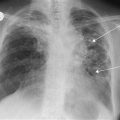
A diagnosis of carcinoma is suggested by a strong family history, nulliparous state, early menarche, late menopause and history of cystic hyperplasia. The patient may have found a breast lump (carcinoma is painless in 85% of cases) or may have noticed nipple retraction, skin dimpling, axillary swelling. Jaundice may have occurred due to liver secondaries or porta hepatis node involvement. Bone pain may be due to secondary deposits and pathological fractures. Breathlessness may be due to lung secondaries or a pleural effusion. Personality change, fits, headaches suggest cerebral metastases. A patient who presents in pregnancy or lactation may have mastitis, abscess or galactocele. A history of trauma suggests fat necrosis. A patient in the fifth decade with retroareolar pain, nipple retraction and a thick, creamy nipple discharge suggests duct ectasia. The patient presenting between the ages of 15 and 25 years with a non-tender swelling suggests fibroadenoma. A patient with a history of painful breasts may have one of the conditions described under breast pain (p. 56). Enquiries should be made of a past history of TB, as rarely a tuberculous abscess may form within the breast or erode through the chest wall from the lung.
Examination
Carcinoma
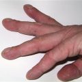
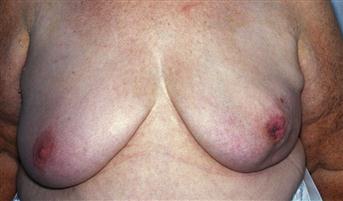
Hard irregular swelling. May be fixed to the skin or fixed deeply. Skin dimpling. Nipple retraction. Peau d’orange. Paget’s disease of the nipple. Axillary lymphadenopathy. Supraclavicular lymphadenopathy. Hepatomegaly. Pathological fractures – spine, ribs, long bones.
Fibroadenoma
Smooth, rounded, mobile mass. Breast ‘mouse’ (so-called because it darts about under the examining fingers).
Cyst
Smooth, mobile swelling. May be tender. May be associated with generalised lumpiness of the breast.
Galactocele
Smooth, mobile swelling occurring in the lactating breast.
Fat necrosis
Hard, irregular swelling. May be overlying bruising of the skin or teeth marks! Occasionally the lesion is fixed to the skin and is difficult to distinguish from carcinoma.
Lipoma
Soft, lobulated swelling. Rare on the breast. ‘Pseudolipoma’ is a bunching of fat between retracted suspensory ligaments of the breast and is associated with an underlying carcinoma.
Duct ectasia
Tender retroareolar area. Retroareolar erythema. Nipple retraction. Thick, creamy nipple discharge.
Sebaceous cyst
Mobile swelling fixed to the skin. May be a punctum. May be infected, with discharge and surrounding redness.
Generalised swellings
Generalised swelling of the breast may occur in pregnancy, lactation and puberty. It may also occur with mastitis, when the breast is enlarged, red, tender and hot.
Swellings behind the breast
These are rare. Tietze’s disease is the most common. There will be prominence and tenderness over the second, third and fourth costal cartilages. With retromammary abscess, chest signs may be obvious on percussion and also on auscultation of the chest.
General Investigations
Specific Investigations
■ FNAC
Benign versus malignant. Cystic versus solid.
■ Mammography
Infiltrating radio-opaque mass or microcalcification indicates malignancy. Dilated retroareolar ducts and skin indentation suggests duct ectasia. Coarse calcification may occur with long-standing fibroadenomas.
■ Open excision biopsy
e.g. fibroadenoma, fat necrosis.
■ US
US of breast is better than mammography in young women. US of liver for metastases. US guided FNA/core biopsy/Mammotome®.