Lumbar and sacral plexus anatomy

Published on 27/02/2015 by admin

Filed under Anesthesiology

Last modified 22/04/2025

Print this page

rate 1 star rate 2 star rate 3 star rate 4 star rate 5 star
Your rating: none, Average: 0 (0 votes)

This article have been viewed 1778 times

CHAPTER 23 Lumbar and sacral plexus anatomy

The anterior divisions of the lumbar, sacral, and coccygeal nerves form the lumbosacral plexus. The plexus is usually divided into three parts: the lumbar, sacral, and pudendal plexuses, for ease of description. The lumbar plexus primarily innervates the ventral aspect, whereas the sacral plexus innervates the dorsal aspect of the lower limb.

Lumbar plexus

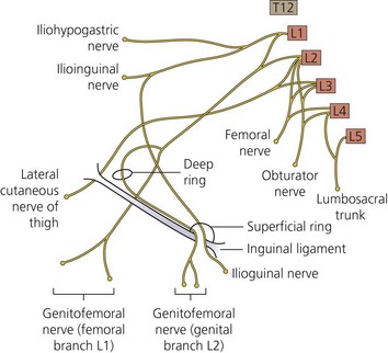
The lumbar plexus (Fig. 23.1) lies deep within the psoas major muscle in front of the transverse processes of the lumbar vertebrae. It is formed by the ventral rami of the first three lumbar nerves and the greater part of the ventral ramus of the fourth nerve. All the branches of the plexus emerge from the substance of the psoas major.

The first lumbar nerve, frequently supplemented by the 12th thoracic, splits into an upper and a lower branch; the upper and larger branch divides into the iliohypogastric and ilioinguinal nerves, the lower and smaller branch unites with a branch of the second lumbar to form the genitofemoral nerve.

The remainder of the second nerve, and the third and fourth nerves, divide into ventral and dorsal divisions. The ventral division of the second unites with the ventral divisions of the third and fourth nerves to form the obturator nerve. The dorsal divisions of the second and third nerves divide into two branches, a smaller branch from each uniting to form the lateral cutaneous nerve of the thigh, and a larger branch from each joining with the dorsal division of the fourth nerve to form the femoral nerve.

The femoral nerve

The femoral nerve, the largest branch of the lumbar plexus, arises from the dorsal divisions of the second, third, and fourth lumbar nerves. It descends through the fibers of the psoas major, emerging from the muscle at the lower part of its lateral border, and passes down between it and the iliacus muscle, behind the iliac fascia; it then runs beneath the inguinal ligament into the thigh, and splits into an anterior and a posterior division. In the thigh, the anterior division of the femoral nerve gives off anterior cutaneous and muscular branches. The anterior cutaneous branches comprise the intermediate and medial cutaneous nerves.

The intermediate cutaneous nerve pierces the fascia lata (and generally the sartorius) about 7.5 cm below the inguinal ligament, and divides into two branches that descend in immediate proximity along the forepart of the thigh to supply the skin as low as the front of the knee.

The medial cutaneous nerve passes obliquely across the upper part of the sheath of the femoral artery, and divides in front or at the medial side of that vessel into two branches: an anterior and a posterior. The anterior branch runs downward on the sartorius, perforates the fascia lata at the lower third of the thigh, and divides into two branches. The posterior branch descends along the medial border of the sartorius muscle to the knee, where it pierces the fascia lata, communicates with the saphenous nerve, and gives off several cutaneous branches.

The posterior division of the femoral nerve gives off the saphenous nerve, and muscular and articular branches.

Sacral plexus

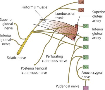
The sacral plexus (Fig. 23.2) is formed by the lumbosacral trunk (L4,L5) and the ventral rami of the first, second, and third sacral nerves. The nerves forming the sacral plexus appear at the medial margin of the psoas major, converge toward the greater sciatic notch, and unite to form a large band located on the posterior wall of the pelvic cavity, in front of the piriformis muscle. From the anterior and posterior surfaces of the band several branches arise. The band itself is continued as the sciatic nerve, which splits on the back of the thigh into the tibial and common peroneal nerves; these two nerves sometimes arise separately in the plexus.

Relations

The sacral plexus lies on the posterior aspect of the pelvis between the piriformis and the pelvic fascia. In front of it are the hypogastric vessels, the ureter, and the sigmoid colon. The gluteal vessels follow the same course as the sacral nerves but in a more anterior plane. The pelvic fascia is fixed medially on the anterior sacral foramina, where the sacral nerves emerge. Through this fascia, the sacral plexus lies near the rectum. Laterally, the sacral plexus lies close to the greater sciatic foramen, sandwiched by the obturator internus muscle. The sacral plexus runs in a fascial plane limited by the pelvic fascia ventrally, the piriformis dorsally, and medially and laterally by the obturator internus muscle. Hypogastric vessels are located near the sacral plexus as well as the superior gluteal artery, which passes between the lumbosacral trunk and the first sacral nerve. The inferior gluteal vessels run between the second and third sacral nerves. Collateral and terminal branches of the sacral plexus include:

From the lower margin of the periformis, the sciatic nerve passes into the buttock on the posterior surface of the ischium. From midway between the greater trochanter and the ischial tuberosity, deep to the gluteus maximus, the nerve passes vertically downward into the hamstring compartment. It lies posterior to the obturator internus, gemelli, quadratus femoris, and adductor magnus, but it is crossed posteriorly by the long head of biceps femoris. The sciatic nerve usually divides into the tibial and common peroneal nerves at the upper angle of the popliteal fossa. It occasionally divides into these components within the pelvis, and the common peroneal part pierces the piriformis as it leaves the pelvis.

From the apex of the popliteal fossa, the tibial nerve (L4, 5, S1, 2, 3) passes vertically down deep to the heads of gastrocnemius but superficial and lateral to the popliteal vein and artery. In the popliteal fossa it supplies skin, the knee joint, calf muscles, and popliteus; it gives the sural nerve, which descends between the two heads of gastrocnemius, accompanied by the small saphenous vein to the back of the lateral malleolus and the lateral border of the foot, supplying the overlying skin. The tibial nerve passes down the leg deep to the soleus, supplying the deep muscles, and reaches the medial side of the ankle, between the malleolus and the heel; here it divides into lateral and medial plantar nerves.

From the apex of the popliteal fossa, the common peroneal nerve (L4, 5, S1, 2) passes downward and laterally, medial to the biceps tendon, and turns round the neck of the fibula in the substance of the peroneus longus, where it divides into superficial and deep peroneal nerves. The superficial peroneal nerve supplies the peroneus longus and brevis, and emerges between them to supply the skin of the lower leg and much of the dorsum of the foot. The deep peroneal nerve passes into the anterior compartment of the leg to supply the muscles here, and proceeds to the foot between the two malleoli to supply the skin of the first web space.

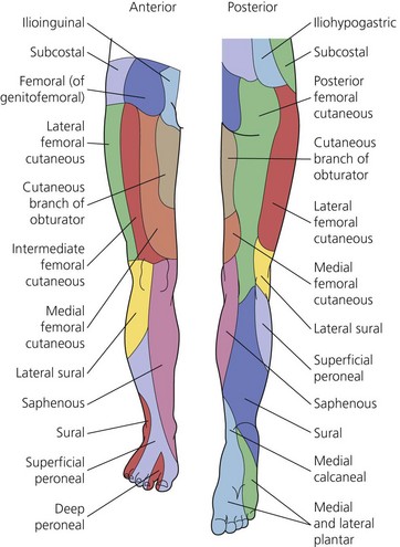
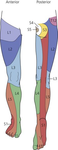
The sensory and motor innervation of the lower limb (Figs 23.3 and 23.4) is clinically important. Knowledge of sensory innervation helps determine which cutaneous nerve distributions within a surgical field require blockade. Motor innervation is clinically relevant as a means of matching a peripheral nerve stimulation response to the particular nerve being stimulated.