[level-membership-for-orthopaedics-category]
6 Lower limb
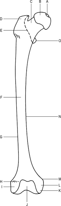
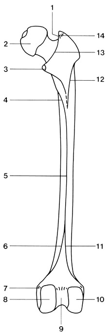
Femur (Figs 6.1 and 6.2)
Type
Main parts
Features of the upper end of the femur
Intertrochanteric line –
anterior line, between the trochanters; marks part of the junction between the neck and shaft.
Features of the lower end of the femur
Medial and lateral condyles –
fused together anteriorly, separated by the intercondylar notch posteriorly.
Medial epicondyle –
most prominent point of the medial condyle; provides attachment for the tibial collateral ligament.
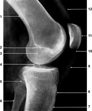
Radiographic appearances of the femur
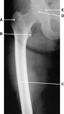
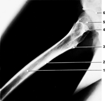
Fig. 6.4 Femur: upper two-thirds; lateral projection.
4 – Femoral neck and greater trochanter superimposed
(From Bryan 1996.)
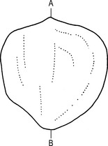
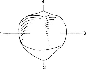
Patella (Figs 6.9 and 6.10)
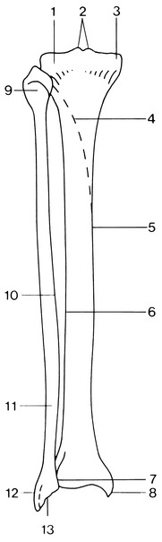
Tibia (Figs 6.13 and 6.14)
Type
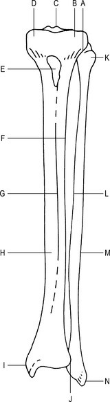
Fig. 6.13 Left tibia (anterior aspect).
C–Tubercles of the intercondylar eminence
Articulations
Tibial condyles with the femoral condyles to form the knee joint.
Lower end of tibia with the talus to form part of the ankle joint.
Fibular notch of the tibia with the lower end of fibula to form the inferior tibiofibular joint.
Main parts
Features of the upper end of tibia
Features of the shaft of tibia
Soleal line –
‘horseshoe-shaped’ oblique line; posterior aspect of the shaft which receives the soleus muscle.
Fibula (Figs 6.13 and 6.14)
Articulations
Lower end of fibula with the fibular notch of the tibia to form the inferior tibiofibular joint.
Lateral malleolus of the fibula with the talus to form part of the ankle joint.
Main parts
Features of the upper end of the fibula
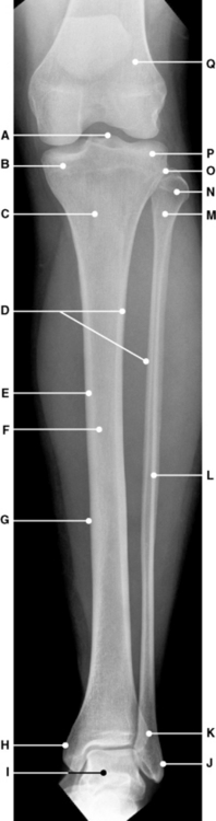
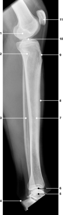
Radiographic appearances of the tibia and fibula
Foot
Tarsal bones (Fig. 6.18)
Type
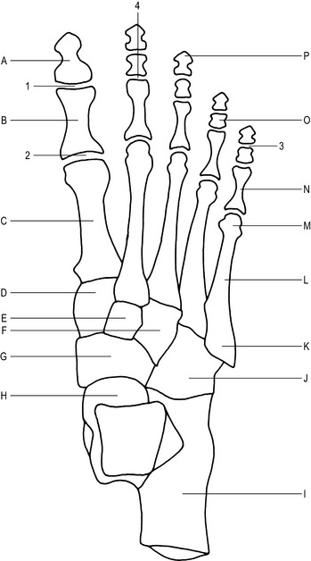
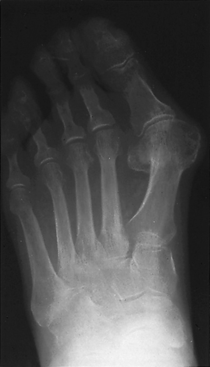
Fig. 6.18 Right foot (dorsal aspect).
A–Shaft of distal phalanx of great toe
B–Shaft of proximal phalanx of great toe
N–Shaft of proximal phalanx of 5th toe
O–Shaft of middle phalanx of 4th toe
P–Head of distal phalanx of 3rd toe
1 – Interphalangeal joint of great toe (synovial hinge joint)
2 – 1st metatarso- phalangeal joint (synovial ellipsoid joint)
3 – 5th distal interpha- langeal joint (synovial hinge joint)
4 – 2nd proximal inter- phalangeal joint (synovial hinge joint).
Articulations
Talus with the tibia, fibula, calcaneus, navicular.
Calcaneus with the talus, cuboid.
Navicular with the talus, cuboid, medial cuneiform, intermediate cuneiform, lateral cuneiform.
Cuboid with the calcaneus, navicular, lateral cuneiform, 4th metatarsal, 5th metatarsal.
Lateral cuneiform with the navicular, cuboid, intermediate cuneiform, 2nd, 3rd and 4th metatarsals.
Intermediate cuneiform with the navicular, lateral cuneiform, medial cuneiform, 2nd metatarsal.
Medial cuneiform with the navicular, intermediate cuneiform, 1st and 2nd metatarsals.
Individual bones
Proximal row
Articulations
The head of the metatarsals with the phalanges to form the metatarsophalangeal joints.
The base of the metatarsals with the tarsal bones to form the tarsometatarsal joints.
1st metatarsal with the proximal phalanx of the hallux and the medial cuneiform.
3rd metatarsal with the proximal phalanx of the 3rd toe and the lateral cuneiform.
4th metatarsal with the proximal phalanx of the 4th toe, the lateral cuneiform and the cuboid.
5th metatarsal with the proximal phalanx of the 5th toe and the cuboid.
The bases of the 2nd–5th metatarsals with the adjacent metatarsal.
Phalanges (Fig. 6.18)
Articulations
The base of the phalanx with the metatarsal to form the metatarsophalangeal joints.
With each other to form the interphalangeal joints.
4 middle phalanges with the corresponding proximal and distal phalanges.
5 distal phalanges with the corresponding middle phalanges and the proximal phalanx of the hallux.
Radiographic appearances of the foot (Figs 6.20, 6.21 and 6.22)
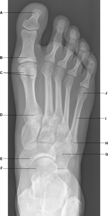
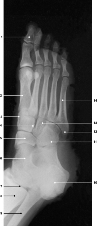
Fig. 6.21 Right foot: dorsiplantar oblique projection.
12 – Base of 5th metatarsal bone
(From Bryan 1996.)
Knee joint (Figs 6.26, 6.27, 6.28 and 6.29)
Type
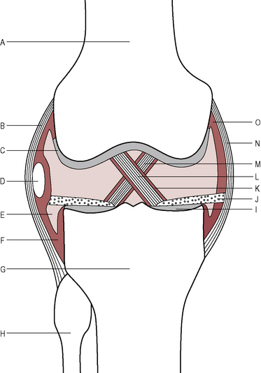
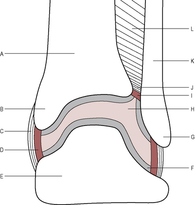
Fig. 6.26 Right knee joint (schematic coronal section).
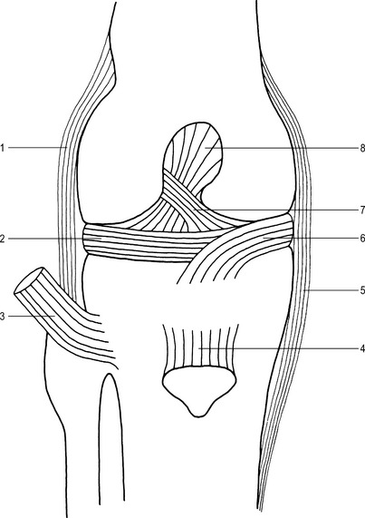
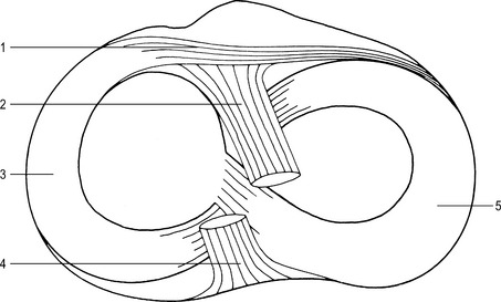
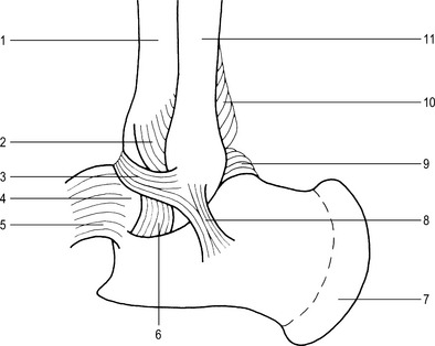
Fig. 6.27 Right knee joint (anterior aspect).
1 – Fibular collateral ligament
5 – Tibial collateral ligament
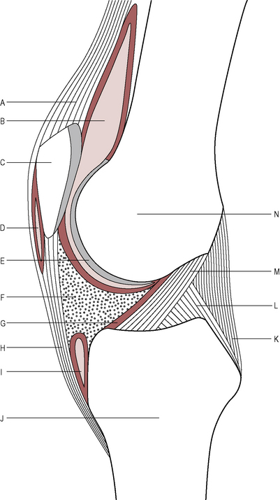
Synovial membrane
Supporting ligaments and tendons
Patellar ligament –
attached to the inferior aspect of the patella and inserted into the tibial tuberosity.
Intracapsular structure
Movements
Flexion by the hamstring muscles aided by the gastrocnemius.
Extension by the quadriceps femoris.
When the knee is in flexion there is a minimal degree of:
Blood supply
Branches of the femoral, popliteal and anterior tibial arteries, forming an anastomosis.
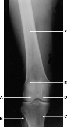
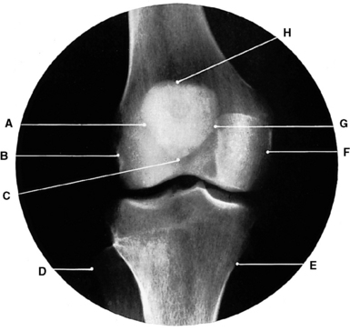
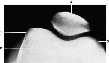
Radiographic appearances of the knee (Figs 6.30 to 6.35)
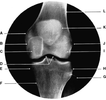
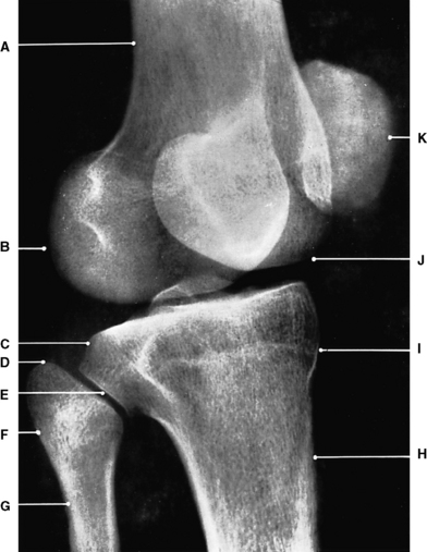
Fig. 6.30 Left knee: anteroposterior projection.
D–Tubercles of intercondylar eminence
(From Bryan 1996.)
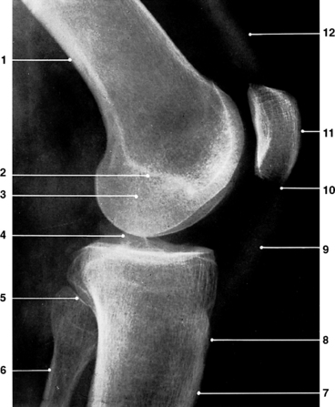
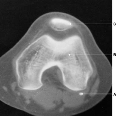
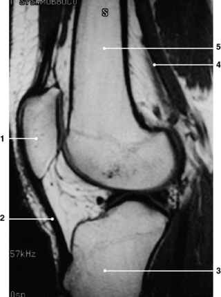
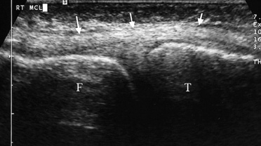
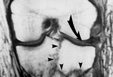
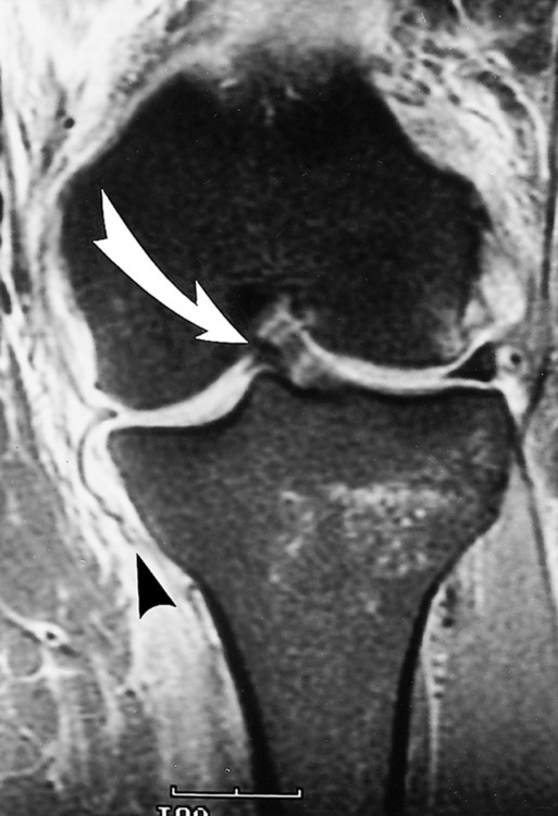
Fig. 6.35 Normal collateral ligaments of the knee (arrowed). Ultrasound scan.
(From Resnick Kransdorf, 2005.)
Superior tibiofibular joint
Inferior tibiofibular joint
Ankle joint (Figs 6.38 and 6.39)
Type
Supporting ligaments
Movements
Accessory movements
 |
slight movement in plantar-flexion. |
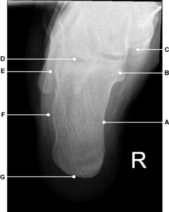
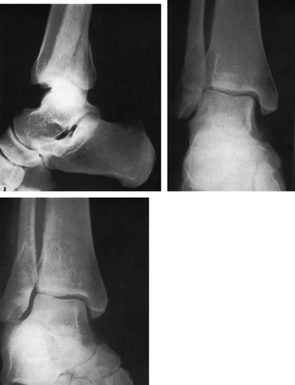
Radiographic appearances of the ankle joint (Figs 6.40, 6.41, 6.42 and 6.43)
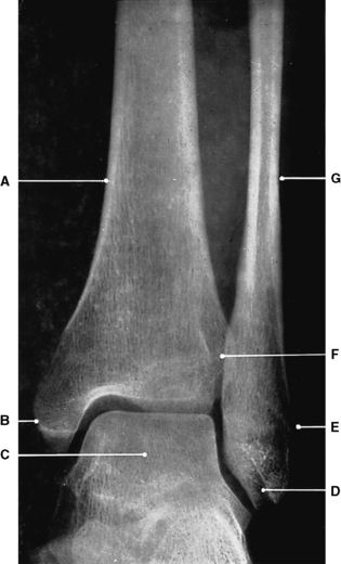
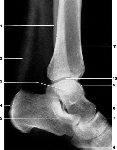
Fig. 6.41 Left ankle joint: lateral projection.
3 – Malleoli medial and lateral
9 – Trochlear surface of talus
(From Bryan 1996.)
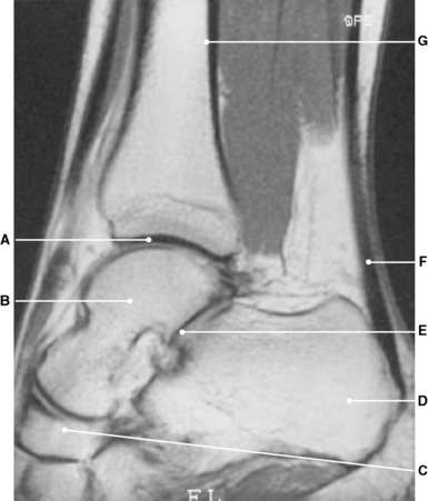
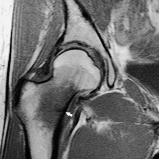
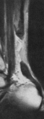
Fig. 6.42 Ankle joint: MR scan, sagittal section.
E–Posterior talo- calcaneal joint
(From Bryan 1996.)
Insight
[/level-membership-for-orthopaedics-category][not-level-membership-for-orthopaedics-category]
6 Lower limb
Femur (Figs 6.1 and 6.2)
Type
Main parts
Features of the upper end of the femur
Intertrochanteric line –
anterior line, between the trochanters; marks part of the junction between the neck and shaft.
Features of the lower end of the femur
Medial and lateral condyles –
fused together anteriorly, separated by the intercondylar notch posteriorly.
Medial epicondyle –
most prominent point of the medial condyle; provides attachment for the tibial collateral ligament.
Radiographic appearances of the femur
Fig. 6.4 Femur: upper two-thirds; lateral projection.
4 – Femoral neck and greater trochanter superimposed
(From Bryan 1996.)
Patella (Figs 6.9 and 6.10)
Tibia (Figs 6.13 and 6.14)
Type
Fig. 6.13 Left tibia (anterior aspect).
C–Tubercles of the intercondylar eminence
Articulations
Tibial condyles with the femoral condyles to form the knee joint.
Lower end of tibia with the talus to form part of the ankle joint.
Fibular notch of the tibia with the lower end of fibula to form the inferior tibiofibular joint.
Main parts
Features of the upper end of tibia
Features of the shaft of tibia
Soleal line –
‘horseshoe-shaped’ oblique line; posterior aspect of the shaft which receives the soleus muscle.
