Lower abdominal pain in a 77-year-old woman

Published on 10/04/2015 by admin

Filed under Surgery

Last modified 22/04/2025

Print this page

rate 1 star rate 2 star rate 3 star rate 4 star rate 5 star
Your rating: none, Average: 0 (0 votes)

This article have been viewed 2017 times

Problem 16 Lower abdominal pain in a 77-year-old woman

There is nothing else from the history that helps towards a diagnosis. Her past history is unremarkable apart from hypertension for which she takes perindopril. The patient is unwell and dehydrated. Her heart rate is 120 bpm and regular, blood pressure 90/70 mmHg and temperature 38°C. Her abdomen is soft apart from an area of localized tenderness in the left iliac fossa. Rectal examination is normal with some soft faeces present.

You decide to perform arterial blood gases to determine the pH and lactate levels as you are worried about the possibility the patient may have ischaemic bowel. The results are shown below (the normal range is in parentheses).

Investigation 16.1 Summary of results

pH 7.2 (7.38–7.43)
pCO2 36 mmHg (35–45)
HCO3 16 (20–24)
Lactate 1.42 (0.50–2.00)
Base excess −9.3 (−3.3–1.2)
K 3.5 (3.8–5.0)

The patient’s condition responds rapidly with the intravenous fluid replacement and after 2 litres of intravenous fluid her blood pressure is 120/85 mmHg. The chest and abdominal X-rays do not show any abnormalities.

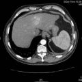
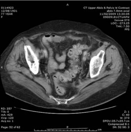
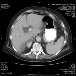
A CT scan of the abdomen is performed. Two representative films are shown in Figures 16.1 and 16.2.

The patient continues to improve and it is decided to pursue a course of conservative management. After 4 days of intravenous fluids and antibiotics her condition is judged satisfactory for her to be discharged home.

Answers

A.1 The story fits well for acute diverticulitis. Other gastrointestinal problems to consider include intestinal obstruction and appendicitis. Pancreatitis may sometimes present with a similar clinical picture. Urinary infection, vascular and gynaecological problems must also be considered although with the nausea, vomiting and constipation, they are less likely. The sudden worsening of her pain raises the possibility of perforation – a well-recognized complication of diverticulitis.

A.2 The patient is hypotensive, probably due to dehydration and sepsis. She has a localized peritonitis and this suggests an inflammatory process, perhaps with abscess formation or free perforation. This would all fit for acute diverticulitis with a possible related complication. The initial management plan and investigations should be as follows:

A.3 The blood gas results indicate a metabolic acidosis, but the normal lactate level makes ischaemic bowel less likely. Metabolic acidosis in this case is probably due to poor tissue perfusion from dehydration and sepsis. Management still lies in urgent resuscitation and rehydration.

A.4 With diverticulitis as the most likely diagnosis a CT scan of the abdomen will be helpful looking for localized thickening of the colonic wall (over 4 mm) in association with any diverticuli and inflammation (’stranding’) of adjacent pericolic fat and mesentery. Evidence of abscess or fistula formation or the presence of free perforation would also be sought. Intraluminal contrast enemas are now outdated in light of the increased sensitivity and specificity of the CT scan in the detection of a disease process which focuses more on the outside rather than the inside of the bowel lumen. Barium enemas are contraindicated in the acute setting because of the risk of spillage of irritant barium into the peritoneal cavity in cases of perforation.

A.5 The CT images show signs of complicated diverticultis with perforation. In Figure 16.1 multiple diverticuli in the sigmoid colon are present, and free air is visible between the liver and diaphragm in Figure 16.2. Other views show inflammation of the pericolic fat and mesentery of the sigmoid colon with locules of free air in the paracolic gutter.

The CT confirms the suspected diagnosis of diverticulitis with perforation.

In some cases where the perforation is contained and the patient is stable, conservative management with intravenous antibiotics and bowel rest may be undertaken. However, most cases of perforated diverticular disease require surgical intervention as a life-saving measure and the safest procedure would be a Hartmann operation where the sigmoid colon containing the perforation is resected, the rectum is oversewn and an end colostomy formed. Formal resection, bowel washout and primary anastomosis with a covering stoma is an acceptable alternative in those patients without gross peritoneal contamination. When consenting a patient for operation, the need for a creation of a stoma should be emphasized. At some later stage the stoma may be reversed but in reality less than half of these patients are suitable for any further surgical intervention because of their poor general state of health.

A.6 Enteric organisms including enterococcus, Gram-negative bacilli (e.g. Escherichia coli) and anaerobes (such as Bacteroides fragilis) are common. Amoxicillin should be used for enterococcus, gentamicin or a third-generation cephalosporin for Gram-negative bacilli and metronidazole for anaerobic organisms.

A.7 The patient will require colonoscopy to confirm the diagnosis and to exclude any colonic malignancy. Normally this is done 4–6 weeks after the acute episode to allow the inflammation to settle and reduce the risk of bowel perforation during colonoscopy.

A.8 The majority of patients will have no further problems and will not need any further treatment. Colonic resection has been advocated for those patients who have had two or more episodes of uncomplicated diverticulitis, but there is little firm evidence to support this approach. Patients are often advised to adhere to high fibre diet and avoid nuts and seeds but there is no evidence to suggest that this strategy will reduce the complications associated with established colonic diverticulae.

Elective resection for diverticular disease is rarely indicated. Patients will uncommonly have recurrent attacks and require elective or emergency surgery. Indications for elective surgery are stricture formation, colovesical fistula or recurrent attacks of diverticulitis. Indications for emergency surgery are perforated diverticular abscess or perforation causing purulent or faecal peritonitis.

Revision Points

The CT scan can rapidly assess the severity of complicated diverticulitis.