CHAPTER 2 Localizing lesions
2.1 The lungs
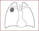
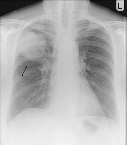
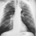
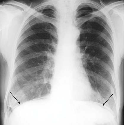
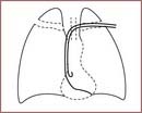
As well as knowing what a lesion is it is often important to know its position within the lung. To accurately localize a lesion on a chest X-ray you need to look at both the PA and lateral films. First look at the PA film:
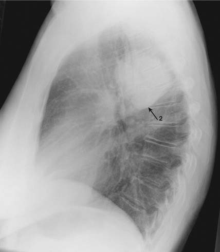
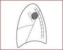
If the lesion is not going to be localized by CT, then a lateral film will be needed.
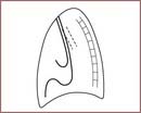
Using the lateral, if the lesion is in the right lung:
If the lesion is in the left lung:
See Chapter 3, which describes localizing using CT scanning.
2.2 The heart
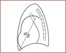
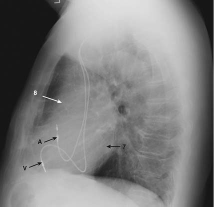
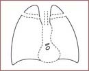
In order to fully assess any abnormalities of the shape of the heart it is important to understand the composition of the heart shadow. Look at the following four films on pages 22–25.
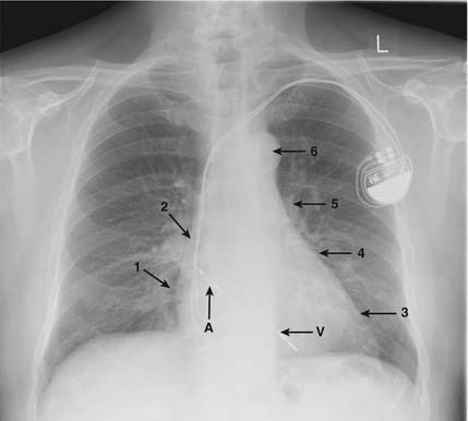
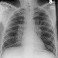
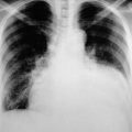
This film is of a patient with an atrial (A) and ventricular (V) pacing wire, with the pacemaker box over the left anterior chest. The atrial wire attaches in the right atrial appendage, and the ventricular wire lies across the tricuspid valve and attaches to the wall of the right ventricle. The numbers are explained in the text on page 21.
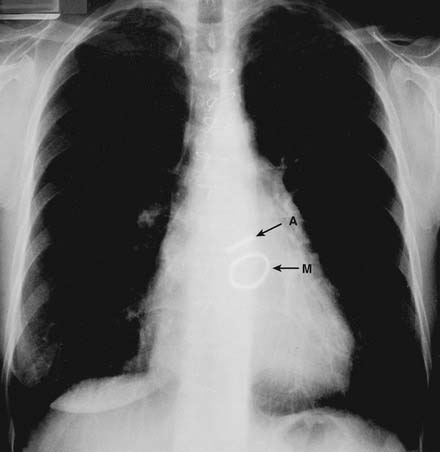
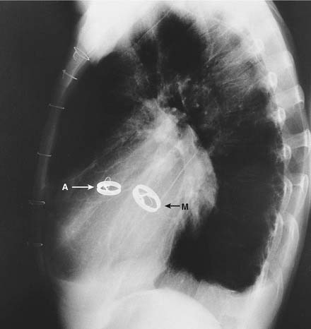
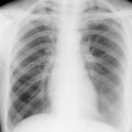
This film is of a patient with prosthetic aortic (A) and mitral (M) valves showing their position in the AP and lateral films.