CHAPTER 10 LIVER PATHOLOGY


HANDLING OF THE LIVER BIOPSY SPECIMEN


PROLONGED NEONATAL CHOLESTASIS
INTRODUCTION


EXTRAHEPATIC BILIARY ATRESIA


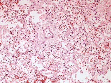
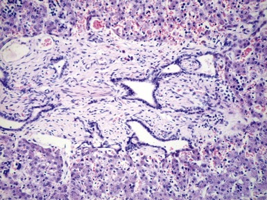
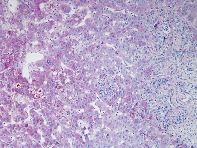
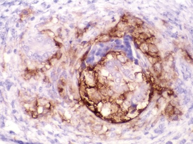
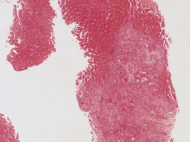
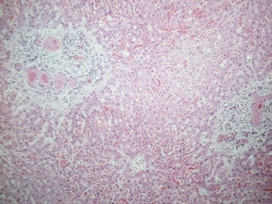
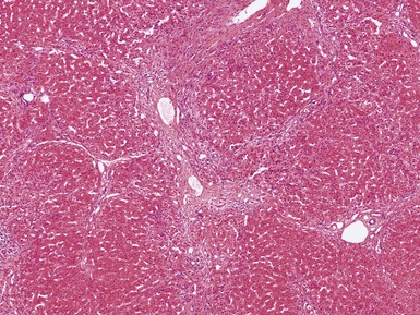
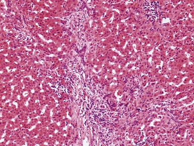
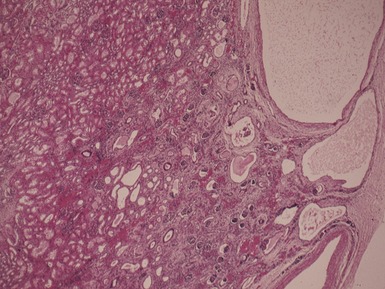
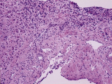
Histopathological features (Figs 10.1–10.3)


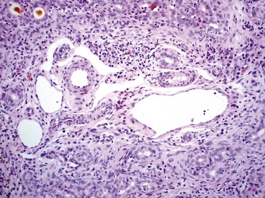
Differential diagnoses and pitfalls


CHOLEDEDOCHAL CYST

Clinical features


NEONATAL HEPATITIS


ALPHA-1-ANTITRYPSIN DEFICIENCY


CYSTIC FIBROSIS


PROGRESSIVE FAMILIAL INTRAHEPATIC CHOLESTASIS (PFIC)


BILE ACID SYNTHESIS DEFECTS


LYMPHEDEMA–CHOLESTASIS SYNDROME


ARTHROGRYPOSIS–RENAL DYSFUNCTION–CHOLESTASIS SYNDROME


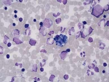
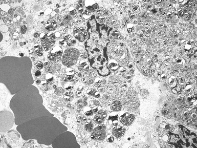
NIEMANN–PICK DISEASE TYPE C


Clinical features


MITOCHONDRIAL CYTOPATHIES
Genetics


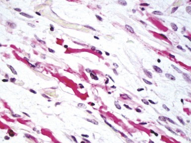
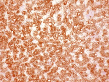
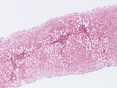
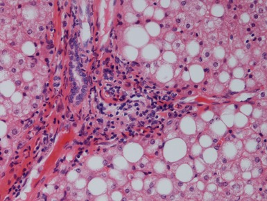
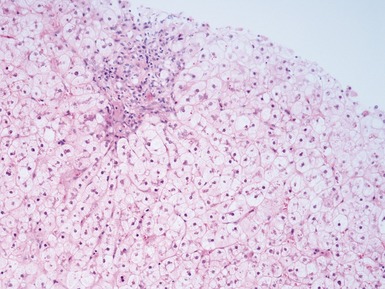
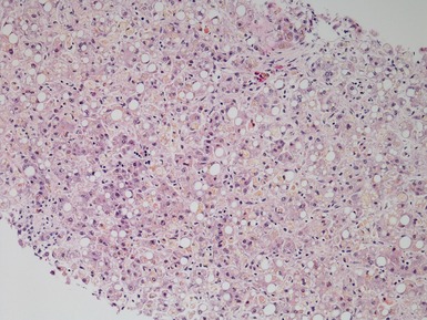
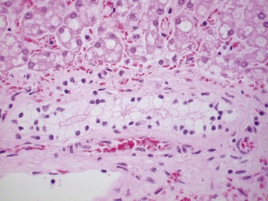
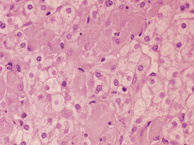
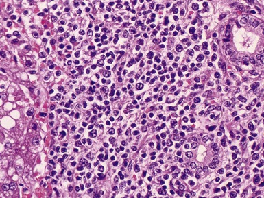
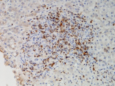
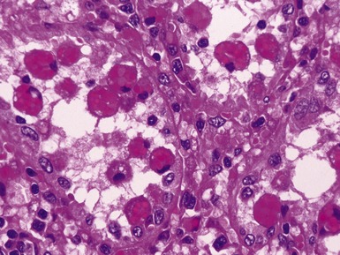
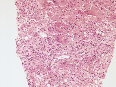
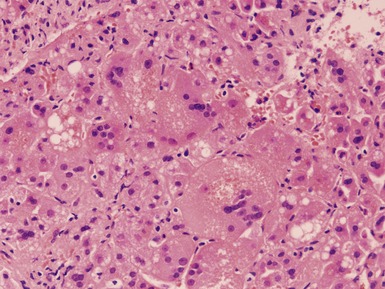
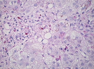
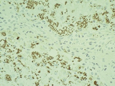
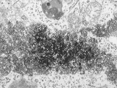
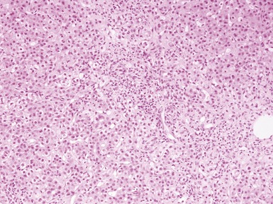
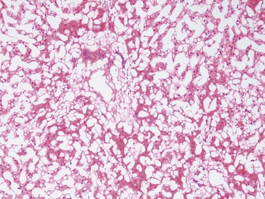
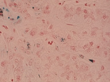
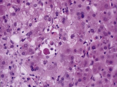
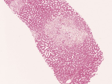
Histopathological features (Figs 10.20–10.25)
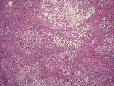
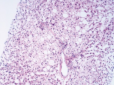
Fig 10.20 Photomicrograph of a liver biopsy from a child with cytochrome oxidase deficiency demonstrating patchy macro- and microvesicular steatosis.
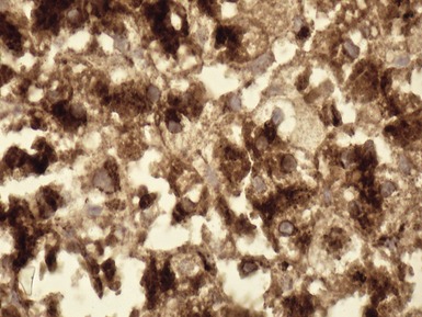
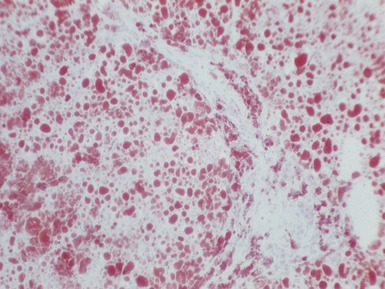
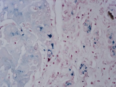
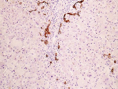
Fig 10.21 Photomicrograph of liver biopsy demonstrating histochemical stain for cytochrome oxidase in which some cells show absent staining in the same case as shown in Fig 10.20.
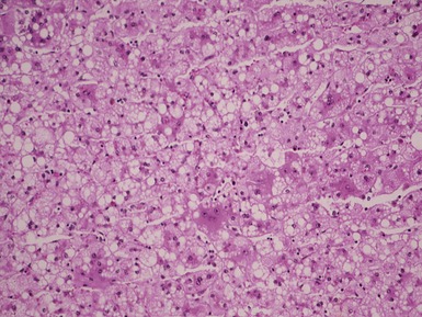
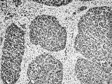
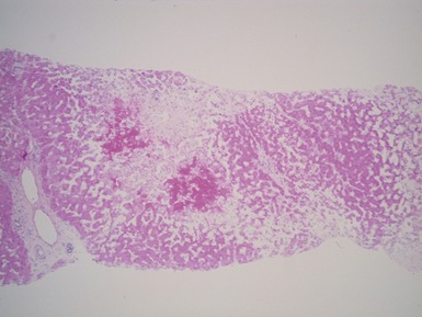
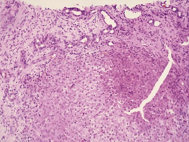
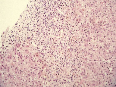
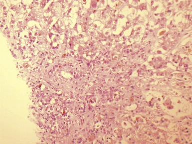
Fig 10.23 Photomicrograph of liver biopsy demonstrating histochemical stain for succinate dehydrogenase in a biopsy from the same patient as shown in Fig 10.22. This biopsy was taken when the child was 10 months old and demonstrates regenerative nodules. The child died shortly afterwards of overwhelming multi-organ failure.


PEROXISOMAL DISORDERS


PAUCITY OF INTRAHEPATIC BILE DUCTS


PRIMARY SCLEROSING CHOLANGITIS


Clinical features


SEPSIS AND TOTAL PARENTERAL NUTRITION-ASSOCIATED LIVER DISEASE


NEONATAL IRON OVERLOAD


INVESTIGATION OF HEPATOMEGALY

CLINICAL FEATURES


GLYCOGEN STORAGE DISEASES


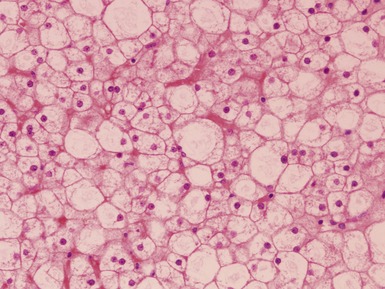
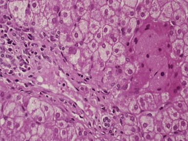
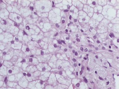
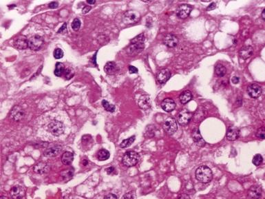
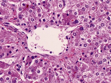
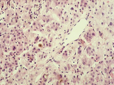
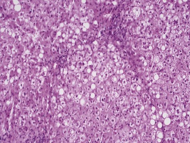
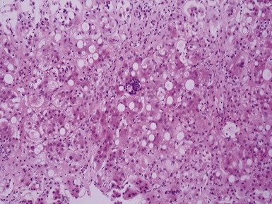
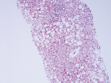
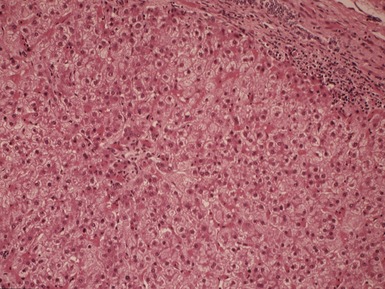
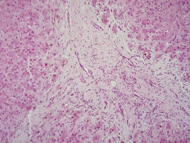
Histopathological features (Figs 10.38–10.42)


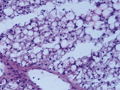
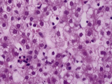
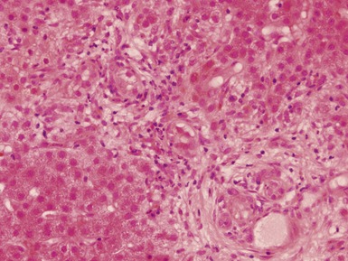
Fig 10.38 Photomicrograph of liver biopsy from a patient with Type I glycogen storage disease demonstrating a combination of enlarged pale hepatocytes together with many hepatocytes containing small fatty vacuoles.
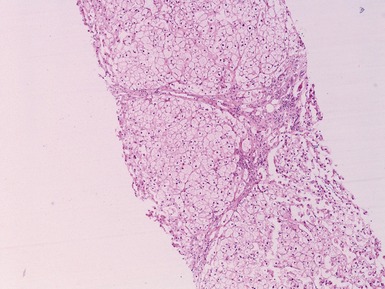
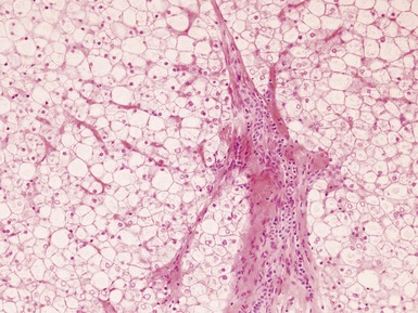
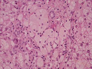
Fig 10.40 Photomicrograph of liver biopsy from the same patient as Fig 10.39 at higher power demonstrating portal fibrosis and enlarged pale hepatocytes.