13 Liver Diseases
Anatomy of the Liver
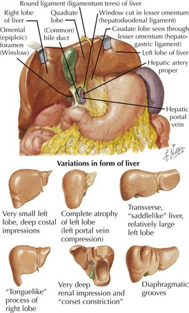
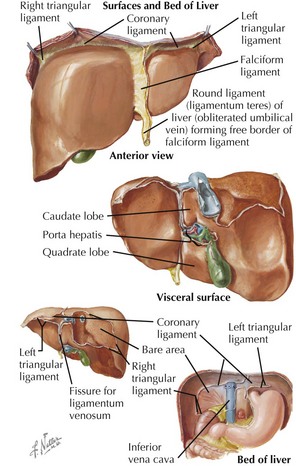
Basic Gross Anatomy


• Hepatoduodenal ligament: peritoneal fold surrounding portal triad (hepatic artery proper, portal vein, bile duct), right edge of lesser omentum
Divisions
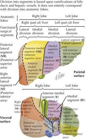
• Right and left hepatic lobes: divided by a plane extending from cystic fossa (anteroinferior) through inferior vena cava (superoposterior)

• Each hemiliver contains its own hepatic artery branch, portal blood supply, venous drainage, and bile duct.


Portal Triads and Bile Duct System
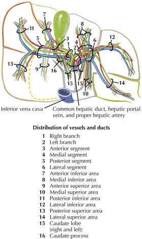
• Portal triads—hepatic artery, bile duct, and portal vein branches—and lymphatics seen in characteristic relationships from microscopic (lobular) to macroscopic (lobar) levels
• Popular functional concepts of liver parenchyma include classic lobules and liver acini organized around vessels.
Innervation of the Liver


• Parasympathetic: vagus


• Sympathetic


Vessels and Lymphatics


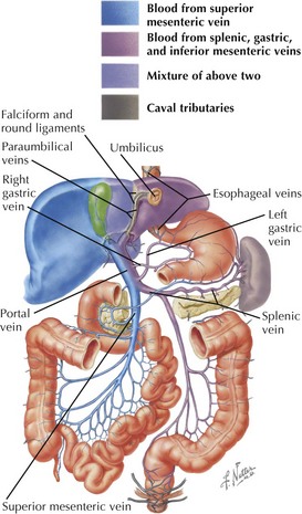
Portal Venous Supply
• Formed by confluence of superior mesenteric vein (right portal) with splenic and inferior mesenteric veins (left portal)
• Left portal drainage goes toward left (hemi) liver, from distal esophagus, lesser curvature of the stomach, spleen, body and tail of the pancreas, and distal half of colon.
Clinical Correlates
Liver Functions


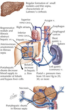
Cirrhosis and Liver Failure
• Mechanism of cirrhosis: hepatocyte destruction → fibrosis and scarring → venous hypertension → portal venous congestion → lymphatic overload → lymph leakage and ascites
Abscesses
• Pyogenic (bacterial)
 Predisposing: biliary diseases or infections in areas with portal drainage (appendicitis, diverticulitis, perforating neoplasms)
Predisposing: biliary diseases or infections in areas with portal drainage (appendicitis, diverticulitis, perforating neoplasms)