CHAPTER 19 Limp
3 Can laboratory tests or imaging studies distinguish transient synovitis from septic arthritis of the hip?
4 Which is a better test for diagnosis and monitoring of bone and joint infections in children: C-reactive protein (CRP) or ESR?
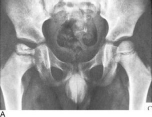
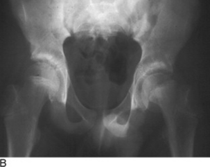
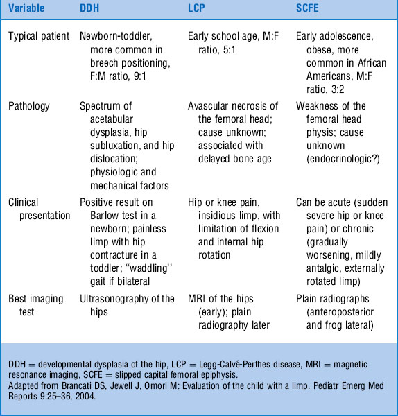
5 Describe the common presentations of the following hip pathologies of children: developmental dysplasia of the hip, Legg-Calvé-Perthes disease, and slipped capital femoral epiphysis (SCFE).
See Figure 19-1A and B. See Table 19-1.
8 What is the best test for distinguishing Legg-Calvé-Perthes disease (avascular necrosis of the hip) from transient synovitis?
9 Describe the benefits of “special” medical imaging tests when compared to plain films in the evaluation of a limping child.
 Bone scanning is useful in detecting early Legg-Calvé-Perthes disease, osteomyelitis, stress fractures, and osteoid osteomas. Scintigraphy is 84–100% sensitive and 70–96% specific for osteomyelitis.


 Magnetic resonance imaging (MRI) can distinguish living from dead bone, which is helpful in studying conditions such as Legg-Calvé-Perthes disease or avascular necrosis, and provides excellent images of the central nervous system, including spinal cord pathology. MRI also has a growing role in the evaluation of infectious processes (see Fig. 19-1).
EMedicine. Herman M: Pediatrics, limp:www.emedicine.com/emerg/topic387.htm
10 A 4-year-old girl presents with limping secondary to knee pain, fever, and a rash. Match the following rashes to the underlying disease.
| 1. Purpuric rash primarily on the lower extremities | A. Lyme disease |
| 2. Salmon-pink evanescent diffuse maculopapular rash | B. Erythema multiforme |
| 3. Multiple large oval papules, some with central clearing | C. Henoch-Schönlein purpura |
| 4. Symmetric papulovesicular rash with target lesions | D. Systemic juvenile idiopathic arthritis |
11 What is the best test for diagnosing juvenile idiopathic arthritis (previously called juvenile rheumatoid arthritis [JIA]) in a child with chronic limp and joint pains?
12 In a limping child with lower-extremity joint pains who meets the clinical criteria for oligoarthritic JIA, which is a more useful serologic test: rheumatoid factor or antinuclear antibody?
14 A 2-year-old child presents with refusal to walk after jumping off of a toy box. He is tender over the distal portion of his left tibia. Plain radiographs of the lower leg are negative. What is the most likely explanation for his pain?
15 An oblique view of the tibia in the previously described case reveals a subtle spiral fracture. The resident wants to consult child protective services given the spiral fracture with minimal trauma. What do you recommend?
1 Look beyond the leg, laboratory values, and x-rays when evaluating the limping child.
2 SCFE is an orthopedic emergency (and may present with referred knee or thigh pain).
3 Limp may be the acute presentation of a more chronic problem, abdominal or back pathology, or even systemic illness. Open physis? Think fracture before sprain.
4 Normal laboratory studies do not exclude the possibility of septic joint or osteomyelitis.
17 A 10-year-old girl presents with limping and pain over the lateral malleolus after twisting her ankle while playing basketball. She is able to bear weight. Plain radiographs of the ankle are negative. Which is a more appropriate management plan: elastic bandage wrap, ankle-strengthening exercises, and family doctor follow-up; or plaster splint, rest, and orthopedic follow-up?
18 A 14-year-old boy presents with limp after twisting his ankle. In performing a complete ankle examination, you include palpation of the lateral aspect of the foot. Why?
19 A 16-year-old female competitive long-distance runner reports insidious but progressive lower-left leg pain and limp over several weeks. After a negative plain film and persistent symptoms, MRI is performed, leading to a diagnosis of stress fracture of the tibia. Why is a detailed nutritional and gynecologic history of particular importance in evaluating this patient?
Brunet II M: Female athlete triad. Clin Sports Med 24:623–636, 2005.
20 A school-aged or young adolescent child presents with a chronically painful flat foot. Radiographs in the past have not been helpful. What condition would you suspect, and what imaging study would be most useful in detecting it?
21 A 15-year-old boy has been waking up with leg pain for two weeks. Initially his symptoms were attributed to “growing pains” and were treated very successfully with aspirin, but his mother is concerned about the duration of the symptoms. What are you concerned about?
22 Match the following potential causes of limp with the location of pain.
| 1. Sever’s disease | A. Patella |
| 2. Sinding-Larsen-Johansson syndrome | B. Calcaneus |
| 3. Osgood-Schlatter disease | C. Tibia |
| 4. Kohler’s disease | D. Metatarsal |
| 5. Freiberg’s disease | E. Navicular |
1 B. Sever’s disease: Insertion of the Achilles tendon on the calcaneus
2 A. Sinding-Larsen-Johansson syndrome: The attachment of the patellar tendon to the inferior patellar pole
3 C. Osgood-Schlatter disease: Insertion of the patellar tendon on the proximal tibial tuberosity
4 E. Kohler’s disease: The tarsal navicular
5 D. Freiberg’s disease: Usually the second metatarsal head, less often in the third and fourth
