130
Leukoplakia
Leukoplakia is a common, sometimes chronic condition of the oral mucosa.
Leukoplakia usually appears after the age of 40, and prevalence approaches 8% after age 70.
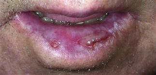
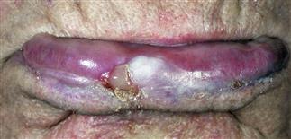
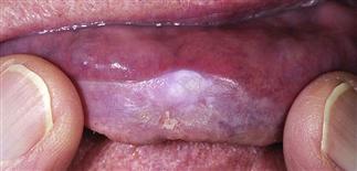
Leukoplakia. The white patches are slightly elevated and well demarcated.
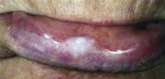
Leukoplakia 2 weeks after treatment with topical 5-fluorouracil.
DESCRIPTION
Leukoplakia is a descriptive clinical term reserved for white patches or plaques occurring on the mucosal surfaces pending definitive diagnosis. It is a descriptive clinical term, not a definitive diagnosis. The term is often misused to designate a premalignant condition.
HISTORY
• A common, sometimes chronic condition of the oral mucosa. • Occurs more frequently in men than in women. • Usually appears after age 40, and the prevalence approaches 8% after age 70. • Most lesions are asymptomatic.
PHYSICAL FINDINGS
• Leukoplakia begins as a single small, well-defined, translucent to white, slightly elevated papule. • Individual lesions may resolve completely, recur, or progress. Multiple papules may coalesce into larger plaques over time. • Uneven hyperkeratosis or small erosions may develop. • Focal red areas or discrete papules termed erythroplakia may develop within plaques, giving a speckled appearance. • Lesions may occur anywhere on the oral mucosa but are most commonly found on buccal mucosa and lower lip. • Differential diagnosis includes candidiasis, oral hairy leukoplakia, white sponge nevus, lichen planus, squamous cell carcinoma.
TREATMENT
• Encourage patients who use tobacco products to stop. • Any area of change within the leukoplakia patch, especially areas of erythroplakia, should be biopsied without delay. • Localized areas of epithelial dysplasia may be treated by cryosurgery, electrocautery, or topical 5-fluorouracil. Combinations of these therapies are sometimes used. • Areas demonstrating squamous cell carcinoma, either in situ or invasive, are best treated with surgical excision. Close clinical follow-up, including lymph node examination, is required. • Indefinite close clinical follow-up is required after treatment to detect recurrence early. This includes inspection of other areas of the oral mucosa, as well as regional lymph node examination. • Progression to squamous cell carcinoma develops in 15–20% of all patients with leukoplakia.


