Leukemic and lymphomatous infiltrates of the skin

Published on 08/03/2015 by admin

Filed under Dermatology

Last modified 22/04/2025

Print this page

rate 1 star rate 2 star rate 3 star rate 4 star rate 5 star
Your rating: none, Average: 0 (0 votes)

This article have been viewed 2859 times

Chapter 46 Leukemic and lymphomatous infiltrates of the skin

Mycosis fungoides

5. How does mycosis fungoides begin?

Typically, mycosis fungoides begins with persistent scaly patches (Fig. 46-1A) that respond poorly to topical therapy with emollients and topical steroids. In the early stages, skin biopsy is frequently not diagnostic. The average time from onset of skin lesions to diagnosis is 7 years. In this early phase of the disease, a diagnosis of parapsoriasis en plaque is often made. In time, the patches thicken and become plaques. Eventually, skin tumors develop (Fig. 46-1B,C) and the lymph nodes can become involved. Visceral disease is a late occurrence in this low-grade lymphoma. Median survival for persons with patch- and plaque-stage disease is 12 years; for tumor-stage disease, it is 5 years; and for nodal or visceral disease, it is 3 years.

Epstein E, Levin D, Croft J, et al: Mycosis fungoides: survival, prognostic features, response to therapy, and autopsy findings, Medicine 51:61–72, 1972.

6. What is parapsoriasis?

The skin diseases included under this diagnosis are poorly understood and encompass a morass of confusing terms. The “splitters” have described over a dozen varieties of parapsoriasis, while the “lumpers” limit this designation to only a few types. This discussion supports the “lumpers” viewpoint.

Small-plaque parapsoriasis is characterized by chronic, well-marginated, mildly scaly, slightly erythematous, and round to oval skin lesions measuring <4 to 5 cm in diameter. The long axes of the lesions are arranged in a parallel configuration, and the lesions occur on the trunk and proximal extremities in a pityriasis rosea–like pattern. The lesions have been likened to fingerprints and reported under the descriptive term of digitate dermatoses. This form of parapsoriasis does not progress to lymphoma.

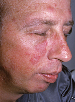
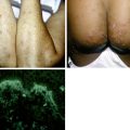
Large-plaque parapsoriasis presents as palm-sized or larger lesions located most frequently on the thighs, buttocks, hips, lower abdomen, and shoulder girdle areas (Fig. 46-2). The lesions may be pink, red-brown, or salmon-colored. They often have fine scale and show epidermal atrophy with cigarette-paper wrinkling. Some patients may have lesions with a netlike or reticular pattern with telangiectasia and fine scale. This clinical type of lesion is referred to as retiform parapsoriasis or poikiloderma atrophicans vasculare. Between 15% and 20% of patients with large-plaque parapsoriasis eventually develop mycosis fungoides.
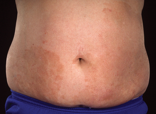
Sehgal VN, Srivastava G, Aggarwal AK: Parapsoriasis: a complex issue, Skinmed 6:280–286, 2007.
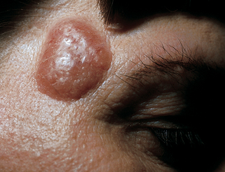
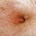
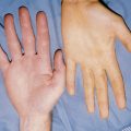
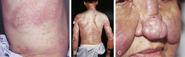
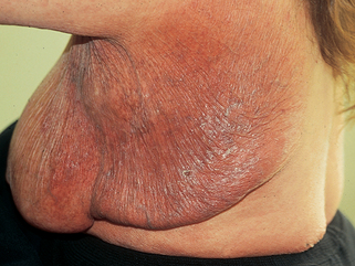
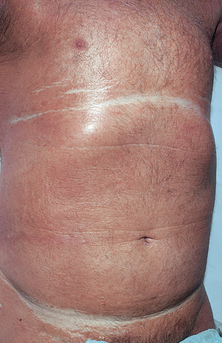
8. Describe the three subtypes of mycosis fungoides.

Sézary syndrome (Fig. 46-4) presents with the classic triad of erythroderma, lymphadenopathy, and atypical circulating mononuclear cells (Sézary cells). These cells are moderately large mononuclear cells with hyperconvoluted nuclei. They resemble activated T cells, and when >15% of circulating lymphocytes are atypical, it is considered significant, with 10% to 15% being considered borderline. However, the finding of circulating Sézary cells must be evaluated in context with the clinical picture and skin biopsy. Severe pruritus, ectropion, nail dystrophy, peripheral edema, alopecia, and keratoderma of the palms and soles are common associated features. The disease tends to wax and wane and generally progresses faster and is more resistant to treatment than typical mycosis fungoides.

Systemic

Combination chemotherapies

EPOCH

National Cancer Institute. Mycosis fungoides and the Sézary syndrome treatment. Available at: http://www.cancer.gov/cancertopics/pdq/treatment/mycosisfungoides/healthprofessional/allpages. Accessed December 6, 2006.

Other lymphomas and leukemias

25. What is lymphomatoid papulosis?

This chronic recurrent skin eruption is characterized by papules and/or nodules that frequently crust or ulcerate and self-heal, often with atrophic scars (Fig. 46-5). There are three histopathologic types. Type A has large Reed-Sternberg–like cells, which are often CD30 (Ki-1) positive. Type B has moderately large atypical cells with cerebriform nuclei similar to the cell type found in mycosis fungoides. These cells are usually CD30 negative. Type C is composed of sheets of cells that resemble the cells of anaplastic T-cell lymphoma. About 15% to 20% of patients with lymphomatoid papulosis will develop a lymphoma.

Table 46-2. Cells Marked by CD Antigens

CD2 T cells (E rosette receptor)
CD3 T-cell receptor
CD4 Helper T cells
CD5 Mature thymocytes, some B-cell subsets
CD7 T cells, natural killer (NK) cells
CD8 T cells, NK cells
CD10 Pre-B cells, lymphoblastic leukemia cells
CD14 Monocytes
CD15 Reed-Sternberg cells, myeloid cells
CD19 Pan-B cells
CD20 Pan-B cells, dendritic cells
CD21 Receptor for complement 2 and Epstein-Barr virus
CD23 Activated B cells, monocytes, eosinophils, and platelets
CD25 Activated T and B cells, monocytes (interleukin-2 receptor)
CD30 Ki-1–related cells, Reed-Sternberg cells, T-cell NHL
CD34 Lymphoid and myeloid precursor cells
CD43 T cells, myeloid cells
CD45 Leukocytes
CD45R T cells, myeloid cells
CD56 NK cells
CD74 HLA-invariant chain
CD75 Follicular center cells

CD, Cluster of differentiation; HLA, histocompatibility locus antigen; NHL, non-Hodgkin’s lymphoma.

image

Figure 46-5. Lymphomatoid papulosis. The lesions occur in crops and self-heal.

(Courtesy of the Fitzsimons Army Medical teaching files.)

El Shabrawi-Caelen L, Kerl H, Cerroni L: Lymphomatoid papulosis: reappraisal of clinicopathologic presentation and classification into subtypes A, B, and C, Arch Dermatol 140:441–447, 2004.

Share this: