Scalp Lesions
Lesions on the scalp are common; the most common are sebaceous cysts, which are often multiple.
Causes
Cystic
Neoplastic
Malignant
History
Traumatic
Check for a history of trauma. A boggy haematoma may overlie a skull fracture. A cephalhaematoma is seen in a newborn baby. It follows a traumatic delivery. The haematoma is below the periosteum of the skull.
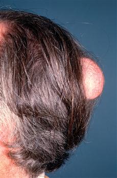
Sebaceous cyst
These may be multiple. The patient usually notices them when combing the hair.
Neoplastic
With an ivory osteoma, the patient may notice a rock-hard swelling on the scalp. The patient is usually a young adult and is asymptomatic. Malignant ulcers may occur on the scalp. The patient will notice a lesion when combing the hair and bleeding may occur. These lesions present earlier in bald patients. Bony tenderness and swelling may be a presenting symptom of secondary deposits. There may be a history of a primary, or a careful history must be taken to establish the site of a primary.
Infective
With Cock’s peculiar tumour, the patient may notice a sore, bleeding lesion on the scalp. Cock’s peculiar tumour is due to a sebaceous cyst suppurating and granulation tissue appearing on its surface. It may be mistaken for a squamous cell carcinoma. Tinea capitis usually occurs in childhood. There is an itchy, red, scaly patch on the scalp and the hairs break easily, leaving patches of stubble.
Other
Psoriasis may affect the scalp. There is usually a history of lesions elsewhere on the body. Seborrhoeic dermatitis presents with a fine, scaly rash on the scalp. Hair growth is usually normal with both these lesions.
Examination
Traumatic
A knock on the head may cause a boggy haematoma. X-ray is required to exclude an underlying fracture. Cephalhaematoma occurs in the newborn; the haematoma spreads beneath the periosteum of the skull and is therefore limited by the skull suture lines.
Sebaceous cyst
Sebaceous cysts are spherical, tender, firm swellings in the scalp. They may be multiple. Rarely is a punctum visible with sebaceous cysts on the scalp.
Neoplastic
An ivory osteoma is a bony, hard, smooth swelling, arising from the outer table of the skull. The skin is freely mobile over it. A squamous cell carcinoma presents as an ulcer with a hard, everted edge. A malignant melanoma is usually pigmented, ulcerated and bleeds. In both these conditions, cervical lymphadenopathy may be present. A basal cell carcinoma is a raised ulcer with a rolled edge with a pearly appearance, often with superficial telangiectasia. Lesions in the skull may be secondaries from lung, breast, thyroid, prostate and kidney. Each of these areas should be examined for a possible primary. Myeloma may present with painful lesions in the skull. There may be areas of localised tenderness.
Infective
Cock’s peculiar tumour presents as an open, granulating sebaceous cyst. It appears angry and swollen and may be mistaken for a squamous cell carcinoma. In tinea capitis, there are red, scaly patches on the scalp, with broken hairs giving a stubbled appearance.
Other
With psoriasis, there are well-demarcated areas of scales heaped up over red plaque. The hair grows normally through the plaques. Check for psoriatic lesions elsewhere. Seborrhoeic dermatitis shows fine scales but the hairs remain intact.
General Investigations
The diagnosis of most of these lesions is made on clinical examination alone.
■ FBC, ESR
Hb ↓ malignancy. WCC ↑ leukaemia. ESR ↑ myeloma and other malignancies.
■ CXR
Primary tumour. Secondary malignancy, e.g. secondaries from malignant melanoma.
■ Skull X-ray
Fracture. Ivory osteoma. Pepper-pot skull – myeloma. Secondary deposits – osteolytic from lung, breast and thyroid. Osteosclerotic from prostate and occasionally kidney.