[level-membership-for-internal-medicine-category]
Finger Lesions
Lesions of the fingers are common. The fingers are important tactile organs and hand function may be impaired. Painful finger lesions are dealt with on p. 153.
Causes
Acquired
Traumatic/Degenerative
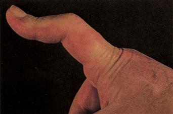
Figure 21 Boutonnière deformity.
This common deformity in advanced rheumatoid arthritis results from the rupture of the central slip of the extensor tendon over the proximal interphalangeal joint. The lateral slips of the extensor tendon mechanism are displaced to the sides and maintain the deformity.
History
Congenital
These lesions will be recognisable at birth. They may be associated with other congenital abnormalities.
Acquired
Traumatic/degenerative
Dupuytren’s contracture
In the early phases, the patient may merely complain of a nodule in the palm near the base of the ring finger. Eventually, the patient complains of being unable to extend fully the MCP joint at the ring, and later, the little finger. It affects the patient’s grip. The patient complains of difficulty dressing – either poke themselves in the eye while brushing hair or catch the finger on a trouser pocket. There may be a family history. Check also for a history of epilepsy, cirrhosis or diabetes.
Implantation dermoid
Cysts occur where skin is forcibly implanted into the subcutaneous tissue as a result of injury. There is likely to be a history of injury, often minor, e.g. pinprick. Implantation dermoid used to be common in women who sewed – hence the use of protective thimbles. The patient complains of small painful swellings on the finger tips.
Pyogenic granuloma
Pyogenic granuloma is a common inflammatory lesion of the skin which arises in response to minor penetrating foreign bodies such as splinters or thorns. Pyogenic granulomas are solitary, reddish-blue fleshy nodules. The surface may be ulcerated in which case the lesion may be clinically indistinguishable from amelanotic malignant melanoma.
Trigger finger
The patient complains that the finger ‘jumps’ or ‘clicks’ as it moves. It may get stuck in the flexed position. There is not usually a history of injury.
Mallet finger
This results from injury to the extensor tendon of the terminal phalanx. There is usually a history of injury. It occurs if the tip of the finger is forcibly flexed during active extension (stubbed), e.g. catching a cricket ball. The finger adopts a position in which the distal phalanx is flexed.
Swan-neck deformity
The patient complains of deformity of the distal finger.
Boutonnière deformity
This is the opposite of swan-neck deformity. Again, the patient merely complains of a deformity of the distal finger.
Heberden’s nodes
The patient complains of swelling close to the distal finger joints, i.e. swelling and deformity of the knuckles. There may be a history of osteoarthritis elsewhere.
Mucous cyst
The patient complains of a swelling over the dorsum of the DIP joint. It may discharge.
Calluses over knuckles (bulimia)
Calluses over the knuckles (Russell’s sign) may arise in bulimics due to repeated self-induced vomiting over a long period of time. The condition arises from the patient’s knuckles making contact with the incisor teeth during the act of inducing the gag reflex at the back of the throat with the fingers.
Metabolic
Tophaceous gout
There may be a history of gout. Deposits of uric acid occur in joints or soft tissues and the patient complains of swellings on the fingers.
Ectopic calcification
The patient complains of hard, whitish, subcutaneous swellings. There may be a history of hyperparathyroidism, hypercalcaemia or chronic renal failure.
Connective tissue disease
CREST syndrome
The patient complains of white deposits in the pulps of the fingers.
Neoplastic
Subungual melanoma
This occurs as a pigmented lesion beneath the nail. There is no history of trauma as with a subungual haematoma, which may also appear pigmented when the bruise organises. Unlike subungual haematoma, a melanoma does not grow out with the nail. Eventually the melanoma may lift the nail and ulcerate.
Enchondroma
The patient notices a bony, hard swelling along the finger. It may occur beneath the nail and result in deformity of the nail (subungual exostosis).
Examination
Congenital
Congenital anomalies of the fingers will be obvious at birth. With camptodactyly, there is painless flexion deformity at the PIP joint of the little finger. Check for other associated congenital anomalies.
Acquired
Traumatic/degenerative
Dupuytren’s contracture
Examination may reveal only a firm painless nodule in the palmar fascia near the base of the ring finger. Puckering of skin of the palm may be obvious. Eventually, the MCP joint and PIP joint become flexed. The DIP joint remains extended. Garrod’s knuckle pads on dorsum of PIP joints. Check for signs of diabetes or liver disease. Occasionally the condition is associated with Peyronie’s disease of the penis.
Implantation dermoid
Examination reveals small firm spherical swellings in the subcutaneous tissues of the finger tips.
Trigger finger
The patient may be able to demonstrate how the finger sticks and then snaps out in extension. Thickening of the tendon and the tendon sheath may be felt over the head of the metacarpal bone.
Mallet finger
The distal phalanx of the affected finger remains flexed at about 20° when the patient tries to straighten the finger.
Swan-neck deformity
There is hyperextension of the PIP joint and flexion of the distal interphalangeal joint.
Boutonnière deformity
This is the opposite of swan-neck deformity. Flexion of the PIP joint occurs, with hyperextension of the DIP joint. It develops when the PIP joint pokes through a rupture of the centre of the extensor expansion.
Heberden’s nodes
These are bony swellings on the dorsal surface of the fingers just distal to the DIP.
Mucous cyst
Swelling on the dorsum of the DIP joint. Discharge may be apparent. May be a groove in the nail due to pressure on the nailbed.
Metabolic
Tophaceous gout
Swellings, often multi-lobulated, occur on the fingers. They contain toothpaste-like infiltrates of uric acid crystals.
Ectopic calcification
Firm to hard, yellow-white deposits of calcium occur in the subcutaneous tissues.
Connective tissue disease
CREST syndrome
Small, hard, subcutaneous nodules in the finger pulps and on the dorsal aspects of the fingers.
Neoplastic
Subungual melanoma
A subungual melanoma is seen as a brown lesion with an indistinct edge. Occasionally it may be quite extensive and have lifted the nail.
Enchondroma
This is a painless, hard swelling on the bone. The surface of it is usually smooth. It may occur under the nail and lift the nail.
General Investigations
The diagnosis of most of these lesions is made on clinical examination alone.
■ FBC, ESR
Hb ↓, ESR ↑ in disseminated malignancy from subungual melanoma.
■ Blood glucose
Diabetes (Dupuytren’s).
■ LFTs
Cirrhosis (Dupuytren’s).
■ Serum calcium
Hypercalcaemia – ectopic calcification. Hyperparathyroidism.
■ Rheumatoid factor
Rheumatoid arthritis – swan-neck deformity. Boutonnière deformity.
■ Finger X-ray
Heberden’s nodes. Enchondroma. Mucous cyst – degenerative/osteoarthritic changes in the DIP joint. Ectopic calcification. Calcinosis. Erosion of phalangeal bone with tophaceous gout.
■ Biopsy
Biopsy to distinguish pyogenic granuloma from amelanotic malignant melanoma. Excision biopsy for malignant melanoma.
[/level-membership-for-internal-medicine-category][not-level-membership-for-internal-medicine-category]
Finger Lesions
Lesions of the fingers are common. The fingers are important tactile organs and hand function may be impaired. Painful finger lesions are dealt with on p. 153.
Causes
Acquired
Traumatic/Degenerative
Figure 21 Boutonnière deformity.
This common deformity in advanced rheumatoid arthritis results from the rupture of the central slip of the extensor tendon over the proximal interphalangeal joint. The lateral slips of the extensor tendon mechanism are displaced to the sides and maintain the deformity.
History
Congenital
These lesions will be recognisable at birth. They may be associated with other congenital abnormalities.
Acquired
Traumatic/degenerative
Dupuytren’s contracture
In the early phases, the patient may merely complain of a nodule in the palm near the base of the ring finger. Eventually, the patient complains of being unable to extend fully the MCP joint at the ring, and later, the little finger. It affects the patient’s grip. The patient complains of difficulty dressing – either poke themselves in the eye while brushing hair or catch the finger on a trouser pocket. There may be a family history. Check also for a history of epilepsy, cirrhosis or diabetes.
Implantation dermoid
Cysts occur where skin is forcibly implanted into the subcutaneous tissue as a result of injury. There is likely to be a history of injury, often minor, e.g. pinprick. Implantation dermoid used to be common in women who sewed – hence the use of protective thimbles. The patient complains of small painful swellings on the finger tips.
Pyogenic granuloma
[/not-level-membership-for-internal-medicine-category]
