167
Leshmaniasis
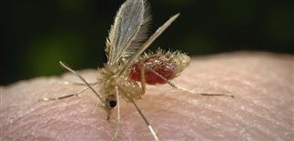
Phlebotomus sandfly vector of Old World leishmaniasis. Courtesy of Centers for Disease Control and Prevention, Atlanta, GA, USA.
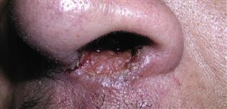
Small single shallow ulceration with rolled border at the edge of the nasal mucosa of L. braziliensis. From Bolognia, JL, Jorizzo JL, Schaffeer, JV, eds. Dermatology. 3rd edn. London, Saunders; 2012. Courtesy, Kalman Watsky, MD.
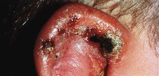
An inflamed and ulcerated ear of L. mexicana, or chiclero ulcer, may lead to cartilage destruction. From James WD, Berger T, Elston DMD, eds. Andrew’s Diseases of the Skin. 11th edn. London, Saunders; 2011.
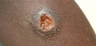
A typical ulcer of L. major, with a shallow ulcer and rolled border. From Magill AJ, Ryan ET, Hill DR, Solomon T. eds. Hunter’s Tropical Medicine and Emerging Infectious Diseases. 9th edn. London, Saunders; 2013.
DESCRIPTION
A parasitic, protozoal infection, caused by several Leishmania species and transmitted by the sandfly. Acquired in tropical and subtropical areas of the world. Variable clinical presentations.
HISTORY
• The intracellular protozoan Leishmania species is considered endemic in tropical and subtropical areas of South America, Asia and Africa, also some Mediterranean countries. • More common in developing countries. • Children commonly affected. • The sandfly (Phlebotomous or Lutzomyia) bite is the major mode of transmission. Leishmania are found in a flagellated promastigote stage within the midgut of the sandfly, transmitted to humans by the bite of the female sandfly. Incubation times range from 1–24 months from time of bite to clinical symptoms. • Classified by geographic area, specific species and clinical presentation. • Old World leishmaniasis (Europe, Asia, Africa): L. tropica, L. major, L. infantum, L. donovania. • New World leishmaniasis (Americas): L. mexicana, L. amozonesis, L. braziliensis.
PHYSICAL FINDINGS
• The major types of clinical presentations: cutaneous (skin only), mucocutaneous (skin and mucosal surfaces), diffuse cutaneous and visceral. • The clinical spectrum depends on the type of species and the cell-mediated immune response of the host.
SKIN FINDINGS
• Cutaneous leishmaniasis. Most common form, usually Old World type. • Red non-tender papule, 2–4 mm, at site of bite. • Multiple lesions may occur. • Papule slowly enlarges over 2–3 months into a 2–4 cm plaque with crust, may ulcerate and develop a firm violaceous border, persisting for many months. • May heal spontaneously with or without a scar. • Mucocutaneous leishmaniasis. New World type. • Nasopharyngeal involvement, extension of cutaneous disease. • Inflammation of the mucosa with hemorrhage and painful discharge. • Diffuse cutaneous leishmaniasis. Numerous parasites within the skin; poor host immune response to parasite. • Many non-ulcerative nodules evolving from satellite papules around initial lesion. • Visceral leishmaniasis (kala azar). Bone marrow, liver and spleen involvement. • Fever, pain, lymphadenopathy, hepatosplenomegaly and pancytopenia. • Slow to very fast progression, poor prognosis.
TREATMENT
• Depends on species of leishmaniasis and infection location. • Early diagnosis, speciation and detection of systemic involvement are important. • L. major (e.g. Iraq) is a self-limited form and may resolve within a year without treatment. L. tropica (e.g. Afghanistan) may cause aggressive or chronic disease and require systemic therapy. • Systemic therapy including antimonials (meglumine antimoniate, sodium stibocloconate) is usually the first choice. • Amphoteracin B, azole antifungals (fluconazole, kenoconazole), dapsone, rifampin and allopurinol are other options. • Proper monitoring for toxicities of these medications is required. Assistance from an infectious disease specialist may also be helpful.


