113
Lentigo, juvenile lentigo,
solar lentigo
Lentigines are common, benign, brown macules on the sun-exposed skin of Caucasians.
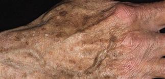
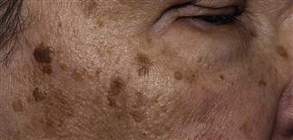
Lentigines (liver spots) occur in sun-exposed areas of the face, arms, and hands.
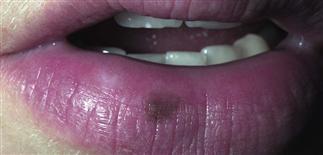
Labial lentigines occur on the vermillion border of the lower lip. They have a smooth border and are benign.
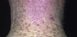
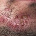
Reactive hyperplasia of melanocytes causes persistent pigmentation in the form of lentigines on the neck and upper back.
DESCRIPTION
Common, benign, brown macules occurring on sun-exposed skin of white people.
HISTORY
Localized hyperpigmentation in three patterns: freckles (ephelides), juvenile lentigo, solar lentigo. While similar in size, distribution, clinical appearance, the three patterns differ in age of onset, clinical course, relation to sun exposure. • Freckles. Appear in childhood. Occur as autosomal dominant trait. Usually confined to face, arms, upper trunk. Increase in number and slightly darken in summer; often fade completely in winter. • Juvenile lentigines. Appear in childhood with mean number of 30 lentigines in each prepubertal child. Lesions do not increase in number or size, or darken, in response to sunlight; do not fade in absence of sunlight. Juvenile lentigines also occur as characteristic feature of certain hereditary conditions, such as Peutz–Jeghers, LEOPARD, LAMB syndromes. • Solar lentigines. Common on sun-exposed skin. Increase in number and size with advancing age. Roughly 75% of white people over 60 have one or more lesions, which occur in setting of actinic damage.
PHYSICAL FINDINGS
• Freckles. 1- to 2-mm, sharply defined, red or tan to light brown macules with uniform color. Number varies from few sparse lesions over nose and malar cheeks to hundreds, with near confluence on sun-exposed skin. Usually limited to face, arms, upper trunk. Freckles show increased melanin within basal layer keratinocytes. • Juvenile lentigines. Round to oval macules, 2–10 mm in diameter, darker than freckles, uniformly tan, or brown or black. Color uniform, although pigment may have lacy or fine grainy pattern. Juvenile lentigo has increased number of non-nested melanocytes along dermoepidermal junction. • Solar lentigines. Tend to be larger (2–20 mm), oval to geometric macules, uniform in color, although pigment may appear as fine grains. May appear blotchy, although borders should be sharply defined. Surrounding skin shows actinic damage. Solar lentigines also show irregular elongation of rete ridges, basal layer keratinocyte hyperpigmentation, and increased numbers of junctional melanocytes.
Juvenile lentigo persists year-round, with little change over years, or may spontaneously resolve. Solar lentigo usually persist; additional lesions may appear elsewhere on sun-exposed skin. Lesions are asymptomatic but may be of cosmetic concern.
TREATMENT
• Monitor existing lesions for interval change. Stable lesions do not require treatment, but it may be requested for cosmetic reasons. • New lesions best prevented with sun-protective measures, including sun avoidance, hats, clothing, sunscreens. • Hydroquinone solutions, tretinoin, azelaic acid creams are helpful self-treatments. • In-office glycolic or trichlroacetic acid peels are of benefit in reducing hyperpigmentation over weeks to months. • Light cryosurgery also effective but requires experience.