Intravenous Leiomyomatosis
Synonyms/Description
Etiology
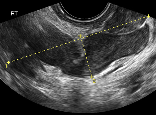
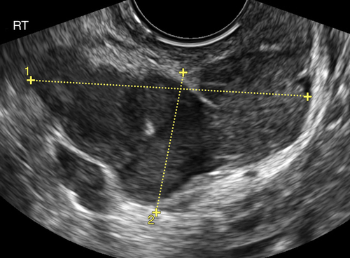
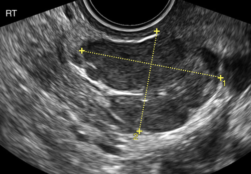
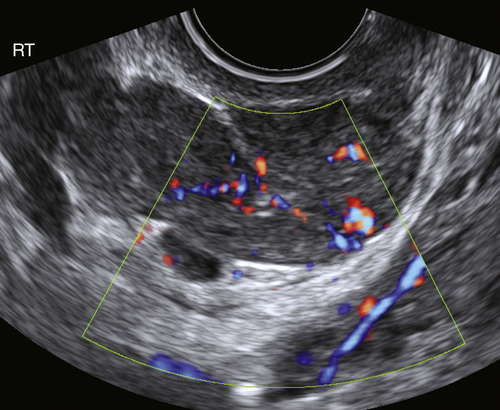
Ultrasound Findings
Differential Diagnosis
Clinical Aspects and Recommendations
Figure
Suggested Reading
Andrade L.A., Torresan R.Z., Sales Jr. J.F., Vicentini R., De Souza G.A. Intravenous leiomyomatosis of the uterus. A report of three cases. Pathol Oncol Res.. 1998;4:44–47.
Diakomanolis E., Elsheikh A., Sotiropoulou M., Voulgaris Z., Vlachos G., Loutradis D., Michalas S. Intravenous leiomyomatosis. Arch Gynecol Obstet. 2003;267:256–257.
Kokawa K., Yamoto M., Yata C., Mabuchi Y., Umesaki N. Postmenopausal intravenous leiomyomatosis with high levels of estradiol and estrogen receptor. Obstet Gynecol. 2002;100:1124–1126.
Lou Y.F., Shi X.P., Song Z.Z. Intravenous leiomyomatosis of the uterus with extension to the right heart. Cardiovasc Ultrasound. 2011;24(9):25.
Low G., Rouget A.C., Crawley C. Intravenous leiomyomatosis with intracaval and intracardiac involvement. AJR. 2012;265:971–974.


