Lattice Degeneration
Clinical Findings:
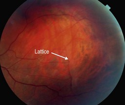
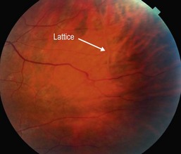
The vast majority of affected patients are asymptomatic with lattice being noted as an incidental finding on a dilated retinal examination. Some patients may complain of photopsias and floaters. Typical lattice consists of sharply demarcated spindle-shaped areas of retinal thinning usually located in the retinal periphery between the equator of the retina and the posterior border of the vitreous base (Fig. 26.1.1). Lattice degeneration occurs more frequently temporally and superiorly in the retina. The retina may be thinned and atrophic. Atrophic holes are the most commonly seen type of retinal break, which typically remain stable, and are rarely associated with retinal detachments. Occasionally, vitreous traction over lattice can cause formation of horseshoe retinal tears which may result in retinal detachment.
OCT Features:
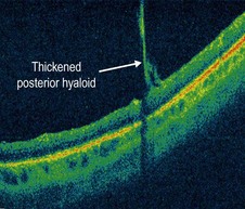
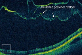
Posterior vitreous separation may be noted over the area of the lattice. Alternatively, adherence of the vitreous over the area of the lattice with separation of vitreous anterior and posterior to the lattice may cause a U-shaped appearance to the vitreous (Fig. 26.1.2). This area of U-shaped traction may be associated with focal retinal detachments with hyper-reflective areas within the retina representing disruption of normal cell structure or pigment migration. Retinal thinning may also be seen in the area of lattice with the inner retina most severely affected. Areas of atrophic holes may be seen within these areas of lattice. Vitreous membranes and cellular aggregates may also be seen in the vitreous of eyes with lattice degeneration (Figs 26.1.3 and 26.1.4).
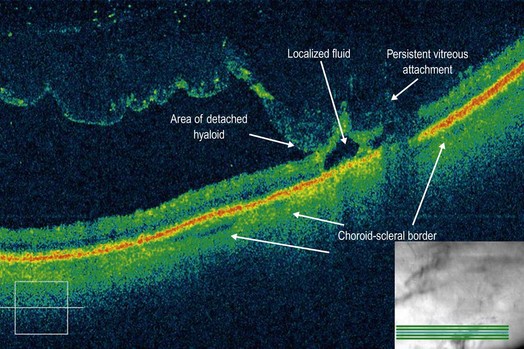
Figure 26.1.2 Line scan over the area of lattice degeneration shows vitreous strongly adherent over the area of the lattice (between white arrows) and detached anterior to the lattice. There is traction with a focal tractional detachment over the area of lattice. There is thinning of the retina especially the inner retina. Also note the thinned choroid that is characteristic of myopia.