L
Labrador keratopathy See keratopathy, actinic.
lacquer cracks See spot, Fuchs’.
lacrimal Relating to tears.
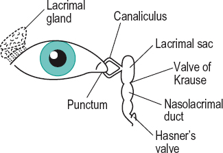
lacrimal apparatus The system involved in the production and conduction of tears. It consists of the lacrimal gland and accessory lacrimal glands (glands of Krause and Wolfring); the eyelid margins; and the two puncta lacrimale. Each punctum is a small round or oval aperture situated on a slight elevation at the inner end of the upper and lower lid margin (lacrimal papilla) and forms the entrance to the canaliculi. Each canaliculus consists of a vertical portion of about 2 mm long and then bends inward for some 8 mm, the upper one being slightly shorter. The canaliculi pierce the lacrimal fascia (i.e. the periorbita covering the lacrimal sac or tear sac) and unite (forming the common canaliculus) to enter a small diverticulum of the sac called the sinus of Maier. The lacrimal sac is closed above and open below where it is continuous with the nasolacrimal duct which extends over some 1.5 cm in length to Hasner’s valve (or Bianchi’s valve or plica lacrimalis) (folds of mucous membrane) at the inferior meatus of the nose. The inferior opening of the duct is called the ostium lacrimale (Fig. L1).
See dacryocystitis; epiphora; fistula, lacrimal; fossa for the lacrimal sac; syndrome, Sjögren’s; tear duct; test, dye dilution; test, Jones II; valve of Krause.
lacrimal artery See artery, lacrimal.
lacrimal bone See orbit.
lacrimal canaliculi See lacrimal apparatus.
lacrimal caruncle See caruncle, lacrimal.
lacrimal crest, anterior The anterior margin of the fossa for the lacrimal sac located on the frontal process of the maxilla.
See fossa for the lacrimal sac.
lacrimal crest, posterior The posterior margin of the fossa for the lacrimal sac situated on the lacrimal bone.
lacrimal duct See tear duct.
lacrimal fascia See lacrimal apparatus.
lacrimal fluid See tears.
lacrimal gland See gland, lacrimal.
lacrimal lake Accumulation of tears in the angle between the eyelids and the inner canthus prior to draining into the lacrimal puncta.
See epiphora; tear meniscus.
lacrimal layer See film, precorneal.
lacrimal lens See lens, liquid.
lacrimal nerve See nerve, ophthalmic.
lacrimal papilla A small elevation at the inner canthus of each eyelid containing a puncta lacrimale.
lacrimal prism See tear meniscus.
lacrimal punctum A small, round or oval, opening of the lacrimal canaliculus (duct) on the margin of each eyelid near the inner canthus and situated in the middle of a small elevation the lacrimal papilla. A lacrimal punctum is normally visible only if the lid is everted. Plural: lacrimal puncta. Syn. lacrimal point.
lacrimal reflex See reflex, lacrimal.
lacrimal sac See lacrimal apparatus.
lacrimal tubercle A small bump on the frontal process of the maxilla situated near the lower orbital and the anterior lacrimal crest, to which the medial palpebral ligament attaches. Syn. papilla lacrimalis.
See ligament, palpebral; lacrimal crest, anterior.
lacrimation 1 . Secretion and flow of tears. 2. Synonym for weeping.
See epiphora; reflex, lacrimal; tear secretion.
lacrimation, paradoxic See tears, crocodile.
laevoclination Rotation of the upper pole of an eye towards the subject’s left. Syn. laevocycloduction; laevotorsion.
laevocycloduction See laevoclination.
laevocycloversion Rotation of the upper poles of the vertical meridians of both eyes towards the subject’s left.
See dextrocycloversion.
laevodeorsumversion Movement of the eyes down and to the left.
laevoduction Rotation of one eye to the left. Note: also spelt levoduction.
See duction.
laevophoria A tendency of the visual axes of both eyes to deviate to the left, in the absence of a stimulus to fusion.
See dextrophoria; heterophoria.
laevosursumversion Movement of the eyes up and to the left.
laevotorsion See laevoclination; torsion.
laevoversion Movement of both eyes to the left. See version.
lag of accommodation See accommodation, lag of.
lagophthalmos Failure of the upper eyelid to close the eye completely. During sleep, about one-third of the population has a slight lagophthalmos.
See ectropion; keratopathy, exposure.
Lagrange’s law See law, Lagrange’s.
lambert Unit of luminance equal to 3183 candelas per m2. Symbol: L.
Lambert’s cosine law See diffusion.
lamellar keratoplasty See keratoplasty.
lamina Thin sheet or layer. Example: the lamina vitrea of Bruch’s membrane.
lamina cribrosa See cribriform plate.
lamina elastica See membrane, Bruch’s.
lamina elastica anterior See, layer, Bowman’s.
lamina elastica posterior See membrane, Descemet’s.
lamina fusca See choroid.
lamina papyracea Synonym for the orbital plate of the ethmoid bone, which forms part of the medial wall of the orbit. It is thus named because it is as thin as paper and this may contribute to an infection of an ethmoidal sinus spreading into the orbit and resulting in orbital cellulitis.
See cellulitis, orbital.
lamina vitrae See membrane, Bruch’s.
laminated lens See lens, laminated.
lamp Any device that produces light or heat. Burton l. Ultraviolet lamp, including some short wavelengths from the visible spectrum (e.g. Wood’s light), mounted with a magnifying lens in a rectangular frame. It is used primarily in the evaluation of the fit of a hard contact lens, in conjunction with the instillation of fluorescein into the eye.
See staining.
filament l. A lamp in which light is produced by electrically heating a filament, usually of tungsten. The filament is contained in a bulb in which there is either a vacuum or an inert gas. The emitted spectrum is continuous.
See spectrum, continuous.
fluorescent l. Discharge lamp in which most of the light is emitted by a layer of fluorescent material excited by the ultraviolet radiation from the discharge (CIE).
See fluorescence.
halogen l. A tungsten filament lamp in which the glass envelope is made of quartz and is filled with gaseous halogens. This permits a higher filament temperature and consequently provides a higher luminance and a higher colour temperature as well as a longer operating life than a conventional filament lamp of the same input power. Halogen lamps are used in some ophthalmoscopes and retinoscopes and as very bright sources for people with low vision. Syn. tungsten-halogen lamp.
incandescent electric l. Lamp in which light is produced by means of a body (filament of carbon or metal) heated to incandescence by the passage of an electric current (CIE).
See incandescence; luminescence.
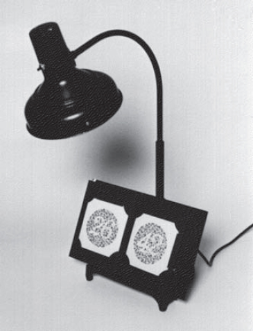
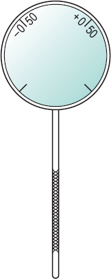
Macbeth l. A lamp used in testing colour vision. It contains a powerful tungsten filament bulb with a blue filter of specific absorption properties such that it produces a source of a colour temperature of about 6800 K, thus approximating the spectral characteristics of natural sunlight. The lamp is also fitted with a stand to hold the colour vision booklet (Fig. L2). Syn. Macbeth illuminant C.
See illuminants, CIE standard; plates, pseudoisochromatic; test, Farnsworth. tungsten-halogen l. See lamp, halogen.
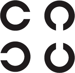
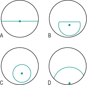
Landolt ring A test object used for measuring visual acuity consisting of an incomplete ring resembling the letter C. The width of the break and of the ring are each one-fifth of its overall diameter. The subject must indicate where the break is located, the break being positioned in any direction. The minimum angle of resolution corresponds to the angular subtense of the just noticeable break at the eye (Fig. L3). Syn. Landolt broken ring; Landolt C; Landolt test type.
See chart, Landolt broken ring.
Lang stereotest See stereotest, Lang.
Langerhans’ cells See cells, Langerhans’.
lantern test See Edridge–Green lantern; test, lantern.
Lanthony desaturated D-15 test See test, Farnsworth.
Lanthony tritan album See plates, pseudoisochromatic.
LASEK A surgical procedure on the cornea aimed at correcting ametropia. An alcohol solution is applied to the cornea (usually for less than 30 seconds) to loosen the epithelium. A trephine is used to make an incision in the epithelium leaving a hinge of 2–3 clock hours of intact margin. The loosened edges of the epithelium are lifted along the trephine mark and the epithelium is folded or rolled back exposing Bowman’s layer. Excimer laser ablation is then performed on the anterior stromal surface and Bowman’s layer is ablated away over the treatment area. The epithelial flap is repositioned and a bandage soft contact lens is worn for 3–4 days to minimize discomfort and to protect the epithelium. The method is less invasive than LASIK and appears to give rise to fewer complications. LASEK is an acronym made from the following italic letters ‘laser assisted epithelial keratomileusis’.
See keratectomy, photorefractive; pachometer.
laser An intense luminous source of coherent and monochromatic light. The term is an acronym for light amplification by stimulated emission of radiation. Lasers are used in the treatment of a variety of ocular conditions, especially of the cornea, the retina (e.g. detached retina, diabetic retinopathy), glaucoma and refractive errors.
See cyclodiode; iridotomy; keratectomy, photorefractive; LASEK; LASIK; ophthalmoscope, scanning laser; photocoagulation; trabeculoplasty.
argon l. A laser with ionized argon gas as the active medium, which emits a bluegreen light beam with a wavelength of 514 nm. It may be used to perform iridectomy, iridoplasty, iridotomy, photocoagulation or trabeculoplasty.
excimer l. A gas laser that emits pulses of light in the ultraviolet region (at 193 nm). All the energy is absorbed by the superficial layers (e.g. the corneal epithelium), which are then exploded away or ablated without any change to the underlying or adjacent tissue or material.
See keratectomy, photorefractive; LASEK; LASIK.
l. interferometry See maxwellian view system, clinical.
l. iridotomy See iridotomy.
krypton l. A laser with krypton gas ionized by electric current as the active medium, which emits a light beam in the yellow-red region of the visible spectrum (521 nm, 568 nm or 647 nm). It may be used to perform photocoagulation or trabeculoplasty.
neodymium-yag l . (Nd-Yag) A solid-state laser whose active medium is a crystal of yttrium, aluminium and garnet doped with neodymium ions. It emits an infrared light beam with a wavelength of 1064 nm. It is typically used with a slit-lamp and in conjunction with a helium-neon laser which produces a red beam of light (633 nm) to allow focusing. It may be used to perform capsulotomy, iridotomy or trabecular surgery. Yag is an acronym for yttrium–aluminium–garnet.
l. refraction See refraction, laser.
l. refractive keratoplasty See keratectomy, photorefractive.
l. trabeculoplasty See trabeculoplasty, laser.
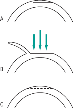
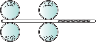
LASIK A surgical procedure on the cornea aimed at correcting ametropia. A suction ring is applied to the globe and an increase in intraocular pressure to approximately 65 mmHg is induced for a maximum of two minutes. During that time an automated microkeratome advances across the cornea creating a corneal flap of about 8.5 mm in diameter, which contains the epithelium, Bowman’s layer and a portion of the anterior stroma. The vacuum is then switched off and the suction ring removed. The corneal flap, which is hinged on one side of the cornea, is turned round onto the conjunctiva and the exposed stroma is ablated with the excimer laser. On completion of the laser ablation, the corneal flap is repositioned and left to adhere without sutures. There are some complications associated with this procedure, but it gives rise to less postoperative pain and more rapid visual rehabilitation than other similar surgical procedures (Fig. L4). LASIK is an acronym made from the following italic letters ‘laser in situ keratomileusis’ or ‘laser assisted intrastromal keratoplasty’.
See ectasia, corneal; epikeratoplasty; Intacs; keratome; keratomileusis; keratophakia; keratectomy, photorefractive.
latanoprost See prostaglandin analogues.
latent hyperopia See hyperopia, latent.
lateral geniculate body See geniculate bodies, lateral.
lateral inhibition See inhibition, lateral.
lateral rectus muscle See muscle, lateral rectus; muscles, extraocular.
lateral rectus paralysis See paralysis of the sixth nerve.
lathe-cut contact lens See lens, lathe-cut contact.
lattice degeneration of the retina See retina, lattice degeneration of the
lattice dystrophy See dystrophy, lattice.
lattice theory See theory, Maurice’s.
Laurence–Moon–Bardet–Biedl syndrome
See syndrome, Laurence–Moon–Bardet– Biedl.
law In science, a statement of facts or principles which is considered invariable under the given conditions having been tested and tried.
Abney’s l. The total luminance of an area is equal to the sum of the luminances that compose it.
Alexander’s l. An increase in the intensity of a jerk nystagmus when the eyes move in the direction of the fast phase.
all or none l. The response in a nerve fibre to any stimulus strong enough to produce a response is always of the same amplitude. However, different nerve fibres have action potentials with different amplitudes. An increase in the intensity of the stimulus yields only an increase in the frequency of nerve impulses (or action potentials). Syn. all or nothing law.
Aubert–Förster l. See phenomenon, Aubert–Förster.
Bloch’s l. The luminance L of a stimulus required to produce a threshold response is inversely proportional to the duration of exposure t of the stimulus, i.e.
< ?xml:namespace prefix = "mml" />

where C is a constant. This law is only valid for exposure t below about 0.1s.
Bunsen–Roscoe l. In photochemistry, the product of the intensity of the light stimulus and the duration of exposure is a constant. Syn. law of reciprocity.
cosine l. See diffusion.
Descartes’ l. See law of refraction.
Donders’ l. For any determinate position of the line of fixation with respect to the head there corresponds a definite and invariable angle of torsion.
Draper’s l. An effect is produced in a medium only by that portion of the spectrum which is absorbed by the medium. The effect may be thermal, chemical or the production of fluorescence. Syn. Grotthus’ law.
Emmert’s l. The apparent size of a projected after-image varies in proportion to the distance of the surface on which it is projected. The law can be expressed by the following relationship h/H = d/D, where h is the linear size of the object, H the apparent size of the projected after-image, d the object’s distance from the observer and D the distance between the observer and the surface on which the after-image is projected. It follows from the above expression that H = hD/d, i.e. the greater the distance of the projected image the larger its apparent size.
l. of equal innervation See law of equal innervation, Hering’s.
Fechner’s l. The intensity of a sensation S varies as the logarithm of the intensity I of the stimulus, i.e.

where k is a constant. However, in some conditions this law is not valid and Stevens’ law (or power law) is more appropriate. This stipulates that the intensity of a sensation S varies as the intensity of the stimulus I to the power of x, i.e.

where x is a constant which depends on the stimulus.
See magnitude estimation.
Fermat’s l. The path taken by a light ray in going from one point to another is that route which takes the least time. Syn. Fermat’s principle.
Ferry–Porter l . The critical flicker frequency F is directly proportional to the logarithm of the luminance L of the stimulus, i.e.
where a and b are constants. See frequency, critical fusion,
Granit–Harper l. The critical fusion frequency increases with the logarithm of the retinal area stimulated.
Grassmann’s l’s. Laws of colour mixture. 1. The first law states that any colour C of the visible spectrum can be matched in appearance by a mixture of three primary colours, such as red R, green G and blue B, provided that none of these can be matched by a mixture of the other two, i.e.
where α, β and γ are the relative proportions of the chosen primaries.
2. Additive property: if a colour is added in an identical manner to two equivalent mixtures (or single colours) the two new mixtures will appear identical, i.e. if A + B = C + D, then A + B + X = C + D + X, or if A = B, then A + X = B + X.
3. Scalar property: if the brightness of each of two equivalent mixtures is increased or decreased by the same factor the two new mixtures will appear identical, i.e. if A + B = C + D, then k (A + B) = k (C + D).
4. Associative property: if a colour is substituted in one of the mixtures by an equivalent colour the two new mixtures will appear identical, i.e. if A + B = C + D and X = B, then A + X = C + D.
Grotthus’ l. See law, Draper’s.
Helmholtz’s l. of magnification See law, Lagrange’s.
Hering’s l. of equal innervation
Innervation to the extraocular muscles is equal to both eyes. Thus, all movements of the two eyes are equal and symmetrical. Syn. Hering’s law; law of equal innervation.
See muscles, yoke.
l. of identical visual directions An object stimulating corresponding retinal points is localized in the same apparent monocular direction in each eye. Syn. law of oculocentric visual direction.
See line of direction; retinal corresponding points.
inverse square l. of illumination The illuminance E of a surface by a point source is directly proportional to the luminous intensity I of a point source and to the cosine of the angle θ of incidence and inversely proportional to the square of the distance d between the surface and the source, i.e.
Syn. law of illumination.
See illuminance.
Imbert–Fick l . Applied to applanation tonometry, this law states that the intraocular pressure P (in mmHg) is equal to the tonometer weight W (in g) divided by the applanated area A (in mm2), hence,

This law is correct only for infinitely thin, dry, elastic, spherical membranes.
Kirschmann’s l. The greatest contrast in colour is seen when the luminosity difference is small.
Knapp’s l. A correcting lens placed at the anterior focal plane of an axially ametropic eye forms an image equal in size to that formed in a standard emmetropic eye. Knapp’s law applies to the relative spectacle magnification but not to the spectacle magnification. Syn. Knapp’s rule.
Kollner’s l. See rule, Kollner’s.
Lagrange’s l. In paraxial optics, the product of the index of refraction of image space n’, the image size h’ and the half-angle of the refracted cone in image space u’ is equal to the product of the index of refraction of object space n, the object size h and the half-angle of the incident cone in object space u, i.e.
Syn. Helmholtz’s law of magnification; Lagrange’s relation; Smith–Helmholtz law. See sign convention.
Lambert’s l. See diffusion.
Listing’s l. When an eye moves to any position from the primary position, it may be considered to have made a single rotation about an axis that is perpendicular to both the initial and final lines of fixation at their point of intersection.
l. of oculocentric visual direction See law of identical visual directions.
Planck’s l. Law giving the energy distribution of a black body as a function of wavelength, for a specified temperature.
Prentice’s l. The prismatic effect P in prism dioptres at a point on a lens is equal to the product of the distance c in centimetres of the point from the optical centre of the lens, and the dioptric power F of the lens, i.e.

Syn. Prentice’s rule.
See effect, differential prismatic; power, prism; prism, induced.
l. of reciprocity See law, Bunsen–Roscoe.
l. of reflection The incident and reflected rays and the normal to the surface at the point of incidence lie in the same plane and the angle of incidence is equal to the angle of reflection (Fig. L5).
l. of refraction The incident and refracted rays and the normal to the surface at the point of incidence lie in the same plane and the ratio of the sine of the angle of incidence i to the sine of the angle of refraction i’ is a constant for any two media, i.e.
where n and n’ are the refractive indices of the first and second medium, respectively. This constant (n’/n) is called the relative index of refraction for the two media. Syn. Descartes’ law; Snell’s law.
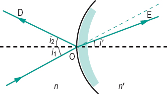
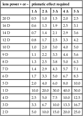
Table L1
Approximate amount of spectacle lens decentration (in mm) of its optical centre away from the pupillary centre of the eye to produce five prismatic effects (in prism dioptres) for distance vision. The results ignore the effect of spherical aberration
| lens power + or – | prismatic effect required | ||||
| 1 Δ | 2 Δ | 3 Δ | 4 Δ | 5 Δ | |
| 20 D | 0.5 | 1.0 | 1.5 | 2.0 | 2.5 |
| 16 D | 0.6 | 1.3 | 1.9 | 2.5 | 3.1 |
| 14 D | 0.7 | 1.4 | 2.1 | 2.9 | 3.6 |
| 12 D | 0.8 | 1.7 | 2.5 | 3.3 | 4.2 |
| 10 D | 1.0 | 2.0 | 3.0 | 4.0 | 5.0 |
| 9 D | 1.1 | 2.2 | 3.3 | 4.4 | 5.6 |
| 8 D | 1.3 | 2.5 | 3.8 | 5.0 | 6.3 |
| 7 D | 1.4 | 2.9 | 4.3 | 5.7 | 7.1 |
| 6 D | 1.7 | 3.3 | 5.0 | 6.7 | 8.3 |
| 5 D | 2.0 | 4.0 | 6.0 | 8.0 | 10.0 |
| 1 D | 10.0 | 20.0 | 30.0 | 40.0 | 50.0 |
| 4 D | 2.5 | 5.0 | 7.5 | 10.0 | 12.5 |
| 3 D | 3.3 | 6.7 | 10.0 | 13.3 | 16.7 |
| 2 D | 5.0 | 10.0 | 15.0 | 20.0 | 25.0 |
See index of refraction; sign convention.
Ricco’s l. The product of the absolute threshold of luminance L and the image area A is a constant, i.e.

This law is valid for small images subtending an angle of a few minutes of arc in the fovea and to one degree in the near macular region. For larger images in the macular area, Piéron’s law applies; it states that the product of the luminance L of the image at threshold and the cube root of the retinal area A stimulated is a constant, i.e.
In the peripheral retina, Piper’s law becomes valid. This law states that the product of the luminance of the stimulus L and the square root of the area A is a constant, i.e.

In the far periphery of the retina, L tends to become independent of A.
Sherrington’s l. of reciprocal innervation The contraction of a muscle is accompanied by simultaneous and proportional relaxation of its antagonist. For example, if the superior oblique muscle contracts, its antagonist, the inferior oblique muscle, relaxes. The validity of this law has been established by electromyography.
Smith–Helmholtz l. See law, Lagrange’s.
Snell’s l. See law of refraction.
Stevens’ l. See law, Fechner’s.
Talbot’s l. See law, Talbot–Plateau.
Talbot–Plateau l. The brightness of a light source presented at short intervals above the critical fusion frequency is equal to that which would be produced by a constant light source of an intensity equal to the mean value of the intermittent stimuli. Syn. Talbot’s law.
Weber’s l. The just noticeable difference (or difference threshold) in intensity of a stimulus ΔI varies as a constant ratio of the initial intensity of the stimulus I, i.e.

where k = {ΔI/l} is a constant called Weber’s fraction (Weber’s constant).
Example: if the initial stimulus was a light source of 1000 cd/m2 and k = 0.01 (or 1%), ΔI = 0.01 × 1000 = 10 cd/m2. Syn. Weber–Fechner law.
See threshold, differential.
Weber–Fechner l. See law, Weber’s.
layer A sheet of one thickness lying over or under another and distinguished from it by a difference in composition or colour.
Bowman’s l. Thin layer of the cornea (about 12 μm) located between the anterior stratified epithelium and the stroma. This layer is acellular; it is a modified superficial stromal layer found only in primates. It is composed of a randomly orientated array of fine collagen fibrils, primarily of collagen types I, III and V. Syn. anterior limiting layer; Bowman’s membrane; lamina elastic anterior.
l. of Chievitz, transient A temporary layer found in the developing embryonic retina lying between the inner neuroblastic layer (which will form the ganglion, amacrine and Mueller cells) and the outer neuroblastic layer (which will form the bipolar, horizontal and photoreceptor cells). It contains the inner processes of Mueller’s fibres.
Haller’s l. An outer layer of the choroid lying between Sattler’s layer and the suprachoroid. It contains connective tissue and large vessels, mainly veins.
l. of Henle, fibre Located in the macular region, it is formed by the cone and rod fibres that run parallel to the retinal surface within the outer molecular layer of the retina.
See Haidinger’s brushes; macular star.
retinal l’s. See retina.
Sattler’s l. An inner layer of the choroid lying between the choriocapillaris and Haller’s layer. It contains small blood vessels.
leaf room See room, leaf.
Leber’s congenital amaurosis A hereditary, bilateral blindness present at birth or in early childhood. It is caused by mutations in the gene encoding the retinal guanylate cyclase (GUCY2D) on chromosome 17. Initially, the ocular fundus appears normal, although the ERG is markedly reduced. A salt and pepper fundus and optic atrophy appear later. The condition is often accompanied by nystagmus and photophobia.
Leber’s disease See Leber’s hereditary optic atrophy.
Leber’s hereditary optic atrophy A mitochondrial inherited bilateral condition, which appears suddenly in healthy people, primarily males, of about the age of 20 and results in a marked loss of vision and ultimately optic atrophy. A very small percentage of people recover some visual acuity in one or both eyes after the disease has run its course (Fig. L6). Syn. Leber’s disease; Leber’s hereditary optic neuropathy.
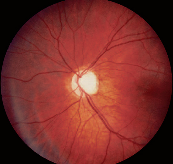
See atrophy, optic; inheritance; mitochondrion.
Leber’s miliary aneurysms See disease, Coats’.
legibility Term referring to the difference in the ease of difficulty with which optotypes can be read. Some letters are easier to recognize (e.g. L, T, U, V, Z and C) than others (e.g. S, G, H, F, R and B). Test charts either mix these letters or use letters of similar difficulty; the latter facilitates standardization of charts.
length, equivalent focal In an optical system composed of more than one lens, it is the linear distance separating the principal focus from the corresponding principal point. It is usually the most important quantity in the specification of an optical system as in objectives, eyepieces, etc.
See focus, principal; power, equivalent.
length of the eye, axial The distance between the anterior and posterior poles of the eye. In vivo, it is measured either by ultrasonography or by partial coherence interferometry (PCI). These measurements represent the distance between the anterior pole and Bruch’s membrane. (In young eyes in which there is a refractive index difference at the retina-vitreous interface ultrasonography measures the distance between the anterior pole and the anterior surface of the retina.) The axial length of the eye at birth is approximately 17 mm and reaches approximately 24 mm in adulthood. It is typically longer than 24 mm in myopes and shorter than 24 mm in hyperopes. Each mm of change in axial length of the eye corresponds to approximately 2.5 D
See biometry of the eye; ultrasonography.
length, focal The linear distance separating the principal focal point (or focus) of an optical system from a point of reference (e.g. vertex, principal point, nodal point). The first (anterior) focal length is the distance from the lens (or first principal point) to the first principal focus. The second (posterior) focal length is the distance from the lens (or second principal point) to the second principal focus. Symbol: f.
In a spherical mirror the focal length f, i.e. the distance between the focal point and the pole of the mirror, is equal to half its radius of curvature r

Syn. focal distance. See focus, principal; mirror; Points, cardinal; Points, principal; power, equivalent; power, refractive; sign convention.
lens A piece of transparent glass, crystal, plastic or similar substance (e.g. liquid) having two opposite regular surfaces which can be plane or curved and which alter the vergence of pencils of light transmitted through it. There are many types of lenses, which are described below.
See coquille; glass; moulding; surfacing.
absorptive l. A lens that absorbs a proportion of the incident radiation. Some lenses absorb mostly in the infrared region of the spectrum, others absorb mostly in the ultraviolet region and others absorb more or less equally throughout the visible spectrum.
See CR-39; filter; lens, coated; pterygium.
achromatic l. A compound lens designed to reduce or eliminate chromatic aberration. The most common type is called a doublet. Syn. achromat.
See lens, apochromatic; lens, hyperchromatic.
achromatizing l. Lens aimed at reducing or eliminating the chromatic aberration of the eye. It consists of either a doublet or a triplet that possesses longitudinal chromatic aberration opposite to that of the eye and thereby neutralizes it. Thus the lens system has negative power for short wavelengths and positive power for long wavelengths of an amount similar to that of the eye for those wavelengths.
l. adherence 1. Refers to a contact lens being firmly fixed to the cornea. 2. Attachment of bacteria (e.g. pseudomonas, staphylococcus) to a contact lens, particularly soft lens materials (the amount and strength vary with the material). Syn. lens binding.
See lens, silicone hydrogel; tear stasis.
afocal l. A lens of zero power (Fig. L7).
Syn. plano lens. See lens, aniseikonic.
Alvarez l. A variable power lens composed of two elements that can be moved with respect to each other along two mutually perpendicular axes. When the two lenses are in exact register with each other the Alvarez lens provides zero power. Moving one of the elements either laterally or vertically in relation to the other provides increasing spherical or cylindrical power. Moving both elements produces a combined spherocylindrical power. Such a lens is used in the Humphrey Vision Analyser.
anastigmatic l. 1. A lens that has a single focal point. 2. A lens that is corrected for oblique astigmatism and minimum curvature of field. Syn. stigmatic lens.
See lens, astigmatic; Petzval surface.
aniseikonic l. Lens designed to correct aniseikonia. It can have power like a regular ophthalmic lens but also produces a magnification of the image. A lens which only produces magnification but has zero power is called an overall size lens and if it magnifies in only one meridian it is called a meridional size lens. Syn. iseikonic lens; eikonic lens; size lens.
See lens, afocal; magnification, shape.
anterior chamber intraocular l. See lens, intraocular.
anti-actinic l. A lens that absorbs ultraviolet radiations to a much greater extent than a white spectacle lens.
See actinic.
anti-reflection coated l. See lens, coated.
aphakic l. A lens used for the correction of aphakia. It is of high dioptric power, usually above +10 D. Due to their thickness, these lenses are usually made of plastic to reduce weight and because of the aberrations, aspherical surfaces are used.
See lens, lenticular; phenomenon, jack-in-the-box.
aplanatic l. A lens designed to correct for spherical aberration and coma.
apochromatic l. A compound lens designed to correct chromatic and spherical aberrations. It uses three or more kinds of glass. This lens corrects chromatic aberration more thoroughly than an achromatic lens.
See lens, achromatic.
aspheric l. A lens in which one or both surfaces are not spherical, so designed to minimize certain optical aberrations.
astigmatic l. A toric or cylindrical lens that produces two separate focal lines at right angles, instead of a single focal point. Hence it has two principal powers. One of these powers may be zero (cylindrical lens).
See axis, cylinder; lens, anastigmatic; lens, spherocylindrical; protractor; Sturm, interval of; transposition.
back toric contact l. A contact lens used to correct corneal astigmatism in which the surface of the lens facing the cornea is not spherical but toroidal in order to obtain a good physical fit on the cornea. To create better stability of the lens on the eye it usually incorporates a prism ballast.
See ballast.
Bagolini’s l. See glass, Bagolini’s.
balancing l. A lens fitted to a spectacle frame or mount, to balance the weight of the other lens, its power being unspecified or unimportant.
bandage l. See lens, therapeutic contact.
best-form l. A curved lens whose curvatures are calculated to eliminate or minimize aberrations when viewing through the peripheral portions of the lens. Syn. corrected lens; point-focal lens.
See ellipse, Tscherning.
biconcave l. A lens that has two concave surfaces.
biconvex l. A lens that has two convex surfaces.
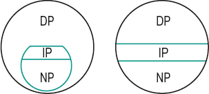
bifocal l. A lens having two portions of different focal power. Usually the upper portion is larger and is used for distance vision while the lower portion is smaller and used for near vision. There are, however, many types of bifocals: those in which the use of the two portions is the opposite of that described above, others in which the shape of the near portion (or segment) differs; in certain types the near segment is fused onto the surface of the glass (fused bifocal); in others the near portion is produced by grinding or moulding a different curvature on one surface (solid bifocal or one-piece bifocal). There is also a one-piece bifocal in which there is a gradual transitional zone between the two portions instead of a clear line of demarcation (blended bifocal). In addition, several other bifocals are known by their trade name (Fig. L8).
See button; jump; lens, progressive; monocentric; segment height; segment of a bifocal lens; wafer.
l. binding See lens adherence.
biomimetic contact l. A hydrogel contact lens made up of a material that imitates the chemistry of natural cell membranes to minimize changes in ocular physiology.
l. blank A moulded piece of ophthalmic glass before completion of the surfacing processes.
See glass; lens, semi-finished.
l. capsule See capsule; lens, crystalline.
l. carrier See lens, lenticular; telescope, bioptic.
cast-moulding contact l. A contact lens produced by pressing a concave female mould filled with liquid monomer against a male mould. The concave mould determines the front surface of the lens. The edge is formed when the two sides of the mould come together. The assembled moulds are irradiated with ultraviolet light to cause polymerization and a dry contact lens, which is then hydrated in saline water. Cast-moulding has been used to manufacture hard and soft lenses, especially mass production of the latter.
See lens, lathe-cut contact; lens, spin-cast contact.
Chavasse l. A lens with an irregular surface used to depress the visual acuity while permitting the eye to be seen from the front (British Standard).
ChromaGen l. Trade name of a tinted, soft contact lens worn with the aim of enhancing colour perception in individuals with colour vision deficiencies, especially red-green defects. It is fitted on either one or both eyes. The lenses exist in a variety of tints and are chosen by the patient by trial and error. Some patients with dyslexia or migraine have also claimed to benefit from wearing these lenses.
l. clock See lens measure.
coated l. A lens upon which is deposited an evaporated film consisting of a metallic salt such as magnesium fluoride, about one-quarter as thick as a wavelength of light. This film reduces, by interference, the amount of light reflected by the surfaces and to some extent the amount of stray light reflected inside the lens. With multilayer coating the lens can selectively reflect radiations and increase transmission. All coated lenses show some residual colour. Syn. anti-reflection (AR) coated lens; bloomed lens.
See anti-reflection coating; coating; image, ghost.
cobalt l. A lens that absorbs the central region of the visible spectrum and only transmits the red and blue ends of the spectrum. It is sometimes used in the testing of ametropia, since a light source located at 1.4 m from the eye will form, in an emmetropic eye viewing it through a cobalt lens, two equal circles superimposed on the retina and the subject will report seeing a purple circle. A hyperope will see a blue spot surrounded by a red annulus and a myope will see a red spot surrounded by a blue annulus. An appropriate spherical lens placed in front of the eye, which changes the appearance to a purple circle, represents the spherical refractive correction. Syn. cobalt-blue glass.
collimating l. See collimator.
combination l. A lens made from two different materials; usually a rigid centre portion made from gas-permeable material surrounded by a soft peripheral flange of HEMA material. It is used in the management of keratoconus, irregular astigmatism, etc. Few such lenses exist at present.
composite l. A contact lens composed of two or more different materials.
compound l. 1. A lens that functions as a combination of a spherical lens and a cylindrical lens. Example: a spherocylindrical lens. 2. A system composed of several refracting surfaces or lenses placed along the same axis. Examples: doublet; triplet.
See lens, spherocylindrical.
concave l. A lens that causes incident rays of light to diverge. Syn. diverging lens; minus lens; negative lens.
condensing l. See condenser.
contact l. A small lens usually made of a plastic material, worn in contact with the cornea or sclera and used to correct refractive errors of the eye. There are many types of contact lenses. Lenses that rest on the sclera are called scleral (or haptic) contact lenses whereas lenses that rest on the cornea are called corneal contact lenses, or, more commonly, contact lenses. Lenses that are made of a hard plastic material which is impermeable to oxygen, such as those made of polymethyl methacrylate (PMMA), are called rigid contact lenses. Almost all rigid contact lenses nowadays transmit oxygen. They are called gas permeable contact lenses (GP, GPL, GPCL, HGP or RGP), or merely rigid contact lenses and they usually contain silicone acrylate or fluorosilicone acrylate.
Other lenses made of a soft plastic material which transmit a certain amount of oxygen are called soft (or hydrophilic or hydrogel or gel or flexible) contact lenses (SCL) whose water content varies; the greater the water content the more oxygen is transmitted (for equal thickness). Very high water content lenses are used for extended wear (EW). There are also bifocal contact lenses consisting of two segments with different focal powers, which provide either simultaneous vision (light from both the distance and near portions enters the eye at the same time) or alternating vision (the lens must be moved to see through either portion). Other bifocal contact lenses have a zone of variable power between the two portions and others are diffractive in which light from both distance and near objects can be focused on the retina (without moving the lens) owing to diffraction produced by a series of rings in the centre of the back surface of the lens (the higher the near addition the greater the number of rings). There are also toric contact lenses in which the back optic surface is toroidal; they are used to improve the physical fit. Bitoric contact lenses are lenses in which both surfaces are toroidal; they are used to improve the physical fit and correct the induced astigmatism. Disposable contact lenses are worn for one day, one week, two weeks or one month and then discarded.
See acidosis; hypercapnia; lens adherence; lens, back toric contact; lens, biomimetic contact; lens, cast-moulding; lens, ChromaGen; lens, cosmetic contact; lens, extended wear; lens, fenestrated; lens, flare; lens, flat; lens, flexure; lens, lathe-cut contact; lens, liquid; lens, ortho-k; lens, piggyback; lens, reverse-geometry contact; lens, scleral contact; lens, sealed scleral contact; lens, silicone hydrogel; lens, spin-cast contact; lens, steep; lens, therapeutic contact; lens, X-Chrom; modulus of elasticity; oxygen permeability; water content.
contact l. deposits See deposits, contact lens.
converging l. See lens, convex.
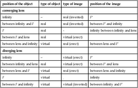
Table L2
Relationship between object and image formed in a converging and in a diverging lens. The object is moved from left to right. F and F’ are the first and second focal points, respectively
| position of the object | type of object | type of image | position of the image |
| converging lens | |||
| infinity | – | real (inverted) | F’ |
| between infinity and F | real | real (inverted) | between F’ and infinity |
| F | real | – | infinity between infinity and lens |
| between F and lens | real | virtual (erect) | |
| between lens and infinity | virtual | real (erect) | between lens and F’ |
| diverging lens | |||
| infinity | – | virtual (erect) | F’ |
| between infinity and lens | real | virtual (erect) | between F’ and lens |
| between lens and F | virtual | real (erect) | between lens and infinity |
| F | virtual | – | infinity |
| between F and infinity | virtual | virtual (inverted) | between infinity and F’ |
convex l. A lens that causes incident rays of light to converge. Syn. convex lens; plus lens; positive lens.
corrected l. See lens, best-form.
l. cortex See lens, crystalline.
cosmetic contact l. A contact lens designed to improve the appearance of the eye, to conceal a disfigurement (e.g. a scar), or to change the colour of the eye.
Examples: a tinted lens; an opaque lens with an artificial pupil and iris.
See aniridia.
cross-cylinder l. An astigmatic lens consisting of a minus cylinder ground on one side and a plus cylinder ground on the other side, the two axes being located 90° apart. The dioptric power in the principal meridians is equal. Usual cross-cylinder lenses are provided in three powers: ±0.25 D, ±0.37 D and ±0.50 D (higher values are also available for use with low vision patients). This lens is used in the subjective measurement of the power and axis of astigmatism, or to refine the cylindrical correction determined otherwise (Fig. L9). Syn. Jackson cross-cylinder lens.
See test for astigmatism, cross-cylinder.
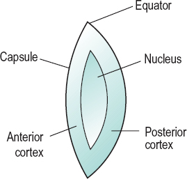
while maximum accommodation alters these values to about 6 mm and –5.3 mm respectively. The crystalline lens displays a complex gradient of refractive index (averaging 1.42), and a power of 21 D. It consists of the capsule which envelops the lens, the epithelium which consists of a single sheet of cells and lines the anterior and equatorial capsule and the cortex which surrounds the nucleus, the latter two containing the lens fibres. The lens has the highest protein content of any other tissue in the body. The most numerous proteins in the lens are the soluble proteins dominated by the crystallins
crystalline l. The biconvex, usually transparent body situated between the iris and the vitreous body of the eye and suspended from the ciliary body by the zonular fibres (zonule of Zinn), which are attached to the equator of the lens. The diameter of the lens is equal to 9–10 mm and its thickness 3.6 mm, being greater when the eye accommodates. The radii of curvature of the anterior and posterior surfaces are 10.6 mm and –6.2 mm, respectively in the unaccommodated eye, followed by the insoluble proteins especially cytoskeletal proteins. With age, there is an increase in light scatter originating in the nucleus, as well as some light absorption and yellowing of the nucleus (Fig. L10). Syn. lens of the eye.
See constants of the eye; disease, Wilson’s; fibres, lens; fissure, optic; lens, intraocular; lens paradox; lens sutures; luxation; myopia, lenticular; phakic; shagreen; Zinn, zonule of.
curved l. See lens, meniscus.
cylindrical l. A lens in which one of the principal meridians has zero refractive power. It usually consists of one plano surface and one cylindrical surface (Fig. L11).
See lens, astigmatic.
l. dislocation See luxation of the lens.
disposable l. See lens, contact.
diverging l. See lens, concave.
eikonic l. See lens, aniseikonic.
equi-concave l. A lens having two concave surfaces of the same power.
equi-convex l. A lens having two convex surfaces of the same power.
equivalent l. See equivalent, spherical.
l. exfoliation See exfoliation of the lens.
extended wear l. A contact lens designed to be worn continuously for more than one day and, usually, no more than seven days before cleaning and sterilization. It is, typically, a soft lens, with high oxygen transmissibility.
See cornea guttata; corneal infiltrates; lens, silicone hydrogel; microcysts, epithelial; microscope, specular; pannus; ulcer, corneal.
l. extraction See cataract extraction.
fenestrated l. A hard contact lens having one or more small holes to aid tear exchange and corneal oxygenation. It was essential with PMMA scleral contact lenses, but with the advent of gas permeable materials, fenestration is rarely necessary. Syn. ventilated lens.
finished l. A spectacle lens that has been surfaced on both sides to the required power and thickness and is still in uncut form. syn. uncut lens.
See lens, semi-finished; surfacing.
fisheye l. A camera lens with a very wide angle of view. The angle can be as wide as 220°. To achieve this the lens has a large diameter that produces image distortion (especially barrel-shaped distortion); thus the image magnification varies across the picture causing some fishbowl effects.
See lens, wide-angle.
l. flare Type of blur characterized by the presence of a secondary or ghost image. Flare may be caused by a contact lens with an optic zone diameter that is smaller than the pupil diameter or when the lens decentres so that part of the edge of the optic zone is within the pupil area. Flare is usually more apparent under conditions of reduced illumination as the pupil is larger. Management consists in refitting the patient with a lens with a larger optic zone diameter or with better centration.
See image, ghost.
flat l. 1. Any lens that is not of curved form. 2. A contact lens in which the back optic zone radius is longer than the flattest meridian of the cornea. This definition may not be valid for a soft lens that may have a back optic zone radius longer than the flattest meridian of the cornea and yet not be flat fitting. Syn. loose lens (it is preferable to use this term for soft lenses).
See fitted on K; lens, steep.
l. flexure Characteristic of a spherical contact lens to adopt a toroidal curvature when placed on an astigmatic cornea. Flexure depends upon the material and its thickness, being more common with thin lenses. As it occurs during blinking, it sometimes results in fluctuating vision.
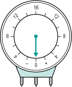
l. flippers Two pairs of lenses mounted on a central bar, one pair on each side. One pair can be held in front of the patient’s eyes and then quickly changed for the other pair by twisting the bar extension handle. The lenses may be one pair of minus lenses of equal power and the other pair of plus lenses of equal power. The most common pairs are ±2.00 D, although ±1.00 D, ±1.50 D and ±2.50 D are also available. These are used in the testing and training of accommodative facility. Prisms may also be used, as for example base-in for one pair and base-out for the other. They are used in the testing and training of vergence facility (Fig. L12). Syn. flippers; flipper bar; flip lenses; flip prisms; prism flippers.
Fresnel l. A plastic lens consisting of one flat surface which can adhere to a clean lens surface and another surface on which there is a series of concentric prismatic rings or elements. The same focusing effect can be obtained in an optical element if the lens surface is divided into small elements and these elements are brought together in a common plane normal to the optical axis. The apical angles of these prismatic elements increase ring by ring towards the lens periphery. Thus, the Fresnel lens has the same power as a continuous spherical lens surface but without the thickness and the weight. However, owing to the imperfect surface of the lens a slight reduction in acuity is obtained. These lenses are primarily used for temporary correction in orthoptics therapy, or following eye surgery and as condensers in overhead projectors, headlights, etc. Fresnel lenses are also made in vinyl material called Fresnel Press-On lenses, which can adhere to a standard lens surface. (Fig. L13)
See prism, Fresnel Press-On.
frosted l. A lens made translucent by having one or both surfaces smoothed but not polished.
See glass, ground; surfacing; translucent.
Goldmann three mirror l See gonioscope; slit-lamp.
gonioscopic l. A lens placed in contact with the cornea for the purpose of viewing the angle of the anterior chamber. It may be a prismatic lens or a lens with mirrors, or merely a thick convex contact lens. Some, like the Goldmann three-mirror goniolens, enable visualization with a slit-lamp of the ciliary body and peripheral retina, as well as the angle of the anterior chamber. Other gonioscopic lenses (e.g. Zeiss) have four mirrors. Syn. goniolens.
See biomicroscope; gonioscopy, indirect; lens, Koeppe; lens, Zeiss.
gradient-index l. A lens with an index of refraction that changes continuously through the whole material, or part of, thus providing an area of progressive power. However, there are still problems in manufacturing these lenses reliably and without unwanted astigmatism.
See lens, progressive.
l. groove An indentation on the edge of a lens designed to accommodate a cord (usually a nylon thread), which retains the lens in a rimless mounting.
See rimless fitting; spectacles, rimless; spectacles, supra.
high index l. A specialized lens made with higher refractive material than crown glass. Included in these are flint and titanium glass in which the refractive index can be as high as 1.8. However, as the index increases there is usually a decrease in the constringence. For example, Zenlite (a trade name) has a refractive index of 1.805 and a constringence of 25.4. Recently, technical advances have made it possible to manufacture high index progressive lenses. High index lenses are used for high prescriptions, as they can be made much thinner than crown glass lenses of equivalent power. As high index lenses have very reflective surfaces it is valuable to have them coated. high water content l. A contact lens whose water content is greater than 50%. honey bee l. A compound magnifying lens consisting of three to six small telescopes mounted in the upper portion of a spectacle lens. The telescopes are so arranged as to resemble the multifaceted eye of the honey bee. They have their axes converging on the eye as well as prismatic objectives to provide an approximately continuous field of view. Hruby l. A spherical diverging lens of –55 D mounted on a slit-lamp and biomicroscope in such a way that it can be placed very close to the patient’s cornea. It is used, coupled with the microscope, to examine internal ocular structures including the retina.
hydrogel l. See lens, contact; lens, silicone hydrogel.
hyperchromatic l. A compound lens designed to have a large amount of chromatic aberration. It may be of use in the correction of presbyopia by extending the depth of focus of the eye; the eye receives a clear red image of distant objects and a clear blue image of near objects without having to change its focus. However, because of the reduced information in the retinal image, such a lens would be more beneficial with high contrast objects.
See lens, achromatic.
immersion l. Objective of a high power microscope in which the space between its front lens and the cover plate of the microscope slide is filled with an immersion liquid, e.g. water, cedar wood oil, etc.
l. implant See lens intraocular.
intraocular l. (IOL) A lens inserted in the eye to replace the crystalline lens after cataract surgery. To obtain the correct lens power it is necessary to measure corneal curvature and axial length of the eye. Several formulae exist to arrive at the power of the intraocular lens. Intraocular lenses are mostly made of silicone or acrylic (propenoic acid) and can be easily folded to be inserted into the eye through a very small opening. There are many types, including monofocal and multifocal ones. Accommodative intraocular lenses are monofocal lenses which are moved by the ciliary muscle, thus providing clear focus at a range of distances. Anterior chamber intraocular lenses (ACIOL) are placed anterior to the iris. Fitting is usually carried out following intracapsular cataract extraction. Posterior chamber intraocular lenses (PCIOL) are placed within the capsular bag or less commonly anchored into the ciliary sulcus. The lens can be folded and inserted into the eye through a very small opening made during phacoemulsification. A PCIOL can also be inserted through a larger incision made during extracapsular cataract extraction. Multifocal intraocular lenses have progressive change in power over the whole surface, thus providing clear focus at various distances. This is usually accomplished by a series of rings, using the principles of refraction and diffraction to change the direction of light propagation to focus at different distances. Short-wavelength filtration intraocular lenses absorb short wavelengths (ultraviolet and blue light). They may be valuable in preventing retinal damage (e.g. age-related macular degeneration). Toric (astigmatism-correcting) intraocular lenses are designed to correct astigmatism. They need to be perfectly positioned. Syn. intraocular lens implant.
See ‘bag’ capsular; biometry of the eye; eye, pseudophakic; SRK formula; sulcus, ciliary.
Irlen l. See syndrome, Meares–Irlen.
iseikonic l. See lens, aniseikonic.
isochromatic l. A tinted lens which absorbs all radiations equally.
Koeppe l. A diagnostic goniolens designed to be placed on the anaesthetized cornea for a direct view (without a mirror) of the angle of the anterior chamber. It consists of a high-plus (50 D) lens with a back concave surface and a flange that retains the lens in place. Like all goniolenses, its overall power more or less neutralizes the refractive power of the cornea. Its image magnification is about half that of goniolenses with mirrors but its field of view is wider. The lens is typically used with a hand-held biomicroscope, in surgery and with children in whom it can be used to examine the fundus as well. Syn. Koeppe contact lens; Koeppe gonioscopic lens.
See gonioscopy, direct; lens, gonioscopic.
laminated l. A lens consisting of a thin layer of plastic (e.g. cellulose acetate) cemented between two layers of glass. Such a lens protects the eye because in case of breakage the glass pieces remain attached to the plastic layer.
See glass, safety; lens, safety.
lathe-cut contact l. A contact lens in which the optic radii of the surfaces are cut into a block of plastic mounted in a lathe and then polished. Hydrophilic polymers are cut in a solid state with shorter radii and thinner centre thickness than is desired in the hydrated state, and they are polished with an oil-based compound, as water cannot be used.
See lens, cast-moulding contact; lens, spin-cast contact.
lenticular l. An ophthalmic lens with a central zone finished to prescription surrounded by a supporting margin (carrier), generally made in order to reduce the weight of lenses of high power. They can be made either as one solid piece or the power element may be cemented on a plano carrier.
See aperture of a lenticular lens; lens, aphakic.
l. liner See lens washer.
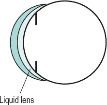
liquid l. The lens formed by the tear layer lying between the back surface of a rigid contact lens and the cornea. It must be taken into account when fitting contact lenses. If the back surface of the lens is steeper than the cornea, the liquid lens is positive and the eye is made more myopic. If the back surface of the lens is flatter than the cornea, the liquid lens is negative and the eye is made more hyperopic. (Fig. L14) Syn. fluid lens; lacrimal lens; tear lens.
low water content l. A contact lens whose water content is less than 50%.
luxation of the l. See luxation.
magnifying l. A converging lens used to magnify an object without image inversion. Syn. magnifying glass.
See magnifier.
l. measure Instrument for determining the radius of curvature of a spherical or cylindrical surface, based on measuring the sag of the curve. The instrument shaped like a pocket watch consists of two fixed, pointed prongs attached to the edge and a movable one located halfway between the two. The sag of the surface in the meridian containing all three prongs is measured by the linear displacement of the central one. The instrument is calibrated to give a reading in dioptres of the surface power for that meridian which has been calculated usually using the refractive index of crown glass (n = 1.523). If the material has a different refractive index, the true surface power, FT, in dioptres, is given by the following formula
where n is the refractive index and FLM is the reading, in dioptres, shown on the lens measure (Fig. L15). Syn. lens clock; spherometer (the old models had three outer fixed prongs instead of two).
See focimeter; neutralization; vertex depth.
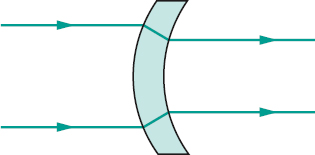
meniscus l. Lens with one spherical convex surface and the other spherical concave. Meniscus lenses often have a base of 6 D for the surface of lesser curvature. Syn. curved lens; bent lens.
meridional size l. See lens, aniseikonic.
microscopic l. See lens, telescopic.
mid-water content l. A contact lens whose water content is between 50% and 65%.
monocentric l. See monocentric.
multifocal l. A lens with various dioptric powers such as a bifocal, a trifocal, or a progressive lens.
negative l. See lens, concave.
l. nucleus See lens, crystalline.
objective l. See objective.
ophthalmic l. Any lens used to correct refractive errors of the eye. Sometimes it also includes lenses used to measure the refractive error.
ortho-k l. A rigid gas permeable contact lens used temporarily to reshape the cornea and change its power resulting in a reduction of myopia, or hyperopia. Most ortho-k lenses for myopia are based on a reverse-geometry design in which the central portion of the posterior surface of the lens is flatter than the average normal cornea and the peripheral portion is steeper. The design is the opposite if the ametropia is hyperopia. The lens is usually worn only at night. Significant improvement occurs within a month. The process is reversible when contact lens wear ceases.
See lens, reverse-geometry; orthokeratology.
orthoscopic l. A lens corrected for peripheral aberrations.
See ellipse, Tscherning.
l. paradox A hypothetical decrease in the power of the crystalline lens due to a continuum of refractive index changes in the layers of the ageing crystalline lens, such that it compensates for an increase in lens power which would otherwise occur due to an increase in lens thickness and lens curvatures (and therefore myopia). This is proposed as an explanation for the fact that the refractive state of the eye actually shifts towards hyperopia (or less myopia) with age.
l. pattern See former.
periscopic l. A spherical lens in which the minus lenses have base curves of +1.25 D and the plus lenses have base curves of –1.25 D. Meniscus lenses are more curved.
photochromatic l. See lens, photochromic.
photochromic l. A lens used either in sunglasses or as an ophthalmic lens. It is made up of a glass material, which changes in colour and/or in light transmission as a result of changes in incident light intensity or heat. The changes are reversible and relatively rapid. There exist several types, which are known by their trade name (e.g. Photogray Extra, Reactolite Rapide). Syn. photochromatic lens.
See vignetting.
piggyback l. A combination of two contact lenses; usually a rigid contact lens over a hydrogel lens. The soft lens is used for comfort and the rigid lens for best visual results. Piggyback lenses can be used after corneal surgery, corneal scarring and in the management of severe keratoconus or irregular astigmatism. Syn. combination system.
See lens, combination; lens, therapeutic contact.
plano l. See lens, a focal.
planoconcave l. A lens with one plane and one concave surface.
planoconvex l. A lens with one plane and one convex surface.
plastic l. Lens made of transparent plastic. It is approximately 50% lighter than a glass lens of equal power but more liable to scratching. Plastic lenses do not shatter like glass lenses and therefore give better protection.
See CR-39; glass, safety; lens, safety.
point-focal l. See lens, best-form.
polarizing l. A lens that transmits light waves vibrating in one direction only. In the other direction perpendicular to it, the light waves are absorbed. In this way reflected glare is reduced. Common polarizing materials include herapathite crystal, calcite
See light, polarized.
posterior chamber intraocular l. See lens, intraocular.
prismatic l. A lens with prism power.
prism ballast l. See ballast.
prismatic effect of a l. See convergence, correction induced; prism, induced.
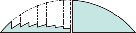
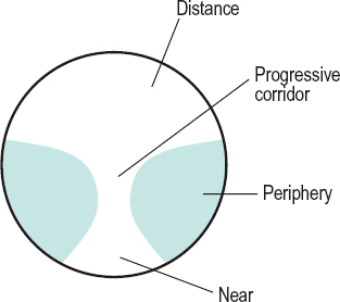
progressive addition l. (PAL) A spectacle lens having a gradual and progressive change in power either over the whole lens or over a region intermediate between areas of uniform power. The progression is produced by a complex aspheric shape of one of the surfaces. This lens is used to correct presbyopia. (Fig. L16) Syn. varifocal lens.
See distance, interpupillary; lens, gradient-index; lens, multifocal.
reading l. A lens prescribed for near vision (or a magnifying glass).
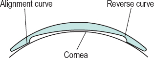
reverse-geometry contact l. A contact lens in which the back optic zone radius (BOZR) is flatter than the cornea. This zone is surrounded by a mid-peripheral curve, which is steeper than the cornea (some lenses omit this reverse curve), a peripheral curve. which is aligned with the peripheral cornea, and edge clearance. This lens design can be used in orthokeratology to control myopia, in the management of keratoconus, after keratoplasty or photorefractive surgery in which the corneal front surface is flatter than normal and there is partial alignment with the BOZR (Fig. L17).
See lens, ortho-k; optic zone radius, back.
1. rim artifact An apparent reduction in the extent of the visual field when perimetry is carried out with the patient’s correction. The greatest reduction is usually found with the standard trial case lens. It is even more noticeable in the elderly, possibly because many of them have deep-set eyes that increase the distance from the lens to the eye. With the patient’s own spectacles the artifact is less extensive. It is usually nonexistent when wearing contact lenses.
safety l. 1. A lens made of safety glass.
2. A general term referring to any lens that protects the eyes against injury due to impact and which is more resistant to fracture and less likely to splinter than an ordinary glass lens. Examples: plastic lenses (especially polycarbonate); toughened lenses; laminated lenses. Plastic lenses have the greatest impact resistance of all these lenses.
scleral contact l. A contact lens that fits over both the cornea and the surrounding sclera. It is divided into an optical zone and a scleral (or haptic) zone. When made of PMMA material it includes small holes (called fenestrations) to aid tear exchange and corneal oxygenation. Modern scleral contact lenses are made of gas-permeable material and do not require fenestration. Scleral contact lenses are used mainly therapeutically for corneal protection, tear retention and pain relief in ocular surface disorders, vision improvement as in keratoconus and after penetrating keratoplasty or occasionally for theatrical and sports purposes.
sealed scleral contact l. A scleral contact lens made up of gas permeable material. The optic portion of the lens has a curvature such that there is no contact between the lens and the cornea and that space (called corneal clearance) is filled with saline water forming a fluid pre-corneal reservoir. There is no tear exchange and corneal oxygenation is essentially provided through the lens matrix, as there is no fenestration. This lens is used in patients with corneal ectasia, such as keratoconus, keratoglobus and pellucid marginal degeneration, dry eyes of various aetiologies (e.g. Stevens–Johnson syndrome), etc.
semi-finished l. An ophthalmic lens of which only one surface is completely polished. The other side can be surfaced to any required curvature. If it is a bifocal lens, the side with the segment is usually the one that is completely surfaced. Syn. semi-finished lens blank.
See lens blank; lens, finished.
silicone hydrogel l. A contact lens made of elements of silicone and a hydrogel polymer to form a co-polymer that has the properties of both. A hydrogel-forming monomer such as HEMA is combined with a modified silicon-containing monomer such as TRIS (tris (trimethylsiloxy) silylpropyl methacrylate) or a derivative of TRIS. Such a lens has a high oxygen permeability, adequate wettability, good optical properties and acceptable lens movement on the eye (unlike basic silicone rubber material, which tends to adhere to the cornea). It can be used for extended wear as contact lens-induced oedema is virtually eliminated. Permeability (Dk) of such lenses exceeds 100.
See lens, therapeutic contact; modulus of elasticity.
single-vision l. (SV) An ophthalmic lens having only one power.
size l. See lens, aniseikonic.
slab-off l. A lens in which a portion of one surface has been ground with the same radius of curvature as the rest of the surface, but with separated centres of curvature. That portion of the lens produces a prismatic effect. Slab-off lenses are used most commonly to correct an induced vertical imbalance as, for example, in a patient with anisometropia looking through the lower part of the spectacle lenses when reading.
spectacle l. Any lens used as a correction or protection and mounted a short distance from the eye, usually in a frame but also as a lorgnette, pince-nez, monocle, etc.
See lens, ophthalmic.
l. speed See f number.
spherical l. A lens in which the two surfaces are spherical.
spherocylindrical l. A lens with one surface spherical and the other cylindrical. It has two different refractive powers in its two principal meridians.
See lens, astigmatic; lens, cylindrical; lens, toric.
spin-cast contact l. A contact lens produced by placing a quantity of liquid mixture into a concave spinning mould with polymerization taking place during rotation. The curvature of the mould determines the front optic zone radius, whereas the speed of rotation and other factors determine the curvature of the back optic zone.
See lens, cast-moulding contact; lens, lathe-cut contact; moulding.
steep l. A contact lens in which the back optic zone radius is shorter than the flattest meridian of the cornea. This definition may not be valid for a soft lens, which may fit steeply even when its back optic zone radius is not shorter than the flattest meridian of the cornea. Syn. tight lens (it is preferable to use this term for soft lenses).
See blanching, limbal; contact lens acute red eye; fitted on K; lens, flat.
stigmatic l. See lens, anastigmatic.
Stokes l. A lens consisting of two planocylinders of equal and opposite power mounted with their flat surfaces almost in contact with each other in a cell, and geared to rotate equally in opposite directions from a zero setting. At that setting, the two cylinder axes coincide and produce the minimum astigmatic value. When the lenses are rotated so that the two cylinder axes are at right angles to each other the lens produces the maximum astigmatic value. Intermediate settings of the two cylinder axes result in intermediate astigmatic values. This lens is used in some ophthalmic instruments.
subluxation of the l. See luxation of the lens.
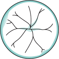
l. sutures Radiating lines in the crystalline lens formed by the meeting of lens fibres. Some of these systems of lens fibres form in the fetus or infant a Y, the arms of which are separated by angles of 120°. The anterior Y is upright and the posterior one is inverted. In the adult eye the systems of lens fibres make up more complicated figures, but the Ys present at birth usually persist throughout life (Fig. L18).
See fibres, lens; lens, crystalline.
telescopic l. A thick lens system forming a galilean telescope used to magnify the image. It is mounted in some form of frame and is lighter than an actual telescope. It is used to help low vision patients for either distance or near vision, although for the latter the lens system (or sometimes a single lens) is often referred to as a microscopic lens. therapeutic contact l. A contact lens, usually hydrogel, which acts as a protective device for the cornea, as in entropion, trichiasis, or damage by application of a tonometer; as a pressure bandage to relieve pain, as in bullous keratopathy; to facilitate corneal healing, as in corneal erosion due to trauma; to improve vision during the healing process; and as a delivery mechanism for drugs, since soft contact lenses placed on the eye can slowly release a drug which was previously absorbed. Silicone hydrogel lenses, which can be worn for up to one month at a time, are an efficient type of therapeutic contact lens. Gas permeable contact lenses are used therapeutically for extremely irregular corneas and some ocular surface disorders (e.g. keratoconus). Because of the risks associated with therapeutic lenses, it is important to follow the patient very assiduously. Syn. bandage lens.
See dystrophy, Cogan’s microcystic epithelial; keratitis, Thygeson’s superficial punctate; lens, combination; lens, ortho-k; lens, piggyback; lens, scleral.
thick l. See lens, thin.
l. thickness caliper See caliper.
thin l. A lens or combination of lenses in which the refracting surfaces are regarded as coincident, that is in which the separation between surfaces does not appreciably alter the total power of the system. Most ophthalmic lenses are thin lenses, but the crystalline lens of the eye is considered to be a thick lens. For optical purposes contact lenses are also regarded as thick lenses.
Thorpe four mirror fundus l. See slit-lamp.
tight l. See lens, steep.
tinted l. An absorptive lens having a noticeable colour and absorbing certain radiations more than others.
See bleaching; lens, absorptive; lens, cosmetic contact; photophobia; sunglasses; transmission curve.
toric l. This is usually a meniscus-type lens with a toroidal convex or concave surface. A toroidal surface is a surface with meridians of least and greatest curvature located at right angles to each other.
See lens, astigmatic; lens, meniscus; lens, spherocylindrical.
toughened l. A lens made of glass that has been either thermally or chemically strengthened.
See glass, safety; lens, safety.
trial l. 1. A lens used in a trial case. 2. A trial contact lens.
trifocal l. A multifocal lens consisting of three portions of different focal power usually for distance, intermediate and near vision (Fig. L19).
See portion, intermediate.
varifocal l. See lens, progressive.
ventilated l. See lens, fenestrated.
Volk l. See slit-lamp.
l. washer A plastic material which may be inserted between a loose lens and the eyewire of a frame to make the lens fit into the frame more securely. Syn lens liner.
wide-angle l. A lens giving good resolution over a wide field of view, usually used in better quality cameras.
Wilson three mirror fundus l. See slit-lamp.
Wollaston l. Lens based on the values provided by Wollaston of the Tscherning ellipse.
See ellipse, Tscherning.
X-Chrom l. Trade name for a dyed hard corneal contact lens aimed at enhancing colour perception in red-green deficient people. It is usually fitted on the non-dominant eye. The lens has maximum transmission above 575 nm with some additional transmission below 480 nm. Wearing this lens shifts the original absorption spectrum of the eye with the contact lens towards longer wavelengths. Objects may thus appear slightly different than with the other eye. This difference between the two eyes may, in certain conditions, improve colour discrimination.
See lens, ChromaGen.
Zeiss l. A type of goniolens, which when placed over the eye allows viewing of the anterior chamber angle. The Zeiss lens differs from other goniolenses in so much that it contains four mirrors, allowing visualization of the entire anterior chamber angle without the need to rotate the lens.
See gonioscopy, indirect; lens, gonioscopic.
zoom l. A lens in which the components can be adjusted to provide continuously variable magnification while the image remains constantly in focus.
Lenscorometer See distometer.
Lensometer See focimeter.
lenticele See phacocele.
lenticonus A conical projection of either the anterior or posterior surface of the crystalline lens of the eye, occurring as a rare congenital anomaly. The anterior lenticonus is the most common and most often associated with Alport’s syndrome. If the bulging is spherical, instead of conical, the condition is referred to as lentiglobus. It produces a decrease in visual acuity and irregular refraction that cannot be corrected by either spectacle or contact lenses.
lenticular astigmatism See astigmatism, lenticular.
lenticular fossa See fossa, hyaloid.
lenticular lens See lens, lenticular.
lenticular progressive degeneration See disease, Wilson’s.
lenticular stereoscope See stereoscope, Brewster’s.
lenticule A disc-shaped piece of corneal tissue or a piece of synthetic material manufactured to produce a given curvature and thickness. It is implanted into or on top of the cornea to change its anterior curvature.
See epikeratoplasty; keratophakia.
lentiglobus See lenticonus.
lentis ectopia See luxation of the lens.
lesion Localized, pathological change in a tissue due to injury or disease.
letter acuity See acuity, visual.
leucocorea See leukocoria.
leucokeratosis See dyskeratosis.
leucoma See leukoma.
leucoplakia See dyskeratosis.
leukocoria A condition characterized by a whitish reflex within the pupil. It is secondary to cataract, Coats’ disease, retinoblastoma, retrolental fibroplasia, persistent hyperplastic primary vitreous, etc. Syn. white pupil; white pupillary reflex. Note: also spelt leucocorea, leukocorea or leukokoria.
leukoma Dense, white, corneal opacity caused by scar tissue. A localized leukoma appears as a whitish scar surrounded by normal cornea. A generalized leukoma involves the entire cornea, which appears white, often with blood vessels coursing over its surface. Visual impairment depends on the location and extent of the leukoma. If the opacity is faint, it is called a nebula. Note: also spelt leucoma.
See hyperacuity; ulcer, corneal.
levator aponeurosis; palpebrae superioris
See muscle, levator palpebrae superioris.
levo Terms with this prefix can be found at laevo-.
levobunolol hydrochloride See beta-blocker.
levocabastine See antihistamine.
levofloxacin See antibiotic.
library spectacles See spectacles, library.
lid See eyelids.
lid eversion See eversion, lid.
lid retractors Term used to refer to the muscles that open the eyelids. In the upper eyelid it is the levator palpebrae muscle and its two divisions; the striated levator aponeurosis and the smooth Müller’s muscle (or superior tarsal muscle). In the lower eyelid it is the smooth fibre bundle derived from the inferior rectus muscle that forms the inferior tarsal muscle.
See muscle, levator palpebrae superioris; muscles, Müller’s palpebral.
lidocaine hydrochloride (lignocaine hydrochloride) A local anaesthetic of the amide type used in eye surgery. It is used in 1–4% solution. Its action starts in less than 1 minute and lasts about 1 hour.
ligament A tough, flexible band of white fibrous tissue that connects the articular extremities of bones, or supports an organ in place.
check l. A strong band of connective tissue which leaves the surface of the sheath of the extraocular muscles and attaches to the surrounding tissues, so as to limit the action of the muscle. The medial rectus is attached to the lacrimal bone (medial check ligament) and the lateral rectus to the zygomatic bone (lateral check ligament). There are also check ligaments restricting the vertical movements but the expansions of these muscles are thinner and less distinct than those of the horizontal recti muscles.
hyaloideocapsular l. See ligament of Wieger.
l. of Lockwood The lower part of the capsule of Tenon’s capsule and parts of the tendons of the inferior rectus and oblique muscles which are thickened to form a hammock-like structure on which the eyeball rests.
palpebral l. Strong connective tissue attaching the extremities of the tarsal plates of the upper and lower eyelids to the orbital margin. There are two sets: (1) the lateral palpebral ligament (lateral canthal tendon) about 7 mm long and 2.5 mm wide which constitutes the deeper portion of the lateral palpebral raphe of the orbicularis muscle and attaches the tarsal plates to the lateral orbital tubercle (Whitnall’s tubercle) on the zygomatic bone and (2) the medial palpebral ligament (medial canthal tendon) which attaches the medial ends of the tarsal plates to the frontal process of the maxilla and another insertion into the posterior lacrimal crest. It lies anterior to the canaliculi and the lacrimal sac.
suspensory l. A ligament whose principal function is to support another structure, e.g. ligament of Lockwood, the zonule of Zinn.
l. of Wieger An attachment of the anterior surface of the vitreous humour to the posterior lens capsule in the shape of a ring about 8–9 mm in diameter. It forms a line called Egger’s line. This adherence is strong in youth but weakens with age enabling intracapsular cataract extraction without pulling the vitreous. Syn. hyaloideocapsular ligament.
light Electromagnetic vibration capable of stimulating the receptors of the retina and of producing a visual sensation. The radiations that give rise to the sensation of vision are comprised within the wavelength band 380–780 nm. This band is called the visible spectrum or visible light. The borders of this band are not precise but beyond these radiations the visual efficacy of any wavelength becomes very low indeed (less than 10–5).
See coherent sources; infrared; lens, absorptive; spectroscope; spectrum, electromagnetic; spectrum, visible; Table C4; theory, quantum; theory, wave; ultraviolet; wavelength.
achromatic l. See achromatic light stimulus.
l. adaptation See adaptation, light.
artificial l. Any light other than natural light.
beam of l. A collection of pencils arising from an extended source or object. Syn. bundle of light.
bundle of l. See light, beam of.
l. chaos See light, idioretinal.
cold light Any visible light emitted by a process other than incandescence such as lasers, glow worms, certain chemical reactions, etc. Cold light is free of infrared.
compound l. Light composed of more than one wavelength.
diffuse l. Light coming from an extended source and having no predominant directional component. Illumination is thus relatively uniform with a minimum of shadows.
See diffusion; source, extended.
fluorescent l. Light emitted by fluorescence as in a fluorescent lamp. Electricity excites a gas that produces ultraviolet light, which in turn causes a phosphor coating on the inner surface of the fluorescent tube to fluoresce and emit visible light. Examples: mercury vapour lamp, neon and argon lamps, sodium vapour lamp, xenon flash lamp.
frequency of l. See hertz; spectrum, electromagnetic; wavelength.
idioretinal l. A visual sensation occurring in total darkness that is attributed to spontaneous nervous impulses in the neurons of the visual pathway. Syn. intrinsic light; light chaos.
incandescent l. Light emitted by incandescence as in an incandescent lamp. An electrical current passes through a thin filament (e.g. tungsten) enclosed in a sealed oxygenfree glass bulb. The filament is heated and photons are released.
See lamp, filament; lamp, halogen.
infrared l. See infrared.
intrinsic l. See light, idioretinal.
monochromatic l. Light consisting of a single wavelength or, more usually, of a narrow band of wavelengths (a few nanometres).
natural l. Light received from the sun and the sky.
pencil of l. A narrow cone of light rays coming from a point source or from any one point on a broad source after passing through a limiting aperture. A pencil of light may be convergent, divergent or parallel. The ray passing through the centre of the aperture is the chief ray. Syn. homocentric bundle of rays; homocentric pencil of rays.
polarized l. Ordinary light is composed of transverse wave motions uniform in all directions in a plane perpendicular to its direction of propagation. Polarized light is composed of transverse wave motions in only one direction, called the plane of vibration. Polarized light can be obtained by using a polarizer (e.g. tourmaline crystals, polarizing material such as Polaroid, Nicol prism, etc.).
See analyser; angle of polarization; crystal, dichroic; lens, polarizing; polarizer; prism, Wollaston; vectogram.
quantity of l. Product of luminous flux and its duration. Unit: lumen-second.
See lumen.
l. reflex See reflex, corneal; reflex, pupil light.
solar l. Light from the sun or having identical properties as the sun.
See blindness, eclipse; light, white.
l. source Any source of visible radiant energy such as natural light (e.g. daylight, moonlight, sunlight) or artificial light (e.g. a candle flame, an incandescent lamp, a discharge lamp, a fluorescent lamp).
See coherent sources; illuminants, CIE standard.
speed of l. The currently accepted figure is 299 792.5 km/s (in a vacuum). This velocity decreases, differentially with wavelength, when the radiation enters a medium.
See index of refraction; spectrum, electromagnetic.
l. stop See diaphragm.
stray l. Light reflected or passing through an optical system but not involved in the formation of the image such as that reflected by the surfaces of a correcting lens. Syn. parasitic light.
l. threshold See threshold, light absolute.
ultraviolet l. See ultraviolet; light, Wood’s.
visible l. See light; spectrum, visible.
white l. Light perceived without any attribute of hue. Any light produced by a source having an equal energy spectrum will appear white after the eye is adapted. Some of the CIE illuminants are often used as a source of white light, e.g. B, C and D. Sunlight is a source of white light.
See chromaticity diagram; spectrum, equal energy.
Wood’s l. Ultraviolet light near the visible spectrum which, when used with certain dyes such as fluorescein, causes fluorescence. It is produced by a special type of glass (called Wood’s glass or Wood’s filter), which contains nickel oxide and transmits ultraviolet radiations near the visible spectrum. It is used to detect corneal abrasions and to evaluate the fit of hard contact lenses. It is available in a slit-lamp or in a Burton lamp.
See fluorescein; fluorescence; lamp, Burton.
light-stress test See test, photostress.
lightness Attribute of visual sensation in accordance with which a body seems to transmit or reflect diffusely a greater or smaller fraction of the incident light. This attribute is the psychosensorial correlate, or nearly so, of the photometric quantity luminance (CIE).
See luminance.
lignocaine hydrochloride See lidocaine.
limbal furrow See furrow, limbal.
limbus, corneal The transition zone, about 1.5 mm wide, between the conjunctiva and sclera on the one hand, and the cornea on the other. Syn. corneoscleral junction.
See corneal epithelium; blanching, limbal; Vogt’s white limbal girdle.
limen See threshold.
liminal Pertaining to a threshold.
limit of resolution See resolution, limit of.
line 1. The connection between two points. 2. In anatomy, a long narrow band or streak that is distinct from the surrounding tissues by colour or texture.
base l. The line joining the centres of rotation of the two eyes. It is approximately equal to the interpupillary distance (Fig. L20).
See distance, interocular.
demand l. The line in Donders’ diagram that represents the perfect amount of convergence required for each level of accommodation, for single binocular vision. Syn. orthophoria line; Donders’ line.
l. of direction Line joining an object in space with its image on the retina (allowing for the optical properties of the eye). The line joining the fixation point to the fovea is called the principal line of direction. However, the object appears to lie along a visual direction and that direction in visual space associated with the fovea is called the principal visual direction. All other visual directions associated with other retinal points are called secondary visual directions. The principal line of direction and the principal visual direction coincide, but the former indicates the direction towards the eye, while the latter indicates the direction away from the eye.
Table L3
Common spectral lines of the visible spectrum
| designation | origin | wavelength (nm) |
| A | oxygen | 759.4 |
| C | hydrogen | 656.3 |
| C’ | cadmium | 643.8 |
| D | sodium | 589.3 |
| d | helium | 587.6 |
| e | mercury | 546.1 |
| F | hydrogen | 486.1 |
| F’ | cadmium | 480.0 |
| G | calcium | 430.8 |
| h | hydrogen | 410.2 |
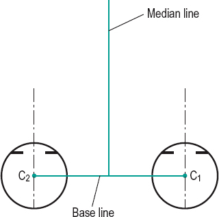
See law of identical visual directions.
Egger’s l. See ligament of Wieger.
Ferry’s line A line containing iron located in the corneal epithelium and adjacent to limbal filtering blebs. It may occur as a result of trabeculectomy.
l. of fixation See axis, fixation.
Fleischer’s l. See ring, Fleischer’s.
focal l. Any astigmatic optical system produces two mutually, usually perpendicular, focal lines of a point object. The focal lines are situated at different image distances.
Each focal line lies parallel to its associated cylinder axis. Syn. image line; line focus; Sturm’s line.
See circle of least confusion; Sturm, conoid of; Sturm, interval of.
Fraunhofer’s l’s. Fine dark lines distributed throughout the length of the solar spectrum due to the absorption of specific wavelengths by elements in the atmosphere of the sun and the earth. Fraunhofer observed about 600 of these lines and denoted the most prominent ones by letters from A in the extreme red to K in the violet. Examples: A corresponds to 759.4 nm, C to 656.3 nm, D to 589.3 nm, F to 486.1 nm, etc.
See constringence; spectrum, solar.
l. of Gennari A distinctive white stripe in the middle of the fourth layer of the visual cortex containing the termination of the optic radiation fibres, as well as intracortical connections. The stripe appears above and below the calcarine fissure except in the most anterior part of the fissure where it only appears below. Syn. area striata; stria of Gennari.
See area, visual; fissure, calcarine.
Hudson–Stähli l. A yellowish-brown, more or less horizontal line containing iron which runs across the cornea below the centre. It occurs in normal corneas, more frequently in the elderly, or in association with corneal opacities.
iron l. Deposits of iron within the corneal epithelium appearing as a brown rust line. They often subside with time leaving a nebulous scar. Several eponymous types have been described depending on the cause and location.
See line, Hudson–Stähli; line, Stocker’s; ring, Coat’s white; ring, Fleischer’s; siderosis bulbi.
median l. Line formed by the intersection of the median plane and the plane of regard (Fig. L20). Syn. midline.
orthophoria l. See line, demand.
phoria l. On Donders’ diagram, it is the line joining all the points representing the passive position of the eyes corresponding to various levels of accommodation.
principal l. of vision See line of sight.
pupillary l. See axis, pupillary.
retinal l. Operationally, the collection of retinal elements that are activated in response to a line stimulus.
Sampaolesi’s l. A pigmented, wavy line anterior to Schwalbe’s ring and found mainly in the periphery of the inferior cornea. It may be noted with gonioscopy in pigmentary dispersion syndrome and pseudoexfoliation syndrome.
l. of Schwalbe See ring of Schwalbe, anterior limiting.
l. of sight Line joining the point of fixation to the centre of the entrance pupil. This line is more practical than the visual axis. Syn. principal line of vision.
l. spectrum See spectrum, line.
Stocker’s l. An abnormal line containing iron located in the corneal epithelium which may appear in front of the advancing edge of a pterygium.
line-spread function See function, line-spread.
linear magnification See magnification, lateral.
linear perspective See perspective, linear.
lipid droplet degeneration See keratopathy, actinic.
lipofuscin Yellowish-brown pigment granules formed as a result of oxidation of protein and lipid residues, and found in various tissues (e.g. liver, kidney, heart muscle, adrenals, nerve cells). It normally accumulates with age within the lysosomes of cells and its accumulation in the retinal pigment epithelium (RPE) is a major risk factor of age-related macular degeneration as it may damage RPE cells and lead to the formation of drusen and RPE atrophy. In albinos the pigment granules are immature and colourless.
liquid paraffin See tears, artificial.
Lisch nodule See nodule, Lisch.
lissamine green A vital stain with dyeing quality similar to that of rose Bengal, but which causes less discomfort. It stains dead or degenerated epithelial cells green and is used to facilitate the diagnosis of keratoconjunctivitis sicca, xerophthalmia, etc. It has a molecular weight of 577.
Listing’s law; plane; reduced eye See under the nouns.
lithiasis, conjunctival See concretions, conjunctival.
lobe, occipital Portion of each cerebral hemisphere posterior to the parietal lobe where visual information is received and processing begins. See area, visual.
local sign See direction, oculocentric.
localization Perception of the location of an object in space with respect to either the eye (oculocentric localization) or the self (egocentric localization).
See egocentre; oculocentre; pointing, past-.
lock, binocular The part of the visual input that is common to both eyes and thus helps to maintain fusion. Syn. fusion lock.
See heterophoria, associated.
Lockwood’s ligament See ligament of Lockwood.
lodoxamide See mast cell stabilizers.
log MAR chart See chart, log MAR.
log MAR crowded test See Glasgow acuity cards.
long ciliary nerve See nerve, long ciliary.
long sight See hyperopia.
loop A circular bend or fold in a vessel or a bundle of nerve or muscle fibres.
Archambault’s l. See loop, Meyer’s.
Axenfeld’s intrascleral nerve l. An anomalous loop of the long ciliary nerve, often accompanied by the anterior ciliary artery, which has pierced the sclera a few millimetres behind the limbus and then reenters the sclera and supplies the ciliary body. It is usually pigmented giving rise to a 1–2 mm black dot on the sclera. It appears only in a small percentage of individuals (10–15%). Treatment is not necessary. Syn. Axenfeld’s pigmented nerve loop.
Meyer’s l. A bundle of inferior nerve fibres that originate from the lateral portion of the lateral geniculate body, extend forward around the anterior tip of the temporal horn of the lateral ventricle and then swing backward toward the occipital lobe. A lesion in this loop may cause a superior homonymous quadrantanopia. Syn. Archambault’s loop.
See radiations, optic.
loratadine See antihistamine.
lorgnette Eyeglasses for occasional use, held before the eyes by a handle, into which the lenses may fold when not in use (British Standard).
See lens, spectacle; spectacles.
lorgnon A spectacle lens for occasional use, mounted on a handle (British Standard).
loteprednol etabonate See antiinflammatory drug.
Lotmar Visometer See maxwellian view system, clinical.
Lotze’s local sign See direction, oculocentric.
loupe See magnifier.
loupe magnification See magnification, apparent.
loupe, Berger’s A binocular loupe fitted with a headband.
low tension glaucoma See glaucoma, normal-tension.
lubricant, ocular See tears, artificial.
lug See endpiece.
lumen 1. SI unit of luminous flux. It is equal to the flux emitted within a unit solid angle of one steradian by a point source with a luminous intensity of one candela. Symbol: lm. 2. The space in the interior of a tubular organ, such as an artery.
See flux, luminous; light, quantity of; lux; SI unit.
Table L4
Approximate luminance (in cd/m2) of some objects
| sun | 109 |
| car headlight | 107 |
| incandescent lamp (tungsten) | 106–107 |
| fluorescent lamp | 104–105 |
| clear sky at noon | 104 |
| cloudy sky at noon | 103 |
| shady street by day | 103–104 |
| full moon | 103 |
| book print under artificial light | >102 |
| photopic vision | >10 |
| street illumination | 1–10–1 |
| mesopic vision | 10–10–3 |
| cloudless night sky with full moon | 10–2 |
| scotopic vision | <10–3 |
| moonless and cloudless night sky | 10–3–10–6 |
luminance Photometric term characterizing the way in which a surface emits or reflects light in a given direction. It is equal to the luminous intensity measured in a given direction divided by the area of this surface projected on a perpendicular to the direction considered. Symbol: L. Units: candela per square metre (SI unit); footlambert; lambert, etc.
See brightness; candela per square metre; footlambert; lambert; lightness; millilambert; nit; photometry; SI unit.
luminaire A complete lighting unit consisting of a light source, a housing, enclosure, etc.
luminescence Emission of light by certain substances resulting from the absorption of energy (e.g. from electrical fields, chemical reaction, or other light), which is not due to a rise in temperature (unlike incandescence). The emitted radiation is characteristic of the particular substance. When the light emitted is due to exposure to a source of light the process is usually called photoluminescence. When the light emitted is due to either a high-frequency discharge through a gas, or to an electric field through certain solids such as phosphor which is used in fluorescent lamps, television picture tubes, etc., it is called electroluminescence.
See bioluminescence; fluorescence; incandescence; lamp, fluorescent; phosphorescence.
luminosity See brightness.
luminous 1. Emitting or reflecting light. 2. Having the capacity of stimulating the photoreceptors.
luminous efficacy; flux; intensity See under the nouns.
luminosity curve See efficiency, spectral luminous.
lumirhodopsin See rhodopsin.
Luneburg’s theory See theory, Luneburg’s.
lupus erythematosus, systemic
See keratitis, peripheral ulcerative; keratoconjunctivitis sicca; scleritis.
lustre 1 . The effect of one colour appearing to be situated behind and through another. This can occur when looking in a haploscope when it is called binocular lustre. 2. Appearance of glossiness on a metallic surface.
lux SI unit of illuminance. It is the illuminance produced by a luminous flux of one lumen uniformly distributed over a surface area of one square metre. Symbol: lx.
luxation of the lens Pathological and complete dislocation of the lens relative to the pupil. If the luxation is incomplete it is called subluxation of the lens (or dislocation or ectopia lentis). Subluxation is one of the causes of monocular diplopia. If the luxation is complete the eye becomes markedly hyperopic and is unable to accommodate. Luxation occurs in contusion of the globe, in many ocular (e.g. buphthalmos) and other diseases (e.g. syphilis) or it can be inherited (e.g. the bilateral, symmetrical, superior subluxation commonly found in Marfan’s syndrome or homocystinuria). It is sometimes associated with ectopic pupils and keratoconus. Unless there are complications (e.g. secondary glaucoma) or monocular diplopia the lens is left in place and management is optical. (Fig. L21)
See corectopia; iridodonesis; pupillary block.
luxometer See photometer.
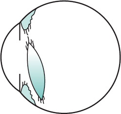
lymphadenopathy An enlargement of a lymph gland. The preauricular lymph node located 1 cm in front of the external ear drains the orbital region and is sometimes involved with eyelid and conjunctival infection (e.g. adult inclusion conjunctivitis, follicular conjunctivitis). Syn. adenopathy (although strictly speaking this term refers to the enlargement of any gland).
lymphoma, B cell A malignant tumour found predominantly within the orbit and conjunctiva. Orbital lymphomas are the most common form of ocular lymphomas and cause proptosis and diplopia. They affect mainly adults and are primarily of the B cell/non-Hodgkin’s type. Conjunctival lymphomas, also mainly of the B cell/non-Hodgkin’s type, are seen in approximately 10% of systemic lymphoma patients. Clinically, conjunctival lymphomas appear salmon-pink in colour and have flat and smooth surfaces. There are several types of B cell lymphoma, one of them being Burkitt’s lymphoma. Treatment is by radiotherapy.
lysozyme An antibacterial enzyme present in the tears (as well as other tissues). In human tears, lysozyme makes up 21–25% of the total protein.


