49. Klippel-Feil Syndrome
Definition
Klippel-Feil syndrome is a rare congenital disorder in which any two of the seven cervical vertebrae are absent or fused, resulting in a short neck with limited range of motion.
Classification of Klippel-Feil Syndrome
| Type I | Massive fusion of the cervical spine |
| Type II | Fusion of one or two cervical interspaces |
| Type III | Thoracic and/or lumbar spine anomalies in conjunction with Type I or II |
Incidence
The true incidence of Klippel-Feil syndrome is not known.
Etiology
The true etiology of Klippel-Feil syndrome is not fully understood. There are various suggestions as to the etiology of this malady, including congenital failure of segmentation of the cervical vertebrae, which results from failure of segmentation of cervical somites during the third through eighth weeks of gestation. Some have suggested the occurrence of some manner of global fetal insult, whereas others suggest the malady may be caused by some manner of vascular disruption.
Signs and Symptoms
For anomalies, see box below.
Anomalies Associated with Klippel-Feil Syndrome
• Brainstem anomalies
• Congenital heart disorder (most commonly ventricular septal defect)
• Congenital scoliosis
• Craniosynostosis
• Dyskinesia
• Renal disease (horseshoe kidney, renal ectopia, bilateral tubular ectasia, hydronephrosis, absent kidney)
• Sprengel’s deformity (elevation of scapula)
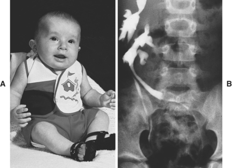 |
| Klippel-Feil Syndrome. A, Short neck (fused cervical vertebrae and nonfunctioning right thumb caused by lack of tendons to this digit. Intravenous pyelogram (B) reveals crossed renal ectopia of the left kidney, whereas the ureter from the left kidney crosses the midline and inserts into the left side of the trigone. |
• Abnormal hand position
• Ataxia
• Blurred vision
• Bruxism
• Decreased cervical range of motion (rotational loss)
• Difficulty swallowing
• Disturbed phonation
• Dizziness
• Duane syndrome (eye contracture)
• Facial asymmetry
• Facial nerve palsy
• Headache
• Hearing loss
• Hydrocephalus
• Hypermobility of unaffected interspaces
• Hypoplastic thumb
• Lateral rectus palsy
• Low hairline
• Micrognathia
• Neck webbing
• Nystagmus
• Oligodontia of deciduous permanent teeth
• Palatal clefts
• Ptosis
• Restrictive mouth opening
• Short neck
• Supernumerary digits
• Syndactyly
• Temporomandibular joint dysfunction
• Torticollis
• Upper extremity hypoplasia
Medical Management
Because of instability of the cervical spine, quadriplegia and even death can occur as the result of relatively minor trauma. The patient is therefore cautioned to strictly avoid contact sports, particularly those that place the neck at particular risk. Mechanical symptoms may be treated with a cervical collar, analgesics, nonsteroidal anti-inflammatory drugs, or even judicial application of cervical traction.
Further medical treatment regimens may be indicated for associated systemic anomalies. Treatment regimens should involve a medical specialist for that particular anomaly (e.g., cardiologist, nephrologist, urologist). Surgical intervention(s) may be indicated for several reasons, such as neurologic deficits, persistent pain, and spinal stenosis.
Complications
• Ataxia
• Death
• Quadriplegia
• Seizures
• Spinal cord compression
• Syncope
Anesthesia Implications
Before anesthesia the patient should undergo a thorough series of x-rays of the head and neck. A complete evaluation of any associated systemic anomaly must be completed as well. Clinical manifestations may result either from compression of the cervical spine, the pons, or medulla or from stretching of cranial nerves. Basilar artery compromise may occur as the result of sudden neck rotation and syncope may ensue.
One of the highest priorities for the anesthetist is positioning, more specifically positioning of the patient’s head and neck throughout anesthesia care. The patient’s head and neck should be maintained in neutral alignment at all times. Quadriplegia and death have been reported with even minor trauma in patients with Klippel-Feil syndrome. The patient should demonstrate his or her neck range of motion to delineate any resulting symptoms that may arise.
The patient scheduled for general anesthesia may prove difficult to intubate because of the decreased neck range of motion. The anesthetist, in conjunction with the patient, must decide preoperatively whether the intubation will be performed with the patient awake or asleep. The advantage of an awake attempt would be the patient’s ability to communicate the onset of any problematic sequelae during the intubation attempt. Placing the patient in the “sniffing” position for intubation may actually cause harm. To maintain neutral alignment it may be necessary to enlist the aid of another anesthetist to apply gentle cervical traction during laryngoscopy. The anesthetist must abort or abandon direct laryngoscopy if the patient’s head and neck cannot be maintained in neutral alignment throughout the intubation attempt. It may be necessary to forgo direct laryngoscopy in favor of the fiberoptic bronchoscopic direct visualization.
For either intubation attempt, the oropharynx should be very well anesthetized to negate the patient’s gag reflex. The vocal cords should be anesthetized using local anesthetic injected via the cricothyroid membrane. Some sedation would be appropriate for this procedure, but the anesthetist should be mindful of the great need for patient cooperation and communication throughout this attempt. Once the airway is secured, the patient may receive the scheduled general anesthetic induction medication doses. Muscle relaxation should be achieved using nondepolarizing muscle relaxants. As with the intubation, patient positioning for the surgical procedure must primarily focus on maintaining neutral axial alignment of the head and neck.
Emergence and extubation for the patient require good communication among anesthetist, patient, and surgeon. Consensus should be obtained before anesthesia concerning this process. The optimal situation is for the patient to be fully in control of airway reflexes and not coughing or “bucking” on the endotracheal tube, thus risking potentially fatal sequelae. On the other hand, extubating the relatively deeply anesthetized patient presents the risk of airway obstruction in the patient with a potentially difficult airway.
Regional anesthesia, specifically continuous subarachnoid anesthesia, has been reported to be successful. Once again, good communication is paramount. Intravenous sedation for anxiolysis may be given with the regional anesthetic, but such administration must be judicious— even “stingy”— to avert airway obstruction. Should airway obstruction occur, the anesthetist must take great care while alleviating that obstruction not to induce extreme movement of the patient’s neck, particularly axial rotation, and so avoid very deleterious sequelae.