Pelvic Kidney
Synonyms/Description
Etiology
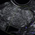
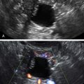
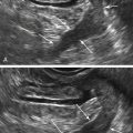
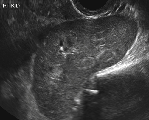
Ultrasound Findings
Differential Diagnosis
Clinical Aspects and Recommendations
Suggested Reading
Cinman N.M., Okeke Z., Smith A.D. Pelvic kidney: associated diseases and treatment. J Endourol. 2007;21:836–842 Review.
Debenedectis C.M., Levine D. Incidental genitourinary findings on obstetrics/gynecology ultrasound. Ultrasound Q. 2012;28:293–298.
Hall-Craggs M.A., Kirkham A., Creighton S.M. Renal and urological abnormalities occurring with Müllerian anomalies. J Pediatr Urol. 2011;28:27–32.
Meizner I., Yitzhak M., Levi A., Barki Y., Barnhard Y., Glezerman M. Fetal pelvic kidney: a challenge in prenatal diagnosis? Ultrasound Obstet Gynecol. 1995;5:391–393.
Yildirim I., Irkilata H.C., Aydur E., Zor M., Basal S., Goktas S. Different clinical presentations of pelvic ectopic kidneys: report of two cases and review of the literature. Urologia. 2010;77:212–215 Review.